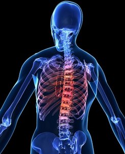
Computed tomography of the lumbar without surgery. Contraindications for the study. Indications for CT of the spine
A healthy spine is the key to a long and active motor activity a person, which is why even with the slightest pain or injury to this part of the body, it is worthwhile to immediately diagnose. One of the most reliable methods that is used for this purpose in modern medicine, - computed tomography of the spine. The data from this study are often used in neurosurgery and neurology. What is this diagnostic method, how is CT of the spine performed, does it require special training - you will find answers to these questions in our article.
However, the main limitation is the lack of interventional tissue differentiation at the cervical and spinal levels. Magnetic resonance imaging is a non-invasive, ionizing, harmless examination that uses radio frequency waves in a strong magnetic field to generate images, and its high spatial resolution allows accurate evaluation of soft tissues within the body. spinal canal, the main asset. This is very safe study which may sometimes require an intravenous injection of a contrast agent, gadolinium.
What are the features of CT diagnostics?
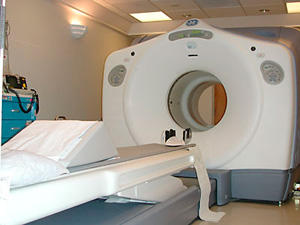
Computed tomography of the spine involves layer-by-layer scanning of this part of the body in order to assess its parameters and identify foci of pathologies. Tomography is based on a computer evaluation of different levels of X-ray radiation, which varies depending on the density of the scanned tissue. During the procedure, you can carefully examine the spine from different angles and in different spatial planes, which allows you to create a three-dimensional image of this part of the body. For this study, an apparatus is used, which is called a tomograph, consisting of a mobile table and a research unit with x-ray sensors.
Its main indications in the examination of the spine are postoperative evaluation, the difference between disk material and tumor of the nervous tissue, dural or intramedullary pathology, evaluation of the metastatic column, spondylodisitis or spinal stenosis.
How is the preparation for CT?
The main contraindications for magnetic resonance are the presence of a pacemaker, some cochlear implants, metallic material in the orbital areas, some catheters or intracranial surgical clamps or heart valves dating back several years, as now the latest surgical equipment can sometimes be a limitation.
To whom is scanning indicated and contraindicated?
CT of the spine today is an indispensable diagnostic method, it is used much more often than ultrasound, MRI or x-rays. A doctor may prescribe a tomography of the spine for prevention purposes, especially for professional athletes and those citizens whose profession is associated with heavy loads on the back. Also, this study is prescribed as a screening if the patient complains of frequent headaches. CT scan of the spine can be used to monitor the condition of this area after punctures, as well as in emergency cases. But still, most often with the help of CT, diseases are diagnosed and pathological processes on this part of the body.
Evaluation of the cervix or dorsal column is markedly superior to magnetic resonance as it allows better differentiation of intra-articular soft tissues; disk, roots, body pouch and spinal cord and can also evaluate the bone marrow. In the presence of a hernia, bone marrow involvement may be a predictor of clinically significant or painful discomfort, in contrast to clinically silent but clinically silent hernias, allowing evaluation of the various components of the hernia. spinal stenosis.
Examination of the lumbosacral region
This is a global analysis, but given its ease of access, it is useful and may reveal clinically significant abnormalities. The choice to proceed with CT or MRI will depend on several factors, including availability. costs, age of the patient, duration of symptoms.
Indications for the appointment of CT:
- Spinal tomography is scheduled for without fail if there is a suspicion of the presence of tumors in this part of the body.
- In the presence of .
- If the physician has concerns about the occurrence of an abscess of the spine.
- In the event that neoplasms in the vertebrae and adjacent tissues were previously diagnosed, but their nature was not established.
- Then, when the location of the vertebrae and their motor function is in doubt.
- When pathologies of the spinal cord are established.
- In the event that a deformation of the processes of the nerve in this area of \u200b\u200bthe back has already been fixed.
- During diagnostic and therapeutic procedures for diseases of the circulatory system.
- If the spine was injured, especially if, subsequently, a fall or blow, the movements began to cause pain.
- Sometimes used to assess condition spinal column after surgery.
- With stenosis of the spinal canal.
- Computed tomography of the head and spine is also used for severe headaches, which are accompanied by vomiting and loss of consciousness.
- In order to scan the bones of those who are sick.
What diseases can this scan detect?
Often the patient may not attach importance aching pains in the back, attributing it to the consequences of physical labor or general fatigue. But if the necessary series of studies is not carried out in time, then the disease can be triggered. Most spinal abnormalities can be corrected medical method or during complex therapy that does not include surgical intervention. But it is relevant only for diseases identified on early stages If you ignore the pathology of the spine for years, then you will not be able to do without surgery. Computed tomography of the spine is effective in identifying such diseases:
In the presence of acute pain in a low back resistant to conservative treatment, both methods may be considered to demonstrate a hernia. In the presence of low back pain with significant clinical signs such as sphincter disorder, extensive neurological deficit, antecedent neoplastic, steroid use, trauma, infection index, magnetic resonance are choices.
When there is chronic low back pain without signs of infection or neoplasia, it is most often degenerative with or without spinal stenosis. Two display options are available depending on availability, cost, or contraindications. Computed tomography is an excellent test for detailing facet joints, ligaments flavum and their hypertrophic changes and their impact on spinal canal gauge and foramen. Spinal stenosis is often multifactorial and evaluation of its various components is importance to guide treatment choices.
- Injury to any part of the spine.
- Various deformations of the vertebrae or changes in their structure.
- At the first scan, it will confirm or refute the diagnoses and.
- It is used to diagnose birth defects of this part of the body.
- Able to show a pathological change in the spinal lumen.
- Recognizes all types of tumors, both benign and malignant.
- Able to show the presence of intervertebral hernias.
Contraindications for the study
Although the patient receives a much smaller dose of radiation during the procedure than with a standard x-ray, such a scan is contraindicated in the following situations:
Magnetic resonance is the criterion of choice for assessing postoperative pain syndromes in the lumbar spine, which allows differentiating recurrent hernia and postoperative fibrosis and assessing the effects of arachnoiditis. The study of medullary pathology, whether cervical, dorsal or conical medullary, is carried out by magnetic resonance. It is the only imaging modality that allows evaluation and has excellent sensitivity. Magnetic resonance allows diagnosing demyelinating lesions, myelomalacia, myelitis various etiologies, intramedullary or dural tumor.
- Pregnancy at any time, as well as breastfeeding.
- Intolerance to iodine-containing drugs, as this component is used in CT with contrast.
- For those who have too big weight or the procedure will not bring harm to the dimensions, but they run the risk of simply not fitting into the device.
- Harmless, but not effective CT for detailed and very detailed scanning of the spinal cord. In this situation, it is better to use MRI diagnostics.
- Such research may weaken general state health of patients and,.
- The method is contraindicated for those who suffer from serious heart diseases.
- Difficult to use for young children, as it requires complete immobility of the body.
How to prepare for a scan?
If you are going to have a CT scan with a contrast agent, then you should refuse any food at least 6 hours before the scan. Tomography of the lumbosacral spine will be difficult if there is an accumulation of gases in the intestines, so it is worth giving up gas-forming products about a day before CT. And on the evening before the procedure, you need to do an enema. When examining the lower part of the spine with ascertaining women, a tampon with this substance is inserted into the vagina. If the scan is performed in the upper part of the spine, then you need to deny yourself not only food, but also fluid intake at least an hour before the tomography.
The production of images requires the intervention of a computerized data processor. The x-ray emitter rotates around the patient, and the detector on opposite side collects an image of the patient's area; the patient's bed flows very precisely and definitely within the scanning tunnel, presenting a different area of the body on each circle. The image sequences along with the viewing angle information are processed by a computer which displays the result on a monitor.
This result consists of a series of sections, not necessarily contiguous with a given thickness: the set of reconstructed sections is the scan volume data, which can be reconstructed with 3D rendering software to generate tomographic images of any spatial plane, or alternatively, to obtain 3D or endoscopic images .

Types of computed tomography of the spine
Depending on which part of the spine is examined, four types of CT are distinguished:
Cervical scan
CT cervical of the spine easily recognizes different tissue densities corresponding to the norm and pathologies. It is in this part of the spine that degenerative-dystrophic processes are most often diagnosed, including protrusions. CT of the cervical spine allows you to assess the condition of the cervical part of the spinal column and identify foci of diseases. On the pictures obtained as a result of the scan, you can see how healthy the body of the intervertebral canal, as well as its processes. it good way for diagnosing the spinal cord, but not the best for scanning ligaments and intervertebral discs. CT of the cervical spine during the first procedure will show tissue injuries and fractures of the bone part.
To obtain tomographic images of the patient from raw scan data, the reconstruction computer uses complex mathematical image reconstruction algorithms. Most important processes obtaining images from the raw data are convolution and rear projection. or vice versa.
The opening images of all sections are usually recorded on the feed system, and the most important sections are sometimes printed on film. Detector high efficiency usually consists of cesium iodide, calcium fluoride, cadmium tungstate. The first generation tomograph was based on the emission of a linear beam of X-rays emitted by an X-ray tube moving in direction and rotation and detected by the detector's motion integral.
Thoracic examination
CT thoracic of the spine is most often prescribed if there are suspicions of tumors in the lungs, as well as with neoplasms of the pleura and mediastinum. It will clearly show whether there are metastases, what structure they have and what is the area of the lesion. Also, CT of the thoracic spine is used to scan the lymph nodes and to detect different types inflammation. This method is often used to assess the nature of the injury, to confirm the presence or absence of foreign objects in the lungs. But this type of CT has the most contraindications, since the radiation is directed to vital organs.
The fourth generation tomographs had fixed sensors and were abandoned. This method allows imaging in a continuous manner: while the table that moves the patient on a sliding plane, the scanning planes describe a "spiral" around the patient, producing a "spiral" scan.
The most common spiroid scanners perform one more or less one second rotation and allow full body volume acquisition in 40 seconds to one minute: this occurs in one apnea, reducing patient movement artifacts. Modern multilayer tomographs can take only a few seconds, giving dozens of scans for each rotation. Super-fast tomographs can allow you to study the heart.
CT scan of the lumbar
CT lumbar spine is exact method study, which will easily determine any abnormalities in the bones and soft tissues this part of the body. Based on the results of the scan, a reliable conclusion can be made about whether there are developmental anomalies, injuries, pathological formations or foci of inflammation in the lumbar region. Effective in detecting tumors and disorders in the circulatory system.
Image diagnostic service. It is indicated for the study and diagnosis of fractures, hernias, arthritis, spondylosis, cysts, benign and malignant tumor lesions, vascular malformations, structural abnormalities and paramorphisms. This can be done with or without contrast material.
There are no absolute contraindications to the performance of this exam. However, since x-rays are used, it is not recommended for pregnant women, presumed or established. For this reason, patients childbearing age should only be used if pregnancy has been excluded. If a contrast agent is used, contraindications are: severe renal, hepatic, and cardiac failure, proteinuria, monoclonal gammopathy, acute myocardial ischemia, out-of-systolic arrhythmia, contrast agent allergy, and uncontrolled diabetes.
Examination of the lumbosacral region
CT scan of the lumbosacral spine allows you to assess whether the lumen of the spinal canal is normal. The pictures of this department show the state of the final part of the spinal cord and nerve roots. This type of scanning is not the most effective for diagnosing diseases of the ligamentous apparatus and intervertebral discs. Most often, it is prescribed after an x-ray to clarify its results and make a more accurate diagnosis. This type of study is often prescribed by a doctor if a patient has a fracture of the spine, tumors of any type are diagnosed. It is used to monitor the dynamics of such diseases: rheumatic diseases of the spine, hemorrhage in the spinal cord, instability of the vertebrae and canal stenosis.
The technology of this sophisticated device has revolutionized diagnostic imaging, allowing for a thorough and detailed evaluation of the anatomical regions being examined. How to perform The exam is neither invasive nor painful. No problem with claustrophobia. The exam may also be performed with a contrast medium that is injected into the patient through a small needle into a vein in the crease of the elbow. Once the exam is completed, the patient does not have to follow specific prescriptions and can immediately resume their normal activities.
Preparation The exam does not require special preparation, except when done with a contrast medium. In this case, you should fast for at least 6 hours and avoid fluid intake. It is also necessary to conduct pre-contrastographic studies to study the function of the kidneys, heart and liver. In addition, any allergies should be reported to the radiologist if a reaction to a contrast agent has occurred in the past.
Tomography is called a research technique human body, at which translucence occurs in various intersecting planes. The first method of tomography was X-ray, but it was not widely used, as it provides a meager amount of information. With the development of technology, a method has been developed computed tomography(CT), which makes it possible to maximize the detail of the resulting image.
It is recommended to pass all examinations made earlier for the comparative assessment of reports and documentation related to the current investigation. Image of the spine. detailed scan and check of the spine. Costs, computed tomography and magnetic resonance imaging.
Computed tomography of the spine
This CT was obtained by irradiating the X-ray skeleton and then digitizing the data. In preparation for the exam, the patient abstains from food - 4 hours before the image of the spine can not be. Immediately before the images, metal objects such as glasses, jewelry, prostheses, etc. must be removed. Those patients who are afraid of confined spaces are advised to take a tranquilizer before the test. At severe pain in the back, they may take pain medications.
Why do you need a CT scan?
CT scan of the spine is indicated in the presence of back pain, with suspicion of pathological changes in the vertebrae, spinal cord, nerve processes and blood vessels, etc.
Both computed tomography and magnetic resonance imaging are used to examine the spine. What type of study - or CT of the spine is suitable in a particular case, is determined by the doctor.
While the images are being scanned on the scanner, the patient moves the CT scan around the spine and photographs in the correct context. After the first series of photographs taken on the patient, contrast and half an hour of reaction are applied. If there is itching, rash, breathing problems, the image of the spine does not continue. If the contrast is not caused by allergies, the second series of photographs.
The result of the study are tomographic images in which the vertebrae are marked with white, soft tissue with gray and cerebrospinal fluid with black. In addition, the density of various tissues and different colors. When taking a spine tomography, the doctor recommends determining the causes of chronic and acute back pain, assessing the condition of the spine before and after surgery in order to diagnose cancer and metastases on the spinal discs.
What diseases can be detected by tomography?
With the help of CT of the spine in Moscow, you can determine the presence of such problems:
- spinal injury;
- structural changes in the vertebrae, that is, various kinds of deformities;
- the development of arthritis and;
- congenital defects of the spine;
- changes occurring in the spinal lumen;
- tumor neoplasms;
- intervertebral hernia etc.
When is research impossible?
It is impossible to make a CT scan of the spine in Moscow if there are one or more contraindications from the list below:
Magnetic resonance of the spine
In order to perform a spinal tomography using the method of a doctor with magnetic resonance, it is necessary to evaluate the condition of the spine as a whole to detect diseases of the spinal cord, postoperative changes in the spine, spinal disc conditions, inflammation and nerve compression, spinal infection, back pain, spinal cord or spinal cord tumors to determine the date of spinal surgery.
The patient is placed in a capsule, an electromagnetic field arises under the action of a molecule in the human body, as well as electromagnetic waves. Then start scanning the spine with radio waves. Continuous imaging of the spine no longer than 20 minutes.
- pregnancy;
- severe renal failure;
- weight over 150 kg;
- inadequacy of the patient;
- presence foreign bodies(prostheses, bandages) made of plaster or metal in the protection of the area under study.
Some aspects of preparation for the study
CT scan of the lumbar spine using contrast materials is contraindicated in the presence of an allergy to iodine or preparations containing iodine.
If a CT scan of the lumbosacral spine is required, then some preparation of the patient is needed to increase the reliability of the result. The day before the study, foods that increase gas formation should be excluded from the menu. On the evening before the tomography, it is necessary to put a cleansing enema. Women should insert a cotton swab, dry or moistened with a contrast agent, into the vagina. A CT scan of the spine should be performed with a full bladder.
CT scan of the cervical spine requires preparation only when contrast material is used. Preparation in this case consists of withholding food for six hours and fluids for one hour prior to the procedure. The same applies to the process of preparing for a CT scan of the thoracic spine.
Research cost
The price for a CT scan of the spine depends directly on the area of study, as well as on the type of irradiation procedure (with or without contrast material). In most cases, computed tomography is done by appointment.
How is tomography performed?
Computed tomography is performed in the supine position on a special moving table of the tomograph. If there is such a need, for the duration of the procedure, fixation with rollers and belts is possible to avoid accidental movements. When using a contrast agent, the drug prescribed by the doctor is administered to the patient intravenously.
During the study, only the patient remains in the room, but he can contact the technologist at any time if there are any problems. The tomography procedure itself consists in moving the table inside the tunnel. At the same time, the tomograph ring rotates around the table. The procedure is accompanied by a slight noise from the tomograph. The patient does not feel any sensation during the tomography.
Interpretation of the result
The results of the study are given in printed form by hand or recorded on a disk. As a rule, the doctor conducting the tomography immediately speaks of the presence of any pathology and advises which narrow specialist should apply for medical care:
- in the presence of fractures, consultation with a traumatologist is necessary;
- at pathological changes spinal cord and its nerve endings, you should contact a neurologist or neurosurgeon;
- any neoplasms require examination by an oncologist to exclude the presence of malignant tumors;
- other spinal lesions require examination by a rheumatologist.
The high popularity of computed tomography for diagnosing diseases of the musculoskeletal system can be explained by the fact that it is optimal for studying the spinal column and surrounding structures, because bone and cartilage tissue is perfectly visualized using x-rays. With this method of examination, patients are less likely to use additional contrasting, therefore allergic reactions on the introduction of a contrast agent do not occur. Radiation exposure to the body during CT is comparable to the radiation dose from a conventional x-ray.
By the way, you may also be interested in the following FREE materials:
- Free book "TOP 7 Bad Morning Exercises You Should Avoid"
- Restoration of knee and hip joints with arthrosis- free video recording of the webinar, which was conducted by a physiotherapist and sports medicine- Alexandra Bonina
- Free lessons for the treatment of lower back pain from a certified physical therapy doctor. This doctor has developed a unique system for the restoration of all parts of the spine and has already helped over 2000 clients with various back and neck problems!
- Want to know how to treat pinching sciatic nerve? Then carefully watch the video on this link.
- 10 necessary components food for healthy spine - in this report you will find out what should be daily diet so that you and your spine are always in healthy body and spirit. Very useful information!
- Do you have osteochondrosis? Then we recommend to study effective methods treatment of lumbar, cervical and thoracic osteochondrosis without medication.