Preparation for MRI head. To part of the doctors, you should address you consultations on magnetic resonance tomography as the cervical spine? What is the study
To get the most detailed and qualitative description of the results of the MRI, a radigenologist must provide all available medical documentation: Medical extracts, including after operations, snapshots and conclusions of previous MRI, ultrasound, MSCT. The more detailed information The radiologist has a radiologist before the study, the more clear task set before him. Also, previous results allow us to assess the dynamics of the course of the disease and the success of the treatment.
In most cases, no special training is required for MRI.
Nevertheless, going to the MRI, you should wear clothes without metal fittings (without lightning, rivets, rhinestones, etc.). If there is a metal on clothes, you will need to change clothes into a disposable shirt. Watch, glasses, decorations, piercing will have to remove. Also on the day of MRI girls are better not to use cosmetics, because there are metal as part of some cosmetics. Electrical appliances and plastic cards may fail under the action magnetic fieldTherefore, they should be left in storage chambers.
Some types of MRI should be prepared in advance.
Preparation for MRI small pelvis men and women
- Provide a doctor-radicalist with your medical records with your medical records before research: postoperative extracts, snapshots and conclusions of previous MRI, ultrasound, MSCT, also desirable direction of the attending physician.
- Women MRI small pelvis is held in the middle menstrual cycle (5-14 days from the first day of menstruation).
- The intestinal gas formation should be reduced. To do this, it is impossible to use gas-forming products, products with fiber within 2 days before MRI (cabbage, fruits, carbonated drinks, black bread, any dairy products. It is desirable to accept Espumizan in accordance with the instructions.
- Do not urinate 2 hours before the study. Additional fluid intake is not needed - bladder There must be moderate filling.
Preparation for MRI abdominal cavity
- A day before the procedure should be excluded from the diet, coarse fiber and products causing excessive gas formation (carbonated drinks, fruits, black bread, cabbage, fermented milk products, etc.).
- It is necessary to stop eating for 6 hours before the study.
- Reception 1-2 NO-SPAP tablets are desirable 30-40 minutes before the start of the study.
Preparation for magnetic resonance enterography (hydro-MRI of the small intestine)
- Within 2 days before the study, a slicing diet should be observed (eliminate fresh fruits, legumes, mushrooms, berries, fermented milk products).
- MRI is performed on an empty stomach or no earlier than 6 hours after the last meal.
- In the evening, the day before the study should make a cleansing enema.
- For 40 minutes, it is necessary to drink 2 liters of mannitol solution (FORTRAS): 150-200 ml each 5 minutes (issued during the study);
- Immediately before MRI intramuscularly introduces 1 ml of glucagon for temporary peristalistic suppression.
Preparation for MRI of the mammary glands
Special preparation for MRI of the mammary glands is not required. But when writing to the study, the date of procedures should be chosen so that it accounted for the first phase (from the 5th to the 15th day) of the menstrual cycle - this will provide even greater informationality of the MRI of the mammary glands.
The gravity of gravity and long non-passing pain in the head area can be a reason for a serious survey. The most accurate and informative method of diagnosis used in such cases is magnetically resonant tomography.
It allows you to get a reliable data array. According to MRI brain, which is needed to prepare for the procedure - this question is worried about many people who need a similar examination.
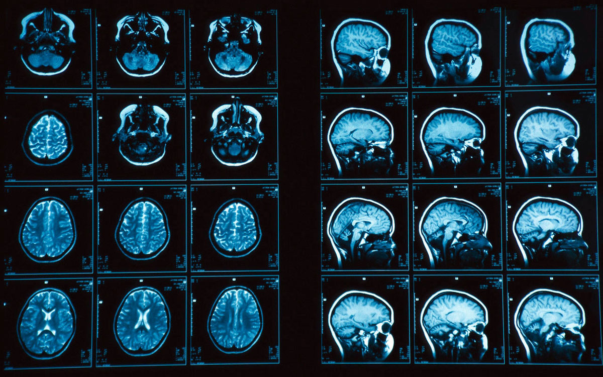
The diverseness of the information obtained during the MRI of the head is due to the fact that with this diagnostics there is a high-precision scanning of two dozen layers of the brain. Tomograph signals are processed and transformed into a three-dimensional image.
Magnetic fields and high-frequency pulses, with which the device operates, do not harm the human body, and it distinguishes tomography, for example, from a radiographic study.
The brain MRI procedure is quite expensive, so it is not conducted "for the sake of interest, just in case," and is strictly targeted.
For its passage, people are usually sent with suspicions of heavy pathology, including:
- tumors of soft tissues, including malignant;
- aneurysm and pathology of vessel shells;
- serious vision problems;
- strokes;
- pituitary diseases;
- chronic neurological diseases;
- head injuries, etc.
The method allows, if necessary, take urgent measures. To see the results, you do not need to wait for snapshots and printouts. An image of what is happening "inside the cranial box," you can immediately consider on the screen. Although the generalized conclusions, the doctor will provide only a few days later.
If the results require further analysis or clarification, the recording can be copied and pick up with them so that other specialists look at it on video.
To whom in the "Camera" can not
Despite the absolute harmlessness, MRI brain is allowed not to all people.
The main contraindication to the study is the use of various electronic equipment to maintain normal life and the presence of such metal objects as:
- dental crowns, prostheses;
- orthopedic corsets;
- pacemakers;
- metal implants inserted during operations on organs of vision or hearing.
On the presence of any of the above or other similar devices, it is necessary to inform the doctor to the diagnostic.
Among those who are not able to calmly postpone tomography, are people who are afraid of a closed space, and the kids under three years. Prolonged fixed presence inside tomograph will be a complex test for them.
Therefore, in some cases, a survey of anesthesia is practiced for this category of persons.
Women "in position" on early timing - low-taking candidates for MRI, but, starting from the 14th week of pregnancy, the ban is gradually losing relevance, and research Research can be done with the permission of the attending physician.
Patients with obesity, whose weight "stepped over" for 100 kg, will not be able to check on the tomograph for purely technical reasons - because of their more than impressive dimensions they simply do not fit inside the apparatus.
Preparing according to the rules
An intricate name diagnostic techniques Causes some prejudice to her from individual people. But their anxiety is in vain: side Effects It almost does not happen here, especially if the preparation for the MRI of the brain was carried out correctly.
No need to be surprised, but the main thing at the preliminary stage is in the positive and calm mood of man. Lack of psychological discomfort - pledge reliable result Surveys.
Preparing for "immersion in tomographic chamber" people with problem kidneys on the recommendation of the doctor can make analyzes - biochemical blood And total urine, in order to avoid unwanted complications.
For all who have a brain MRI, preparation is mainly due to "parting" with all metal objects and devices that enjoy ordinary life. Outside the diagnostic cabinet should be left:
- banking "plastic";
- mobiles, clock;
- glasses;
- lighters, knives;
- metal hairpins and studs, etc.
Even apply makeup to the face of women who are preparing for a survey cannot be. This is explained by the fact that decorative cosmetics Metal particles may be present, and this is fraught with a distortion of final data.
A thorough study will be subjected to the tattoo body, which can also contain metal.
As for the change of nutrition or refusal to receive drugs, this is not required from a person who is preparing for a "deep inspection".
How manipulation is passed
All that is associated with cerebral activity seems to be particularly fragile, vulnerable and sensitive. Therefore, going to any "headstore", patients often cannot get rid of concern. However, there is no reason for anxiety.
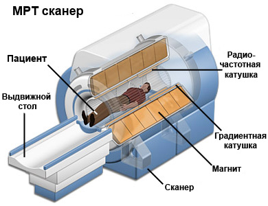
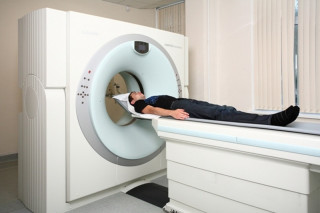
How do MRI brain do? Tune in to the fact that the whole process you have to spend in the lying position.
Showing the patient, as needed to lie on a special couch, doctors with the help of special devices fix the required position of its body. Then the couch "will go" into the tomograph chamber.
This machine is a bulk tube surrounded by magnetic elements. Special sensors that are located near the head will be suppliers of basic information. Data perceived by them immediately appear on the monitor.
It is important that the patient is calm during tomography, relaxed and did not move, - this will help get a high-quality and accurate image.
The procedure passes approximately for 30-45 minutes, after which the patient's couch is extended outward.
In people who passed such a study, no side effects are observed. Patients can return to their usual life without any preparatory actions.
"In contrast"

In some cases, in order to make the MRI brain even more informative, doctors use special contrasts.
What is needed to prepare for MRI brain with contrast? First of all, in detail to inform the doctors about the state of its health - the presence or absence of pregnancy, the inclination to allergies suffered by injuries, etc.
Otherwise, determining how to prepare for the MRI of the brain in a "contrasting format", do not differ from the "fees" for ordinary tomography.
For greater "brightness", research is usually used such a substance as a gadolinium. It has a property of making brain fabrics more sensitive to magnetic effects.
The drug is absolutely safe, and only occasionally made the procedure is accompanied by dizziness, skin item And nausea is a consequence of an allergic reaction.
More precisely
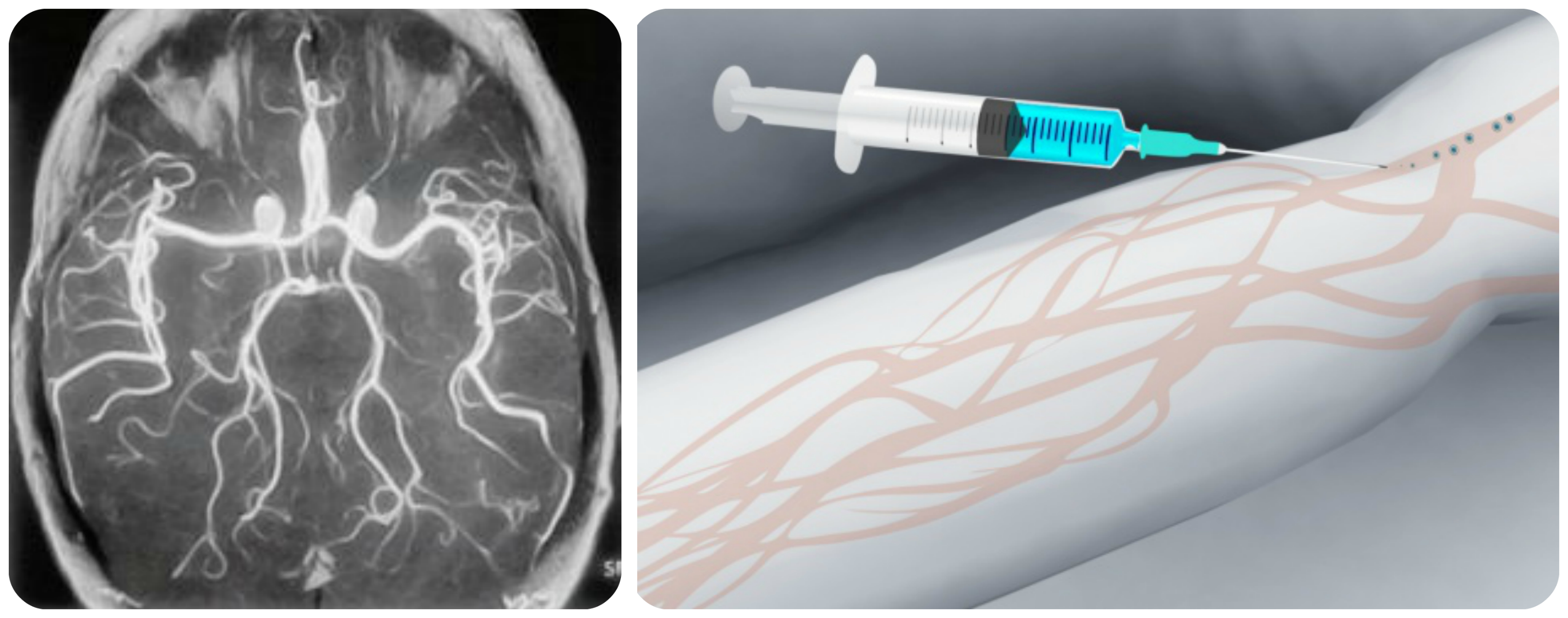
How is the MRI brain with contrast? In the Vienna on the hand of the patient, a special catheter with saline helper is introduced free passage Contrast preparation. Then the dose of gadolinium "on the sample" is introduced.
If everything passes normally, the introduction of this substance before MRI brain continues until the desired volume is reached.
It helps to consider even those tissues in detail, which were previously small.
The drug causes a pleasant chill, some tide of blood to the body, sometimes the feeling of "metal" in the mouth - do not be frightened: it is quite normal reaction organism.
In the appointment of a doctor, people passing tomography, sometimes spectroscopy, which shows biochemical reactions inside the cells.
Otherwise, how the MRI procedure is underway with Gadolinia does not have differences with the procedure for action with a conventional magnetic resonant tomographic study.
If a woman, nursing baby breasts, MRI brain is done with a contrast, for a day she should not give the breast baby.
Why is it important
The results of the MRI brain are always fixed on the digital carrier. This allows, if necessary, to see again, as each stage of the study passes, and do not refer to the patient with a request to pass the procedure again.
However, to track the dynamics of treatment, often conduct repeated studies of the brain. How the processes of recovery or development of the disease are undergoing, the doctor will be able to assess, comparing previous and subsequent visual results.
Experienced specialist who will take into account all the nuances and will take correct solution According to the organization of the procedure, must ensure that MRI passes and its preparatory phase.
To be examined, there are different features: there are not only completely closed tomographs, and and partially open.
With their help, they make tomography even too full people And those who suffer from claustrophobia.
Two methods: which is better
Among modern research methods, two types of tomography are distinguished by their special informativeness: computer and magnetic resonance (by the way, both of the cost are much more expensive than ordinary X-ray and ultrasound). But which one is better, more precisely and safer?
Of course, each of the procedures has its own undeniable advantages, but in terms of brainwriting, MRI is much more preferable to CT.
If in the first case the organs are studied using electromagnetic rays, then in the second - with X-ray.
Therefore, when conducting computer tomography The patient gets a small dose of radiation, which does not occur with a magnetic resonance study.
Today it is possible to sign up for MRI in public clinics, and in private medical centers. The choice of the place of the survey remains only for the patient.
Solving the question: "Where to go?", Always come to this responsibly and take into account the experience and qualifications of doctors, which are planning to contact, as well as the quality and level of equipment on which they work.
Competence and attention even to a minor nuances on the part of diagnostic specialists will raise the correct diagnosis at the very beginning of the disease and, without losing precious time, proceed to its treatment.
Magnetic resonance tomography is modern method Research in which very powerful magnetic fields, high-frequency pulses and a computer, allowing to recreate the clearest image of all tissues, organs and human body systems. This method of diagnostics gives the most accurate information from any site. human body.
The results can be printed, transfer to in electronic format, Move to any media information, which allows doctors to consult with colleagues without losing time. A thorough image of a problem area makes it possible to identifies even microscopic or starting pathology, which is impossible when using ultrasound, x-ray and computed tomography.
MRI is an indispensable method in differentiating diagnoses, early detection of oncology, which significantly increases the effectiveness of treatment and the percentage of survival.
It should be noted that the preparation for the study takes at very little time, and in many cases it is not needed at all.
Indications for MRI brain
MRI is considered to be safe method Surveys, because it does not cause any impacts on the body in the form of exposure. The method gives accurate information about the brain, which is an invaluable invention for the whole world. Most often with the help of magnetic fields, experts exclude or detect the following diseases or status:- ishmemic and hemorrhagic strokes;
- subarachnoid hemorrhage;
- anomalies in the development of the brain;
- aneurysm or others pathological changes brain vessels;
- disorders after the injury of the head;
- the cause of frequent headaches;
- chronic nervous system processes;
- the cause of convulsive syndrome;
- eye diseases, ears.
Clear visualization makes the diagnosis of tissue pathologies and an unmistakable head organs by 98% special preparation Not needed to MRI.
As mentioned earlier, special preparation for the passage of the MRI of the head is not required, but the recommendations and precautions still have:- clothing on the patient should not contain metal elementsotherwise the nurse must help him change to a medical displacement bathrobe;
- as this examination may include the introduction of a contrast agent, the doctor is obliged to find out the patient information about the presence of allergies;
- the fear of a closed space should be reduced by sedatives;
- all metal decorations must be removed;
- pregnant women can take a study only after the first trimester and, in case of emergency;
- the doctor should know all the information about the patient about finding metal implants in his body, because they can distort the accuracy of the research results.
The patient should know that when the magnetic resonance method cannot be stored in order not to distort the read data.
Only if the doctor gives the command to inhale or exhale, the examiner must clearly execute the command. Contraindication for the passage of MRI is the presence of a pacemaker in the body, driver heart Rhythm and other electronic technology built into the human body. Also, staying in the body of foreign objects in the form of clips, pins, prostheses, surgical brackets and other medical devices can cause questions about the passage of diagnostics in this way.
Before MRI brain can not hide from the doctor any important information about foreign objects, especially in the head of the head. A distortion in the diagnosis may be affected by simple seals made from metal or containing metal fragments.
Found a mistake? Highlight it and press Ctrl + Enter
If your doctor sent you to the passage of MRI, you do not need to be afraid, and fall into despair. MRI does not have negative influence on health and does not cause any pain sensations. Before passing the MRI procedure, you need to know some points to prepare.
MRI of the spine is a type of study at which it is not necessary to especially prepare for tomography. Before the passage of the MRI procedure is allowed to conduct the usual image life, no need to sit on special diet And abandon the reception of drugs. All that is needed is just your cheerful attitude to the passage of the procedure in a limited space and completely be fixed for thirty or forty minutes.
There is also no need for any special preparation.
It is not necessary to refuse drug intake, it is only necessary to limit food eating four hours before the survey. The survey is carried out on an empty stomach.
- , I.
This survey requires preparation only for MRI with a contrasting agent. The survey requires the exclusion of food four hours before its start.
For best visualization urinary tractThe bladder must be complete. For these purposes, one liter is required to drink one hour. clean water. Women in the period of menstruation should refrain from the passage of MRI.
Intestinal examination is carried out on an empty stomach. Four days before the examination, it is necessary to switch to a fermented diet, it is necessary to take enzyme preparations.
Preparation for tomography of the stomach and intestines is the most complex and consists of several points:
- A day before the study, it is necessary to refrain from the use of water with gas, dairy products, black bread, confectionery, fruits and vegetables. Such products cause gas formation;
- For four hours it is required to refuse to receive any liquid;
- For half an hour before the procedure, it is necessary to take an antispasmodic medicine.
It is necessary to know that during pregnancy in the first trimester, MRI is recommended to pass only in special casesyou must warn you medical workers O before the start of the survey.
Going to MRI You can not wear clothes with metal inserts: rivets, zippers, buttons. Before the procedure, you need to remove all metal items: hours, jewelry, jewelry, Piercing. Also sometimes you will be asked not to use cosmetics, because some cosmetics Contain metal. You can not take an electrical appliance on the procedure, credit cards, phones.
Health workers are required to notify the presence of metal or electronic objects in the body (implants, prostheses, pins, fragments, bullets, etc.). They may detain the equipment under the influence of the magnetic field, distort the results of the survey, as well as, which is important, is also harmful to the patient. It is also necessary to tell the health workers about the tattoos applied, as there are metal connections in some types of paints, and during the examination it may cause irritation.
Contrast preparations for MRI Research
Some types of intravenously. These substances are used primarily on the basis of gadolinium, they do not contain iodine, and do not cause the risk of adoptive effects and allergies.
Claustrophobia with magnetic resonance tomography
Some patients suffering from claustrophobia are experiencing various inconveniences. But you need to know that during the examination is always in contact with the health worker, which in any case will help you immediately. In particularly severe cases, you can bring a friend or relative with you, so that when passing the survey, it could be next to you.