Anatomy of the scapula and clavicle. Encyclopedia "Anatomy watch online. Human anatomy in pictures". Connections of the bones of the girdle of the upper limb
Scapular region I
Scapular area (regio scapularis)
part of the body, bounded at the top by a line drawn between the clavicular-acromial junction and the spinous process of the VII cervical vertebra, below by a horizontal line running through the lower edge of the scapula, by a medial-vertical line corresponding to the projection of the medial edge of the scapula, outside by the middle axillary line and the posterior edge deltoid. The end of the clavicle at the acromioclavicular joint. In this case, the scapula connects the upper limb to the body. The following diagram explains the relationship of the scapula to the rib cage. Scapula has two surfaces, three boundaries, three corners, and three processes. The scapula serves as an attachment site for many important muscles around the shoulder. Diseases... Often there is a so-called scapular crunch during movements in the shoulder joint, which is accompanied by moderate pain and a feeling of heaviness in the area of the scapula. Often the scapular crunch is due to chronic inflammation of the subscapularis bursa or exostosis of the scapula. In the latter case, surgical treatment is indicated for the removal of exostosis (see Osteochondrodysplasia) .
The coracoid process is a hook-like projection originating from the supolocal surface of the coastal scapula, which lies just below the collarbone. The surface of the pavement is marked with three longitudinal ridges. Another dense ridge adjoins the edge. This part of the bone is almost like a shaft. It acts as a lever for the action of the toothed apron in the upper arm. This surface gives attachment to the spine of the scapula, which divides the surface into a smaller supraspin fossa and a larger deep fossa. The two fossae are connected to each other, located at the root of the spine. The acromion is a projection of the spine that arches over the glenohumeral joint and articulates with the clavicle.
Osteomyelitis of the scapula develops after gunshot and other open injuries of L.O., accompanied by general symptoms of intoxication and local manifestations (dysfunction, etc.). Deep intermuscular leaks often occur, especially when the purulent process spreads to the anterior surface of the scapula (see Phlegmon) .
Surgical treatment in combination with antibacterial and immunostimulating therapy. for function is not always favorable. The pitcher steps back, completes, and the force through the lower body is coordinated by the paddle to throw the ball. This is depicted in the pitcher frame in the above image. The great overhead serve that contributed to your victory was not only made possible through hard work and practice, but also a key bone in your anatomy - which is more commonly referred to as the scapula and is critical to stabilizing and permitting athletic movements such as throwing, swimming, row. It is also important for daily activities such as lifting the box over your head or reaching for a blanket on the top shelf in your closet. Tuberculosis of the scapula is rare and only occurs in adults. More often, the acromial process and scapula are affected (see Extrapulmonary tuberculosis (Extrapulmonary tuberculosis) ,
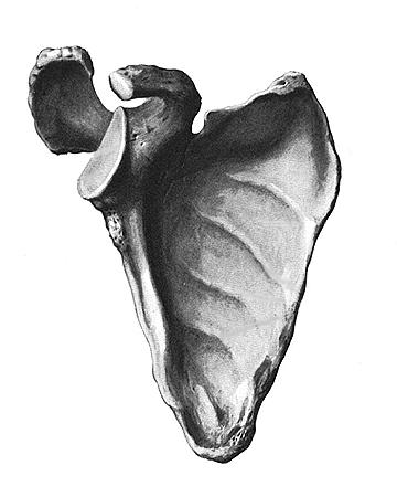
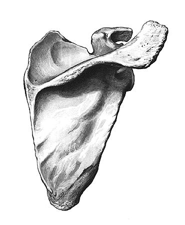
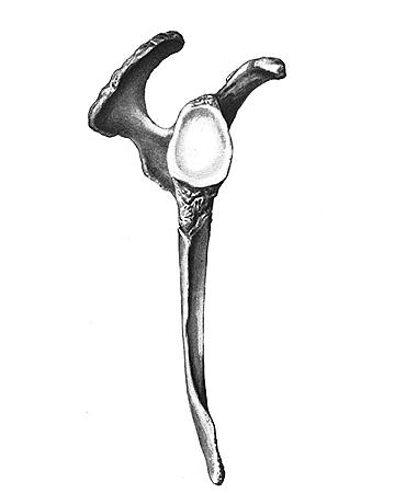
tuberculosis of bones and joints). Tumors... There are benign (, chondroma, osteoblastoclastoma) and malignant (, reticulosarcoma). The main role in their diagnosis is played by X-ray examination. If necessary, perform a puncture or open biopsy. Treatment is operative. In some cases, with malignant tumors, it is possible to perform interscapular-thoracic resection while preserving the upper limb. In general, the scapula coordinates great forces from your back and lower body into effective movement through your arm and arm. The shoulder blade is located on the back and has a triangular shape that looks like a blade, hence the common reference, the shoulder blade. These features make the flap look rather pointed and almost prehistoric - but worry not, the shoulder was designed to be highly functional. The rest of the blade is numbered and listed in the image below if you are interested. The scapula attaches to two other bones, the clavicle and humerus, and several joints and tendons. In addition, it serves as a platform for over 10 chest, arm and back muscles for attachment, including biceps and triceps. In other words, it would be very difficult to pump iron without a spatula! Operations... In the scapular region, the following operations are performed: osteotomy, resection of the scapula, interscapular-thoracic amputation and a number of other surgical interventions. Bibliography: Human Anatomy, ed. M.R. Sapina, vol. 1, p. 103, M., 1986; Volkov M.V. and Dedova V.D. Children's, p. 65, M., 1972, Kaplan A.V. bones and joints, p. 162, M., 1979, Operational and topographic, ed. V.V. Kovanova, s. 4. M., 1985; Friedland M.O. , with. 298, M., 1954; Chaklin V.D. Orthopedics, Vol. 2.c. 350. M., 1957. Together, the shoulder complex allows for a range of motion that is much greater than any other joint in the body. Because of its crucial role in movement, if you injure your scapula it can damage your mobility. Fortunately, scapular fractures are quite rare and most often only occur after blunt force trauma. In addition to being unable to raise your arm, other common fracture symptoms include shoulder swelling, bruising, and severe pain. To assess the extent and location of the injury, the doctor will usually order an X-ray evaluation, such as an X-ray. The fracture is then classified as a layer fracture, acromial fracture, or severity of the coracoid process. Typically, scapular fractures are associated with other injuries such as the spine, collarbone, or ribs; therefore, a doctor will need to do a thorough examination of all related areas, especially if you are involved in an accident. rear view): 1 - upper corner; 2 -; 3 - top edge; 4 - scapula notch; 5 - coracoid process; 6 - acromion; 7 - lateral angle; eight - ; 9 - lateral edge; 10 - bottom corner; 11 - medial edge; 12 - spine of the scapula "> Rice. 2. Right shoulder blade (rear view): 1 - upper corner; 2 - supraspinatus fossa; 3 - top edge; 4 - scapula notch; 5 - coracoid process; 6 - acromion; 7 - lateral angle; 8 - infraspinatus fossa; 9 - lateral edge; 10 - bottom corner; 11 - medial edge; 12 - spine of the scapula. Like most other bone fractures, there is no magic cure. Instead, treatment for a fractured scapula requires immobilization for up to 4 weeks, icing to control swelling, and pain relief to intervene for discomfort. In extreme cases, a doctor may have to perform surgery. Regardless of treatment, scapular fracture rehabilitation may be required. Physical therapy is recommended not only to restore the original range of motion, but also to strengthen the muscles, because strengthening the attached muscles can help reduce remaining pain and help it serve as a supportive cushion for the scapula as it heals. Rice. 4. Radiograph of the shoulder joint area (front projection) with comminuted fracture of the lateral angle of the scapula with displacement of the glenoid cavity. In general, the scapula is a critical bone in the shoulder, attached to several bones, tendons, and joints and serving as a platform for the muscles of the chest, back, and arms. This allows for a wide range of hand movements, such as picking up a child and swinging a tennis racket. Any damage to the scapula must be carefully diagnosed by a healthcare professional. In the event of a fracture, a scapula must be given for proper healing; otherwise, long-term immobility may result. The scapula, also known as the scapula, is a flat, triangular bone located at the back of the torso and located two to seven above the posterior surface. The scapula, along with the clavicle and the manual chest, constitutes what connects the upper limb of the appendicular skeleton to the axial skeleton. The scapula is an important bone as each scapula provides a point of attachment to several muscles that make up the arm and shoulder. It also articulates with the humerus and clavicle to form the acromioclavicular joint, respectively. Rice. 1. Right scapula (front view): 1 - acromion; 2 - the articular surface of the acromion; 3 - coracoid process; 4 - scapula notch; 5 - top edge; 6? upper corner; 7 - subscapular fossa; 8 - medial edge; 9 - muscle attachment lines; 10 - bottom corner; 11 - lateral edge; 12 - subarticular tubercle; 13 - lateral angle; 14 - glenoid cavity. However, since the medial aspect of the scapula is not directly attached to the axial skeleton, but rather is held in place and connected to the ribcage and spinal column by muscles, the scapula is free to move along the posterior chest wall. This allows the shoulder to move with the scapula, providing a wider range of motion and mobility for the upper limb compared to the lower limb. Anatomy, definition, and function of the scapula, also known as the scapula. Like any triangle, the scapula consists of three boundaries: superior, lateral, and medial. The top border is the shortest and thinnest border of the three. The medial border is a thin border and runs parallel to the vertebral column and is therefore often referred to as the vertebral border. The lateral border is often called the axillary border because it runs in the articular region towards the apex of the armpit. It is the thickest and strongest of the three boundaries for muscle attachment. (regio scapularis, PNA, BN A, JNA) - a paired area of the body, highlighted on the posterior surface of the chest, bounded from above by a line connecting the clavicular-acromial - I Shoulder (brachium) proximal segment of the upper limb. Its upper border is a circular line drawn at the level of the lower edges of the pectoralis major muscle and the broadest muscle of the back, the lower one runs along a circular line 5 6 cm higher ... ... Medical encyclopedia It also has a glenoid cavity or socket along this border, a shallow fossa that is pivotally connected to the head of the humerus, forming a glenohumeral joint. There are also three angles to the shoulder blade. The superior border meets the lateral border with the lateral angle and with the medial border with the superior angle. The third corner is the inferior corner where the medial and lateral borders meet. The blade has two surfaces; in the foreground there is a smooth coastal surface, which is concave in shape and is largely absorbed by the subscapular fossa. In the posterior part of the scapula there is a convex and uneven posterior surface that has a protruding bone ridge that unevenly divides it into two parts: the superior supraspin fossa and a much larger, lower vain fossa. SYPHILIS- SYPHILIS. Contents: I. History of syphilis ............... 515 II. Epidemiology ................. 519 III. The social significance of syphilis ........ 524 IV. Spirochaeta pallida ............., 527 V. Pathological anatomy ........... 533 VІ. ... ... VOICE LINKS- VOICE LINKS, two paired folds of the mucous membrane on the side walls of the middle part of the larynx (see), located sagi tagli from front to back one above the other. The upper folds, the so-called false vocal cords (chordae vocales falsae, s. ... ... Great medical encyclopedia Along with the spine, there are two other processes: the coracoid process and the acromion process. The coracoid process is a beak-like bend that protrudes outside the upper border. The surface, in comparison with the coracoid process, is a cavity in the form of a glenoid. The lateral part of the clavicle lies perfectly, and the supracapular notch, which connects the base of the coracoid process to the superior border, is medial to the coracoid process. The crustal process allows different muscles and ligaments to be attached. Coracoid process connections. Coracohumeral ligament - to the larger tubercle of the coracoclavicular ligament - to the clavicle. Coracoacromial ligament - to the acromion process. The acromion process is a palpable lateral and dilated extension of the posterior spine of the scapula, which protrudes the anterior end of the spine. It arches over the glenogumeral joint and is pivotally connected to the lateral acromial end of the clavicle to form the synovial-acromioclavicular joint. This connection is supported by the acromyoclavicular ligament, which attaches to the acromion process at one end and the clavicle to the other. I (thorax, pectus) upper body, limited by the upper and lower apertures of the chest. G.'s bone frame consists of the thoracic spine, ribs and sternum attached to it. This frame is called the chest, and together with ... ... Medical encyclopedia I Klippel Feil disease (M. Klippel, French neurologist, 1858 1942, A. Foil, French neurologist, born in 1884; synonymous with Klippel Feil syndrome) congenital malformation of the spine, characterized by deformity (shortening) of the neck, ... ... Medical encyclopedia Several arteries form an anastomosis to deliver blood to the dorsal scapula. The supracapular artery is a branch of the thyrocervical trunk, which, in turn, arises from the subclavian artery. It runs along the suprascapular nerve and mainly supplies the muscles of the supraspinate and infraspinate. The transverse cervical artery is a branch of the thyrocytic trunk moving along the medial border of the scapula. Because of the large surface area of the scapula, there are many attached muscles that anchor the scapula to the chest wall and allow it to move. I (angulum membri superioris) a set of bones (scapula and clavicle), connected to each other by the acromial clavicular joint, with the thorax by the sternoclavicular joints and muscles holding the scapula, and with a free upper limb ... ... Medical encyclopedia I Sprengel's disease (O.K. Sprengel, German surgeon, 1852 1915) see Scapular region. II Sprengel's disease (O.K. Sprengel) developmental anomaly: short and wide scapula, turned around its sagittal axis and receding from the chest in ... ... Medical encyclopedia These muscles are summarized below and subdivided based on the muscles that arise or are inserted into the scapula. Four of these muscles form the rotator cuff that covers the shoulder capsule. Its actions include flexion and medial rotation, abduction, extension and lateral rotation of the shoulder joint. It is responsible for abduction in the shoulder joint, and it is innervated by the supracapular nerve. Its action includes lateral rotation in the shoulder joint. The muscle is also innervated by the supracapular nerve. It is responsible for extending the elbow, and it is innervated by the radial nerve. Teresa is a minor muscle - it arises from the lateral border of the posterior surface. Its action consists of lateral rotation in the shoulder joint. This muscle is innervated by the axillary nerve. The main muscle Teresa - its origins - the posterior surface of the lower angle and the lower part of the medial border. Its role is to carry out adduction and medial rotation in the shoulder joint. It is innervated by the subscapularis nerve. It performs many actions such as adduction, extension, and medial rotation of the shoulder joint. It receives its innervation through the thoracodorsal nerve. Coracobrachial muscle - Its origin is a coracoid process. Its actions include adduction and flexion of the shoulder joint. It is innervated by the musculocutaneous nerve. This muscle is responsible for flexing the elbow. Subscapular muscle - it arises from the subscapular fossa. It performs adduction and medial rotation in the shoulder joint. The subscapularis nerve innervates it. Omochaeal muscle - Its origin is an excellent boundary and causes depression of the hyoid bone. Trapezius muscle - it is inserted mainly along the spine, acromion process and clavicle. Its actions include raising the scapula and rotating the scapula during its abduction - it is inserted over the scapular spine. Serration anterior muscle - its insertion along the medial border, from the upper angle to the lower angle. This muscle tightens and rotates the scapula. It is innervated by the long pectoral nerve. Rebellious pectoralis muscle - This is inserted into the coracoid process. Its actions consist of tightening and depression of the scapula. The muscle is innervated by the medial pectoral nerve. BLOOD VESSELS- BLOOD VESSELS. Contents: I. Embryology ................. 389 P. General anatomical outline ......... 397 Arterial system .......... . 397 Venous system ...... ....... 406 Table of arteries ............. 411 Table of veins .............. .. ... ... Great medical encyclopedia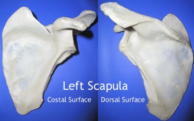
The scapula has two surfaces - the coastal surface, which abuts against the ribcage, and which can be felt when we palpate the bone from behind. The bony surface of the scapula is concave and directed medially and forward. A concave depression on most of its surface is called the subscapular fossa.Blade boundaries, angles and processes

The upper border of the scapula is thin and short. It has a cortical process near the root. The lateral border of the scapula is thick and is an infargenoid tubercle at the upper end. It emerges from the lower third of the rough strip to the border aspect. Inserted along the facet border of the carrier, from the corner to the root of the spine.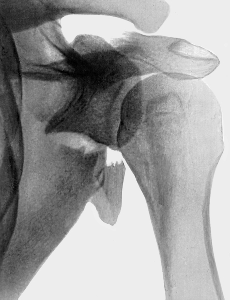

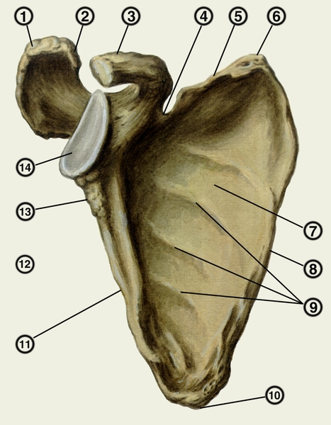 the articular surface of the acromion; 3 - coracoid process; 4 - scapula notch; 5 - top edge; 6? upper corner; 7 - subscapular fossa; 8 - medial edge; 9 - muscle attachment lines; 10 - bottom corner; 11 - lateral edge; 12 - subarticular tubercle; 13 - lateral angle; 14 - glenoid cavity ">
Connections of the bones of the girdle of the upper limb
The most common shape is the wing of the scapula.
The upper edge of the scapula, margo superior scapulae, is thinned, has a notch of the scapula in its outer section, incisura scapulae: above it, the superior transverse ligament of the scapula, lig. transversum scapulae superius, forming, together with this notch, an opening through which the suprascapular nerve passes, nervus suprascapularis.
The outer sections of the upper edge of the scapula pass into the coracoid process, processus coracoideus. The initial part of the process goes up, then it bends at an angle and follows forward and somewhat outward.
The medial edge of the scapula, margo medialis scapulae, is longer and thinner than the upper one. It faces the spinal column and is easily felt through the skin.
The lateral edge of the scapula, margo lateralis scapulae, is thickened, directed towards the axillary region.
The upper corner, angylus superior, is rounded, facing upward and medially.
The lower corner, angulus inferior, is rough, thickened and directed downward.
The lateral angle, angulus lateralis, is thickened. On its outer surface is a flattened glenoid cavity, cavitas glenoidalis, with which the articular surface of the humerus head articulates.
The lateral angle of the scapula is separated from the rest of its part by a small narrowing - the neck of the scapula, collum scapulae.
In the area of the neck above the upper edge of the glenoid cavity, there is a supra-articular tubercle, tuberculum supraglenoidale, and below the glenoid cavity, a sub-articular tubercle, tuberculum infraglenoidale, (traces of the beginning of the muscles).
The front, costal surface, facies costalis, concave, filled with the subscapularis muscle, m. subscapularis, and is called the subscapular fossa, fossa su bscapularis. The posterior, dorsal surface, facies dorsalis, through the spine of the scapula, spina scapulae, is divided into two parts: one of them is smaller, located above the spine and is called the supraspinatus fossa, fossa supraspinata, the other, large, occupies the rest of the posterior surface of the scapula and is called infraspinatus fossa, fossa infraspinata, in these fossae the muscles of the same name begin.
The spine of the scapula, spina scapulae, is a well-developed ridge that crosses the posterior surface of the scapula from its medial edge towards the lateral angle.
The lateral spine of the scapula is more developed and passes into the brachial process, or acromion. acromion, which goes outward and slightly forward and carries on its front edge the articular surface of the acromion, facies arti-cularis acromii. for articulation with the clavicle.

504 rub
Sports anatomy
In an intelligible form, it explains how the human body is arranged and how its individual parts interact with each other, how bones, muscles, tendons and ligaments behave under different loads. Drawings and photographs clearly illustrate the topics discussed and allow readers to look inside their own bodies, as it were. For a wide range of readers.
494 rub

Respiratory system of a person
This book is a short illustrated guide to the anatomy, physiology, health and diseases of the human respiratory system. It has been written and illustrated specifically for students and lay people interested in medicine, health, wellness and first aid. The subject is presented clearly and step by step so that the reader can gradually acquire knowledge and general understanding of the described system. The text includes explanations, diagrams, illustrations, captions, and facts to help the reader capture important information at a glance. A dictionary of scientific and slang words provides definitions of medical terms in everyday language.
The book has five sections. The first section describes the respiratory system and details the breathing mechanism. It also provides breathing exercises and describes the dangerous side effects of smoking. Sections 2, 3 and 4 focus on the main areas of the respiratory system, from the mouth, nose and bronchi to the lungs and their associated circulation. Section 5 describes the non-respiratory functions of these organs, in particular speech. Within each section, a description of the normal structure and function of organs is followed by the principles of maintaining health and wellness. This is followed by the main disorders and diseases affecting these organs. The information is presented as a two-page discussion, divided into subsections.
51 rub
Heart and cardiovascular system
This book is a short illustrated guide to the anatomy, physiology, health and disorders of the human heart and cardiovascular system. It is written and illustrated specifically for students and lay people interested in medicine, health, physical fitness and first aid techniques.
The information is offered in small amounts so that the reader can gradually gain a complete understanding of the subject of study. Explaining texts, diagrams, illustrations, headings and facts provided allow readers to immediately highlight and assimilate the information they need. A dictionary of scientific and jargon terms explains medical terms in a language that is understandable to every reader.
65 rub
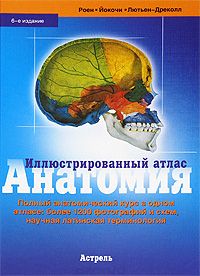
Anatomy. Illustrated Atlas
Complete anatomical course in one atlas:
Publication format: 21.5 cm x 30 cm.
6058 rub

Lower limb. Functional anatomy. Volume 2
It is no longer necessary to represent the world famous in the circles of orthopedic surgeons and kinesitherapists Dr. Adalbert I. Kapandzhi. After a long career as an orthopedic surgeon, who later became involved in hand surgery, he devoted himself entirely to the republishing and updating of three volumes of his work "Functional Anatomy" (previously called "Physiology of the joints. The second volume of this work is devoted to the lower limb and sheds light on such complex concepts as a heterokinetic cardan of the ankles and bones of the rear of the foot, the concept of the lower leg. The book also includes a summary diagram of the factors of knee stability, a table of nerves of the lower extremity, a new chapter on the physiology of walking. As in previous editions, Dr. Adalbert I. Kapandzhi independently prepared and made in color all the diagrams and pictures that help to better understand human biomechanics. By now the book has been translated into 11 languages ...
279 rub

Learning about auditory sensations as a physiological basis for music theory
The readers are invited to the fundamental work of the outstanding German physiologist, psychologist, physicist and mathematician Hermann Helmholtz, in which he attempts to establish a connection between physical and physiological acoustics with musical science and aesthetics. The book consists of three parts (divisions). The first part examines the phenomenon of upper harmonic tones; the essence of this phenomenon is determined, its relation to the differences in shades of sound is proved, a number of shades are analyzed in relation to their upper harmonic tones. The second part is devoted to the study of violations of the simultaneous sounding of two tones, namely combination tones and tremors; the phenomena of consonance and dissonance are described. Finally, the third part of the book deals with the affinity of sounds; the structure of scales and tones is deduced by the author from the research results presented in the first two parts. A review of various principles of musical style in the development of music is carried out, the tonality of homophonic music is investigated, consonant and discordant chords are considered, and the basic laws of voice leading are given.
Publishing binding. The preservation is good.
This publication is the result of many years of work by the scientist, student of Academician I.P. Pavlov, I.T. Kurtsin.
For many years, the scientist studied the role and significance of gastric mechanoreceptors in complex reflex regulation of the activity of the digestive apparatus.
The analysis carried out by I.T. Kurtsin showed that the nature of gastric acid secretion arising from irritation of gastric mechanoreceptors is complex reflex, including both unconditioned and conditioned reflex components.
In connection with these discoveries, the scientist made a number of important conclusions for science.
This monograph is aimed at physiologists, pathologists, histols and other doctors of various fields.
599 rub

The Weirdness of Our Body - 2
The author of "The Weirdness of Our Body", the Australian wizard of "weirdness" - Stephen Juan is at your service again. He, as always, is ready to answer all your questions; cite facts - from those that are heard, to the most surprising and unexpected; dispel your persistent misconceptions about everything about a person - from birth to death and from crown to toes.
Stephen Juan will tell you everything you want to know about the oddities of your body, or rather, everything that he did not have time to tell about in his first book.
255 rub