Bony skeleton of the upper wall in Latin. Human structure. Head bones (skull)
Husband. hard, tough, thin tire, b.h. natural. Skull of an animal, human, bone marrow, bone of the head. The skull is formed as if from three flattened vertebrae, representing a continuation of the ridge. Generations are recognized by the skull ... ... Explanatory dictionary Dahl
SCULL- (cranium), the skeleton of the head of vertebrates, as well as the cartilaginous capsule that protects the brain in cephalopods. In vertebrates, it is formed by cartilage and / or bone. It is subdivided into endocranium, represented by the embryonic cartilaginous skull and its ... ... Biological encyclopedic dictionary
Scull- (cranium), bones of the skull, anatomy of the skull, pictures of the skull Skull (cranium). Front view. 1 frontal bone 2 coronal suture 3 parietal bone 4 ... Human Anatomy Atlas
SCULL- (cranium), i.e. the skeleton of the head of vertebrates, is made up of two main sections: the axial skull and the visceral skeleton. The axial skull is a cartilaginous or bone box that encloses and protects the brain, hearing organ and organ ... ... Great medical encyclopedia
scull- SKULL, a, m. Head. Skull as a House of Soviets who has 1. About a large number problems or a severe headache. 2. Whose l. great mind. Scratch the skull to think, to think. Restoration of facial features on the skull makeup. See also: open; ... ... Dictionary of Russian argo
Scull- a person. SKULL, the skeleton of the head of vertebrates and humans. Protects the brain from damage. In humans, a distinction is made between the cerebral (receptacle of the brain) and the facial (visceral) skull. In an adult, the bones of the skull are connected ... ... Illustrated Encyclopedic Dictionary
SCULL- the skeleton of the head of vertebrates and humans. In an adult, the bones of the skull are connected by sutures. In newborns, at the junction of individual bones of the skull, there are non-ossified areas, the so-called. fontanelles. In the human skull, a brain is distinguished ... ... Big Encyclopedic Dictionary
SCULL- SKULL, bone base of the head. In mammals, the skull consists of the VALA, which covers the brain, as well as the facial and jaw bones (facial skull). The lower JAW is sometimes not considered part of the skull. There are 14 facial bones, most of ... ... Scientific and technical encyclopedic dictionary
SCULL- SKULL, skulls, many others. skull, husband. Head bones in vertebrates. "This skull contained the baron's ponderous brain." Pushkin. "The prince quietly stepped on the horse's skull." Pushkin. ❖ On the skull (hit, stop; vulg.) On the head, on the back of the head. Grab him on the skull. ... ... Ushakov's Explanatory Dictionary
SCULL- Skull: A - cows, B - sheep, C - pigs, D - horses. Skull: A - cows, B - sheep, C - pigs, D - horses, 1 - incisor bone; 2 - upper jaw; 3 - infraorbital opening; 4 - ... ... Veterinary encyclopedic dictionary
scull- teapot, head, skull, bone, tambourine, crock, turnip Dictionary of Russian synonyms. skull n., number of synonyms: 13 tambourine (28) ... Synonym dictionary
Books
- Skull with an arrow, Dmitry Emets. The skull with an arrow is an artifact created hundreds of years ago by one of the first divers. With its help, you can get powerful magic bookmarks, each of which can save someone's life. ... Buy for 266 rubles
- Skull with an arrow, Dmitry Emets. "Skull with an arrow", the sixth book of the "School of Diving" series ("ShNyr"). The skull with an arrow is an artifact created hundreds of years ago by one of the first divers. With its help you can get powerful ...
Open all Close all
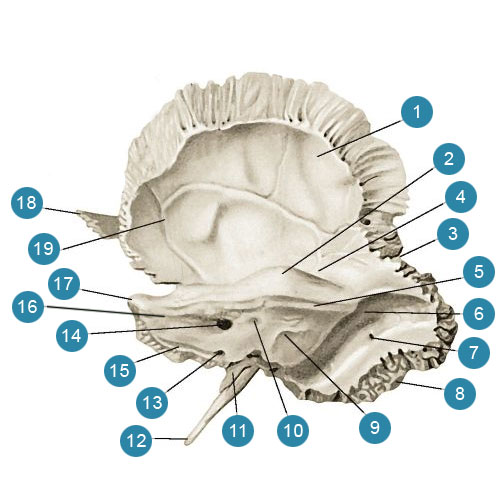
1-temporal bone
2nd parietal bone
3-crown (notched) suture
4-frontal bone
5-frontal tubercle ( tuber frontale)
6-large wing of the sphenoid bone ( ala major ossis sphenoidalis)
7-eye socket
8-lacrimal bone ( os lacrimale)
9-nasal bone ( os nasale)
10-frontal process of the upper jaw ( processus frontalis maxillae)
11-upper jaw
12-alveolar elevations of the upper jaw
13-zygomatic bone
14 chin hole
15-tuberosity lower jaw
16-coronal process of the lower jaw ( processus coronoideus mandibulae)
17-zygomatic arch ( arcus zygomaticus)
18-styloid process ( processus styloideus)
19-articular process of the lower jaw
20-mastoid process temporal bone (processus mastoideus ossis temporalis)
21-external auditory canal ( meatus acusticus externus)
22-scales of the temporal bone
23-occipital bone
24-inferior temporal line
25-superior temporal line.
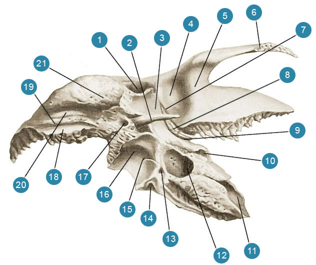
1-frontal bone
2-crown suture ( sutura coronalis)
3rd parietal bone
4-eye socket
5-scales of the temporal bone
6-zygomatic bone
7-upper jaw
8-lug hole
9-lower jaw
10 chin buffiness
11-teeth of the lower jaw
12-intermaxillary suture
13-nasal bone ( os nasale)
14-zygomatic arch ( arcus zygomaticus)
15-lacrimal bone ( os lacrimale)
16-large wing of the sphenoid bone ( ala major ossis sphenoidalis)
17-brow arch
18-glabella (glabella)
19-frontal tubercle.
1-frontal scales ( squama frontalis)
2-frontal tubercle ( tuber frontale)
3-glabella (glabella)
4-zygomatic process ( processus zygomaticus)
5-supraorbital margin ( margo supraorbitalis)
6-nose part (frontal bone)
7-nose spine ( spina nasalis)
8-frontal notch
9-brow arch
10-supraorbital foramen ( foramen supraorbitalis)
11-temporal line
1-parietal margin
2-groove of the superior sagittal sinus ( )
3-frontal ridge ( crista frontalis)
4-zygomatic process ( processus zygomaticus)
5-finger impressions ( impressions digitales)
6-blind hole ( foramen caecum)
7-nose part ( pars nasalis)
8-orbital part ( pars orbitalis)
9-cerebral eminences
10-arterial grooves ( sulci arteriosi)
11-frontal scales.
1-visual channel ( canalis opticus)
2-back saddle
3-posterior oblique process
4-anterior oblique process
5-small wing ( ala minor)
6-superior orbital fissure ( fissura orbitalis superior)
7-parietal angle
8-large wing (brain surface)
9-round hole ( foramen rotundum)
10-pterygoid canal ( canalis pterygoideus)
11-navicular fossa
12-lateral plate (pterygoid process)
13-pterygoid notch ( incisura pterygoidea)
14-pterygoid hook groove
15-vaginal process
16-Wedge Comb
17-body of the sphenoid bone ( corpus ossis sphenoidalis)
18-medial plate (pterygoid process)
19-winged hook ( hamulus pterygoideas)
20-pterygoid fossa ( fossa pterygoidea)
21-groove of the internal carotid artery
1-aperture of the sphenoid sinus ( aperture sinus sphenoidalis)
2-back saddle
3-wedge-shaped shell ( conchae sphenoidalis)
4-small wing ( ala minor)
5-superior orbital fissure ( fissura orbitalis superior)
6-zygomatic edge
7-infratemporal surface, 8th sphenoid bone ( spina ossis sphenoidalis)
9-pterygoid palatine sulcus
10-lateral plate ( lamina lateralis)
11-winged hook ( hamulus pterygoideas)
12-medial pterygoid plate
13-vaginal process
14-Wedge Comb
15-pterygoid notch ( incisura pterygoidea)
16-pterygoid canal ( canalis pterygoideus)
17-round hole ( foramen rotundum)
18 infratemporal crest ( crista infratemporalis)
19-orbital surface of the large wing
20-temporal surface of the large wing
1-groove of the superior sagittal sinus ( sulcus sinus sagittalis superioris)
2-scales of the occipital bone
3-internal occipital protuberance ( )
4-inner occipital crest ( crista occipitalis inferna)
5-large occipital foramen ( foramen occipitale magnum)
6-groove of the sigmoid sinus ( sulcus sinus sigmoidei)
7-muscular canal
8-groove of the lower petrous sinus ( )
9-slope ( clivus)
10-basilar (main) part
11-lateral part ( pars lateralis)
12-jugular notch
13-jugular tubercle
14-jugular process
15-inferior occipital fossa
16-groove of the transverse sinus ( sulcus sinus transversi)
17-superior occipital fossa
1st highest butt line
2-external occipital protuberance ( )
3-upper nuchal line ( linea nachalis superior)
4-lower nuchal line ( linea nuchalis inferior)
5-condylar canal ( canalis condylaris)
6-occipital condyle ( condylus occipitalis)
7-intracranial process
8-pharyngeal tubercle ( tuberculum phanryngeum)
9-basilar (main) part
10-lateral part ( pars lateralis)
11-jugular notch
12-jugular process
13-condylar fossa ( fossa condylaris)
14-large occipital foramen ( foramen occipitale magnum)
15th surface (platform)
16-external occipital crest ( crista occipitalis externa)
17-occipital scales
1-frontal angle ( angulus frontalis)
2-superior temporal line
3-frontal margin ( margo frontalis)
4-inferior temporal line
5-Wedge Angle ( angulus sphenoidalis)
6-scaly edge
7-mastoid angle ( angulus mastoideum)
8-occipital margin ( margo occipitalis)
9-parietal tubercle ( tuber parietale)
10-sagittal edge
1 occipital angle ( angulus occipitalis)
2-occipital margin ( margo occipitalis)
3-arterial grooves ( sulci arteriosi)
4-groove of the sigmoid sinus ( sulcus sinus sigmoidei)
5-mastoid angle ( angulus mastoideum)
6-scaly edge
7-Wedge Angle ( angulus sphenoidalis)
8-frontal margin ( margo frontalis)
9-frontal angle ( angulus frontalis)
10-dimple granulation
11-sagittal edge
12-groove of the superior sagittal sinus.
1-cock comb ( crista galli)
2-orbital plate ( lamina orbitalis)
3-perpendicular plate ( lamina perpendicularis)
4-hooked process ( processus uncinatus)
5-middle turbinate ( concha nasalis media)
6-upper nasal concha ( concha nasalis superior)
7-lattice cells.
1-perpendicular plate ( lamina perpendicularis)
2-middle turbinate ( concha nasalis media)
3-cock comb ( crista galli)
4-lattice cells
5-lattice plate
6-orbital plate ( lamina orbitalis)
7-anterior lattice furrow
8-hooked process
1-scaly part (scales) of the temporal bone
2-zygomatic process ( processus zygomaticus)
3-articular tubercle ( tuberculum articulare)
4-mandibular fossa ( fossa mandibularis)
5-stony-scaly fissure ( fissure petrosquamosa)
6-stony-tympanic (glaserov) slit
7-styloid process ( processus styloideus)
8-drum part of the temporal bone
9-external auditory opening ( porus acusticus externus)
10-mastoid process ( processus mamillaris)
11-mastoid notch ( incisura mastoidea)
12-drum-mastoid fissure ( fissura tympanomastoidea)
13-supraspinous spine (above the ear canal)
14-mastoid opening ( foramen mastoideus)
15-parietal notch ( incisura parietalis)
16-temporal line.
1-scaly part of the temporal bone
2-arc elevation ( eminentia arcuata)
3-parietal notch ( incisura parietalis)
4-roof drum
5-groove of the superior petrosal sinus
6-boroed sigmoid sinus
7-mastoid opening ( foramen mastoideus)
8-occipital margin ( margo occipitalis)
9-outer opening (aperture) of the water supply line of the vestibule
10-subarc fossa ( fossa subarcuata)
11-sheath of the styloid process ( vagina processus styloidei)
12-styloid process ( processus styloideus)
13-outer opening (aperture) of the cochlear tubule
14-internal auditory opening ( porus acusticus internus)
15-groove of the lower petrous sinus ( )
16-posterior surface of the temporal bone pyramid
17-top of the pyramid
18-zygomatic process ( processus zygomaticus)
19-arterial grooves
1-external auditory canal ( meatus acusticus externus)
2-styloid process ( processus styloideus)
3-posture-articular tubercle
4-mandibular fossa ( fossa mandibularis)
5-articular tubercle ( tuberculum articulare)
6-zygomatic process ( processus zygomaticus)
7-stony scaly shell
8-inferior process of the temporal bone pyramid (roof of the tympanic cavity)
9-stony-tympanic (glaserov) slit
10-muscular-tubal canal ( canalis musculotubarius)
11-inner opening of the carotid canal ( foramen caroticum internum)
12-external opening of the carotid canal ( foramen caroticum externum)
13-stony dimple ( fossula petrosa)
14-outer opening (aperture) of the cochlear tubule
15-mastoid tubule
16-jugular fossa
17-awl-mastoid opening ( foramen mastoideus)
18 occipital margin ( margo occipitalis)
19-groove of the occipital artery ( sulcus arteriae occipitalis)
20-mastoid notch ( incisura mastoidea)
21-mastoid process ( processus mamillaris)
1-scales of the temporal bone
2-mastoid cave ( antrum mastoideum)
3-protrusion of the lateral semicircular canal
4-protrusion of the facial nerve canal
5-window vestibule
6-probe in the canal of the facial nerve
7-cleft of the canal of the greater stony nerve ( hiatus canalis nervi petrosi majoris)
8-cleft of the canal of the small stony nerve ( hiatus canalis nervi petrosi minoris)
9-groove of the large stony nerve ( sulcus nervi petrosi majoris)
10-groove of the small stony nerve ( sulcus nervi petrosi minoris)
11-semicanal of the muscle stretching the eardrum
12-half-channel auditory tube
13-inner opening of the carotid canal
14-external opening of the carotid canal ( foramen caroticum externum)
15-cape
16-drum cavity
17-pyramidal elevation
18-awl-mastoid opening ( foramen mastoideus)
19-mastoid cells
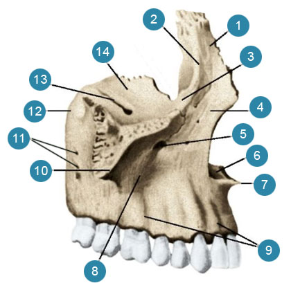
1-frontal process
2-anterior lacrimal crest
3-infraorbital margin
4-front surface
5-infraorbital foramen
6-nose notch
7-anterior nasal spine
8-body of the upper jaw ( corpus maxillae)
9-alveolar eminences
10-zygomatic process ( processus zygomaticus)
11-alveolar holes
12-tubercle of the upper jaw ( tuber maxillae)
13 infraorbital sulcus
14-orbital surface
1-frontal process
2-tear edge
3-lacrimal sulcus
4-maxillary (Haimorova) sinus
5-nasal surface of the upper jaw body
6-large palatine sulcus
7-alveolar process
8-palatine process
9-incisor canal ( canalis incisivus)
10-front nasal spine
11-shell comb
12 mesh comb.
1-frontal process
2-orbital surface ( facies orbitalis)
3-zygomatic-orbital foramen
4-lateral surface
5-temporal process
1-lattice edge
2-left wing of the opener
3-free edge
4-palatal margin
1-nasal seam
2-hole of the nasal bone
3-free edge
1-lacrimal process
2-ethmoid process
3-bottom (free) edge

1-lacrimal sulcus
2-posterior lacrimal crest
3-tear hook
1-orbital process
2-grid comb
3-wedge-palatine notch
4-wedge-shaped process
5-perpendicular plate (nasal surface)
6-shell comb
7-horizontal plate
8-pyramidal process
9-large palatine sulcus
10-posterior nasal spine
11-nose comb
12-maxillary process
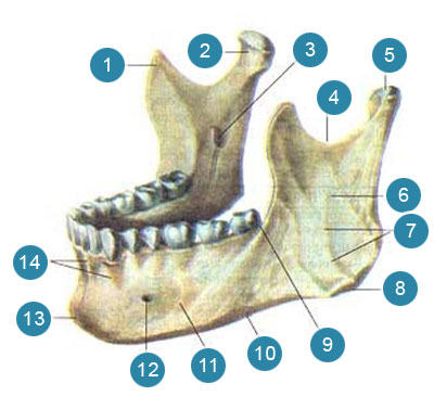
1-coronal process ( processus coronoideus)
2-condylar process
3-hole of the lower jaw ( foramen mandibulae)
4-notch of the lower jaw ( incisura mandibulae)
5-head of the lower jaw ( caput mandibulae)
6-branch of the lower jaw ( ramus mandibulae)
7-chewy buff
8-angle of the lower jaw ( angulus mandibulae)
9-oblique line
10-base of the lower jaw
11-body of the lower jaw ( corpus mandibulae)
12 chin hole
13-chin lip
14-alveolar eminences

1-body hyoid bone (corpus ossis hyoidei)
2-big horn
3-small horn
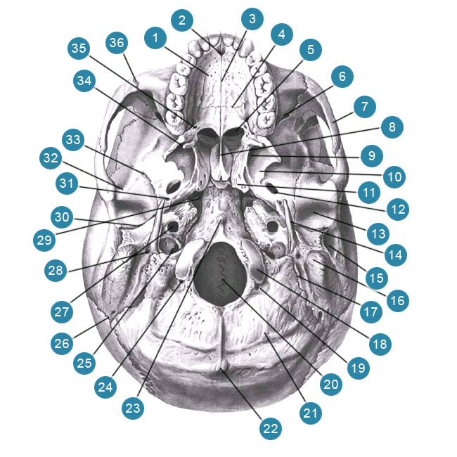
1-palatine process of the upper jaw ( processus palatinus maxillae)
2-incisor hole
3-median palatine suture
4-transverse palatine suture
5-choana
6-inferior orbital fissure ( fissura orbitalis inferior)
7-zygomatic arch ( arcus zygomaticus)
8-wing opener
9-pterygoid fossa ( fossa pterygoidea)
10-lateral plate of the pterygoid process
11-pterygoid process ( processus pterygoideus)
12 oval hole ( foramen ovale)
13-mandibular fossa
14-styloid process ( processus styloideus)
15-external auditory canal ( meatus acusticus externus)
16-mastoid process ( processus mamillaris)
17-mastoid notch ( incisura mastoidea)
18-occipital condyle ( condylus occipitalis)
19-condylar fossa ( fossa condylaris)
20-large (occipital) foramen
21-lower nuchal line ( linea nuchalis inferior)
22-external occipital protuberance ( protuberantia occipitalis externa)
23-pharyngeal tubercle ( tuberculum phanryngeum)
24-muscular canal
25-jugular foramen
26-occipital-suture suture
27-external carotid opening
28-awl-mastoid opening ( foramen mastoideus)
29-torn hole
30-stony-tympanic fissure ( fissura petrotympanica)
31-spinous hole ( foramen spinosum)
32-articular tubercle ( tuberculum articulare)
33-wedge-scaly suture
34-winged hook ( hamulus pterygoideas)
35-large palatine foramen
36-zygomatic-maxillary suture
1-orbital part of the frontal bone
2-cock phoebe
3-lattice plate
4-visual channel ( canalis opticus)
5-pituitary fossa
6-back saddle. 7-round hole ( foramen rotundum)
8 oval hole ( foramen ovale)
9-torn hole
10-spinous hole ( foramen spinosum)
11-internal auditory opening ( porus acusticus internus)
12-jugular opening
13-sublingual canal
14-lambdoid suture ( sutura lambdoidea)
15-slope ( clivus)
16-beard transverse sinus
17-internal occipital protuberance
18-large (occipital) foramen
19-occipital scales ( squama occipitalis)
20-groove of the sigmoid sinus ( sulcus sinus sigmoidei)
21-pyramid (stony part) of the temporal bone
22-scaly part of the temporal bone
23-large wing of the sphenoid bone ( ala major ossis sphenoidalis)
24-lesser wing of the sphenoid bone
1-zygomatic process of the frontal bone ( processus zygomaticus ossis frontalis)
2-large wing of the sphenoid bone (orbital surface)
3-orbital surface zygomatic bone
4-frontal process of the zygomatic bone
5-inferior orbital fissure ( fissura orbitalis inferior)
6-cheekbone-facial disgust
7-zygomatic bone
8 infraorbital sulcus
9-upper jaw (maxilla, infraorbital surface)
10-infraorbital foramen
11-orbital surface of the upper jaw ( facies orbitalis maxillae)
12-nasal cavity
13-orbital process of the palatine bone
14-lacrimal bone ( os lacrimale)
15-eye plate of the ethmoid bone
16-nasal bone ( os nasale)
17-lacrimal groove (lacrimal bone)
18-posterior lacrimal phoebe (lacrimal bone)
19-frontal process of the upper jaw ( processus frontalis maxillae)
20-front lattice hole
21-rear lattice hole
22-frontal notch
23-orbital part (orbital surface) of the frontal bone
24-supraorbital foramen ( foramen supraorbitalis)
25 visual channel ( canalis opticus)
26-small wing of the sphenoid bone ( ala minor ossis sphenoidalis)
27-superior orbital fissure
1-frontal bone (scales of the frontal bone)
2-frontal sinus
3-cock comb ( crista galli)
4-ethmoid plate of the ethmoid bone
5-upper nasal concha ( concha nasalis superior)
6-middle turbinate ( concha nasalis media)
7-sphenoid sinus ( sinus sphenoidalis)
8-wedge-palatine foramen
9-inferior turbinate ( concha nasalis inferior)
10-vertical plate of the palatine bone
11-medial pterygoid plate
12-horizontal plate of the palatine bone
13-palatine process of the upper jaw ( processus palatinus maxillae)
14-incisor canal ( canalis incisivus)
15-lower nasal passage ( meatus nasi inferior)
16-middle nasal passage ( meatus nasi medius)
17-upper nasal passage ( meatus nasi superior)
18-nasal bone.
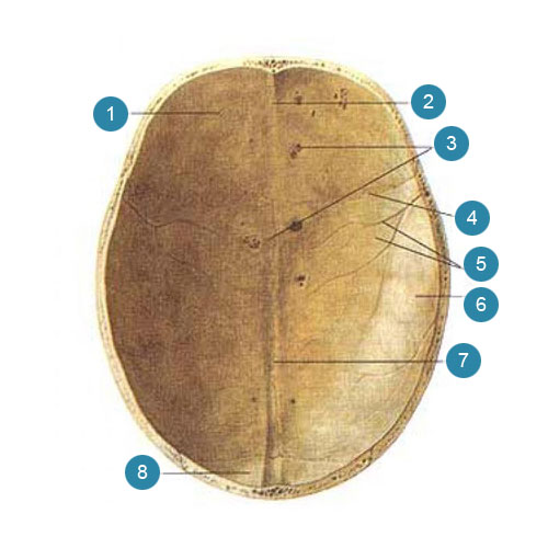
1-crown suture ( sutura coronalis)
2-sagittal suture ( sutura sagittalis)
3-lambdoid suture ( sutura lambdoidea)
4-occipital bone (scales)
5-parietal bone
6-frontal bone
1-frontal bone
2-frontal ridge ( crista frontalis)
3-dimple granulation
4-crown suture ( sutura coronalis)
5-arterial grooves ( sulci arteriosi)
6-parietal bone
7-groove of the superior sagittal sinus ( sulcus sinus sagittalis superioris)
8 occipital bone
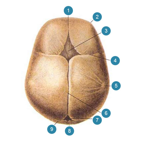
1-frontal suture
2-frontal tubercle ( tuber frontale)
3-anterior (frontal) fontanelle
4-crown suture ( sutura coronalis)
5-parietal tubercle ( tuber parietale)
6-sagittal suture
7-posterior occipital) fontanelle
8 occipital bone
9-lambdoid suture
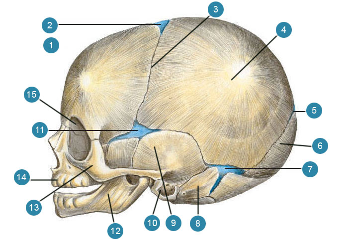
1-frontal bone
2-anterior (frontal) fontanelle
3-crown suture ( sutura coronalis)
4-parietal tubercle ( tuber parietale)
5-posterior (occipital) fontanelle
6-occipital bone (scales)
7-mastoid fontanelle
8-stony part (pyramid) of the temporal bone
9-scales of the temporal bone
10-tympanic bone (tympanic ring)
11-wedge-shaped (anterolateral) fontanelle
12-lower jaw
13-zygomatic bone
14-upper jaw
15-eye socket
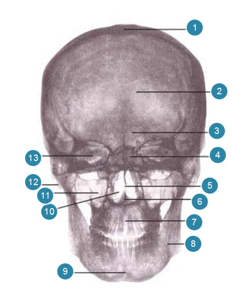
1-roof (vault) of the skull
2-frontal bone
3-frontal sinus
4-cell ethmoid bone
5-bone septum of the nasal cavity
6-anterior nasal spine
7-intermaxillary suture
8-lower jaw
9-chin lip
10-nasal cavity
11-maxillary sinus
12-mastoid process ( processus mamillaris)
13-eye socket
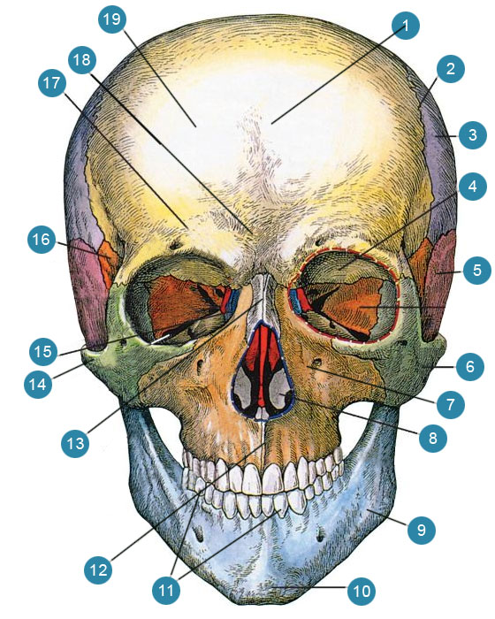
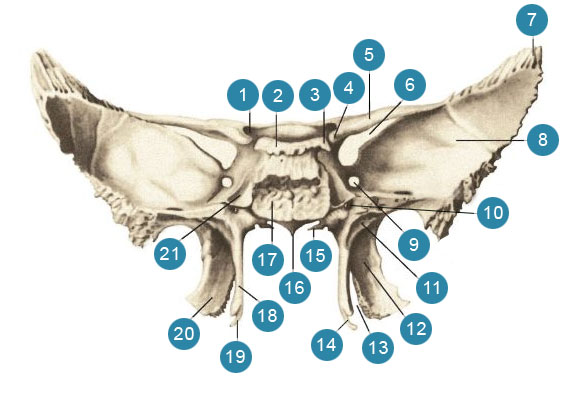
Scull, cranium, - consists of two sections - the bones of the skull, ossa cranium, and the bones of the face, ossa faciei.
The skeleton of the head is a skull, cranium, the individual bones of which are subdivided into the bones of the cerebral section of the skull, which form the cranial cavity, cavitas cranii, a receptacle for the brain and bones of the face, ossa faciei... The skull serves as a receptacle for the brain (cerebral skull) and some of the senses (organs of sight, hearing, and smell).
The bones of the face (facial part of the skull) make up the skeleton of the face, the initial parts of the digestive and respiratory systems.
Both parts of the skull are formed from separate bones, connected together motionlessly by means of sutures, suturae, and cartilage joints, synchondroses, with the exception of the lower jaw, connected to the skull movably by means of the temporomandibular joint, .
The bones of the cerebral skull, based on data on its development, include unpaired bones: occipital, wedge-shaped, frontal, ethmoid, vomer - and paired bones: temporal, parietal, inferior turbinate, lacrimal, nasal.
Facial bones include paired bones: the upper jaw, palatine bone, zygomatic bone - and unpaired bones: the lower jaw and hyoid bone. The latter, although located in the neck region, develops as a bone of the facial region of the skull and is described along with it.
Topographically, the inferior turbinate, opener, lacrimal and nasal bones belong to the skeleton of the face.
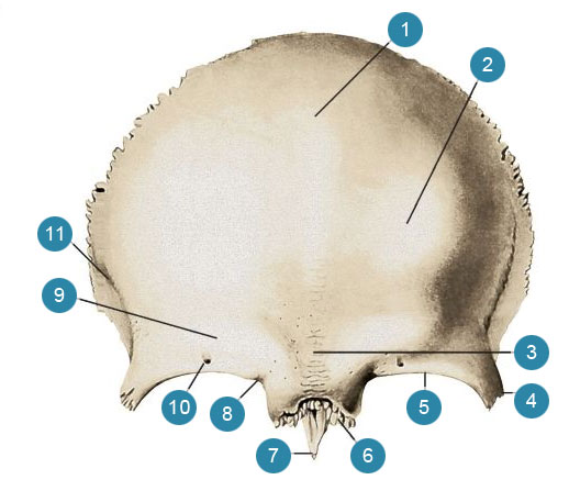
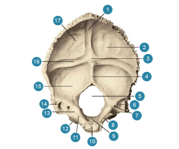
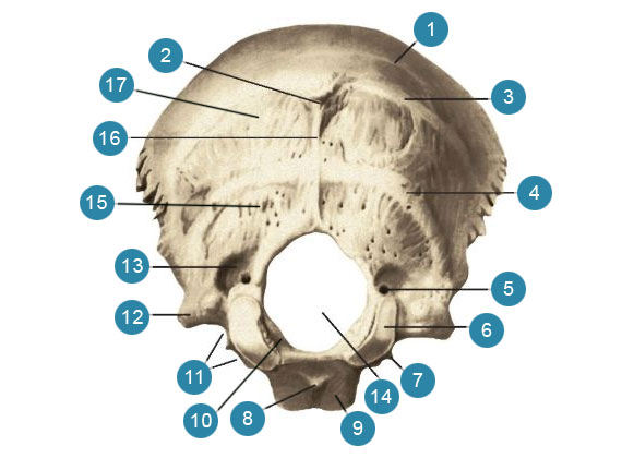
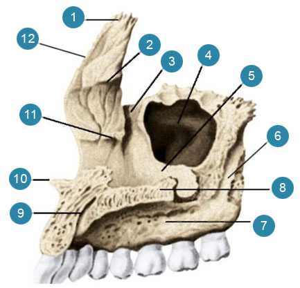
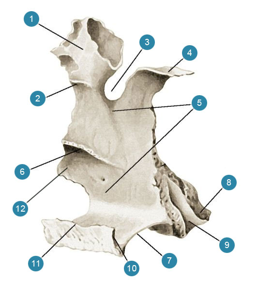
Occipital bone
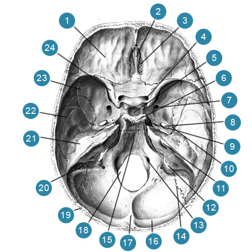
Occipital bone, os occipitale, unpaired, forms the posterior inferior part of the skull. Its outer surface is convex, and the inner, cerebral, concave. In its anteroinferior part there is a large (occipital) foramen, foramen magnum connecting the cranial cavity with the spinal canal. This opening is surrounded by a shallow groove of the occipital sinus, sulcus sinus occipitalis... Based on data on the development of the occipital bone, four parts are distinguished in it that surround the large (occipital) foramen: the basilar part is in front of the foramen magnum (occipital) foramen, paired lateral parts are on the sides of it and the occipital scales located behind.Basilar part, pars basilaris, short, thick, quadrangular; its posterior margin is free, smooth and slightly pointed, bounding a large (occipital) foramen in front; the anterior edge is thickened and rough, connects to the body of the sphenoid bone through cartilage, forming sphenoid-occipital synchondrosis, synchondrosis sphenooccipitalis.
V teenage years the cartilage is replaced by bone tissue and both bones are fused into one. Top surface the basilar part, facing the cranial cavity, is smooth and slightly concave. It makes up a slope with the part of the body of the sphenoid bone in front of it, clivus directed to the large (occipital) foramen (medulla oblongata, bridge and basilar artery of the brain with branches lie on it). In the middle of the lower, outer, slightly convex surface of the basilar part there is a small pharyngeal tubercle, tuberculum pharyngeum, (the place of attachment of the anterior longitudinal ligament and the fibrous membrane of the pharynx), and rough lines (traces of attachment of the straight anterior and longitudinal muscles of the head).
The outer, slightly uneven edge of the basilar part and the lateral parts of the occipital bone is adjacent to the posterior edge of the petrous part of the temporal bone. A stony-occipital fissure is formed between them, fissura petrooccipitalis, on a non-macerated skull, it is made by cartilage, forming a petrosoccipital synchondrosis, synchondrosis petrooccipitalis, which, like the remainder of the cartilaginous skull, ossifies with age.
Lateral parts, paries laterales, somewhat elongated, thickened in the posterior parts, and somewhat narrowed in the anterior; they form the lateral sides of the large (occipital) foramen, fused in front with the basilar part, and behind with the occipital scales.
On the cerebral surface of the lateral part, at its outer edge, there is a narrow groove of the lower stony sinus, sulcus sinus petrosi inferioris, which is adjacent to the posterior edge of the petrous part of the temporal bone, forming a canal with the groove of the temporal bone of the same name, where the venous inferior petrosal sinus lies, sinus petrosus inferior.
On the lower, outer, surface of each lateral part there is an oblong oval convex articular process - occipital condyle, condylus occipitalis... Their articular surfaces approach each other in front, diverge behind; they articulate with the superior glenoid fossa of the atlas. There is a condylar fossa behind the occipital condyle, fossa condylaris, and at the bottom of it there is an opening leading to the unstable condylar canal, canalis condylaris, which is the location of the condylar emissary vein, v. emissaria condylaris.
On the outer edge of the lateral part, there is a large, smooth-edged jugular notch, incisura jugularis, on which a small intracranial process protrudes, processus intrajugularis.
The jugular notch with the fossa of the same name in the petrous part of the temporal bone forms a jugular foramen, foramen jugulare.
The intracranial processes of both bones divide this opening into two parts: a large posterior one, in which the upper bulb of the internal jugular vein lies, bulbus v. jugularis superior, and the smaller anterior, through which the cranial nerves pass: glossopharyngeal ( n. glossopharyngeus), wandering ( n. vagus) and additional ( n. accessorius).
Behind and outside the jugular notch is limited by the jugular process, processus jugularis... On the outer surface of its base there is a small peri-mastoid process, processus paramastoideus, (the place of attachment of the rectus lateral muscle of the head, m. rectus capitis lateralis).
Behind the jugular process, from the side inner surface skull, there is a wide groove of the sigmoid sinus, sulcus sinus sigmoidei, which is a continuation of the eponymous groove of the temporal bone. Anteriorly and medially lies a smooth jugular tubercle, tuberculum jugular... Behind and downward from the jugular tubercle, between the jugular process and the occipital condyle, the hyoid canal passes through the thickness of the bone, canalis hypoglossalis, (the hypoglossal nerve lies in it, n. hypoglossus).
Occipital scales squama occipitalis, limits the posteriorly large (occipital) foramen and makes up most of the occipital bone. It is a wide, curved, triangular plate with a concave inner (cerebral) surface and a convex outer surface.
The lateral edge of the scales is divided into two sections: the larger upper, highly serrated lambdoid edge, margo lambdoideus, which, joining the occipital edge of the parietal bones, forms a lambdoid suture, sutura lambdoidea, and a smaller lower, slightly serrated mastoid margin, margo mastoideus, which, adjoining the edge of the mastoid process of the temporal bone, forms the occipital-mastoid suture, sutura occipitomastoidea.
In the middle of the outer surface of the scales, in the area of its greatest bulge, there is an external occipital protuberance, protuberantia occipitalis externa easily felt through the skin. Paired convex upper nuchal lines diverge from it, lineae nuchae superiores, above which and parallel to them there are additional highest nuchal lines, lineae nuchae supremae.
From the external occipital protuberance to the large (occipital) foramen, the external occipital ridge descends, crista occipitalis externa... In the middle of the distance between the large (occipital) foramen and the external occipital protuberance from the middle of this ridge to the edges of the occipital scales, the lower nuchal lines diverge, lineae nuchae inferiores running parallel to the top. All of these lines are muscle attachment points. On the surface of the occipital scales below the superior nuchal lines, muscles are attached that end in the occipital bone.
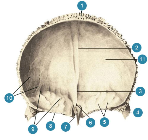
On the brain surface facies cerebralis, the occipital scale is a cruciform eminence, eminentia cruciformis, in the middle of which the internal occipital protuberance rises ( protuberantia occipitalis interna). On the outer surface of the scales, it corresponds to the outer occipital protuberance.
A groove of the transverse sinus departs in both directions from the cruciform eminence, sulcus sinus transversi, upward - the groove of the superior sagittal sinus, sulcus sinus sagittalis superioris, downwards - the internal occipital crest, crista occipitalis interna going to the posterior semicircle of the large (occipital) foramen. A dura mater with underlying venous sinuses is attached to the edges of the furrows and to the internal occipital crest; in the area of the cruciform eminence is the place of confluence of these sinuses.
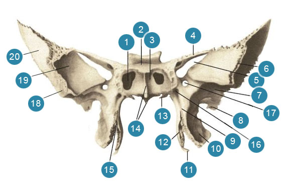
Sphenoid bone
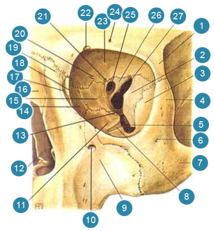
Sphenoid bone, os sphenoidale, unpaired, forms the central section of the skull base.The middle part of the sphenoid bone is the body, corpus, cubic in shape, has six surfaces. On the upper surface facing the cranial cavity, there is a depression - the Turkish saddle, sella turcica, in the center of which is the pituitary fossa, fossa hypophysialis... The pituitary gland lies in it, hypophysis... The size of the fossa depends on the size of the pituitary gland. The border of the Turkish saddle in front is the tubercle of the saddle, tuberculum sellae... Behind it, on the lateral surface of the saddle, there is an unstable middle tilted process, processus clinoideus medius.
Anterior to the tubercle of the saddle, there is a shallow transverse pre-cross groove, sulcus prechiasmatis... Behind her lies the intersection of the optic nerves, chiasma opticum... Laterally, the furrow passes into the visual canal, canalis opticus... In front of the furrow there is a smooth surface - a wedge-shaped elevation, jugum sphenoidale connecting the small wings of the sphenoid bone. The anterior faucet of the upper surface of the body is serrated, protrudes slightly forward and connects with the posterior edge of the ethmoid plate of the ethmoid bone, forming a wedge-shaped ethmoid suture, sutura spheno-ethmoidalis... The rear border of the Turkish saddle is the back of the saddle, dorsum sellae, which ends on the right and left with a small posterior inclined process, processus clinoideus posterior.
A sleepy groove runs along the sides of the saddle from back to front, sulcus caroticus, (trace of the internal carotid artery and its accompanying nerve plexus). At the posterior edge of the groove, from its outer side, there is a pointed process - a wedge-shaped tongue, lingula sphenoidalis.
The posterior surface of the saddle back passes into the upper surface of the basilar part of the occipital bone, forming a slope, clivus, (the bridge, medulla oblongata, basilar artery and its branches lie on it). The back surface of the body is rough; through the cartilaginous layer, it connects to the anterior surface of the basilar part of the occipital bone and forms sphenoid-occipital synchondrosis, synchondrosis spheno-occipitalis... With age, the cartilage is replaced by bone tissue and both bones grow together.
The front surface of the body and part of the lower one face the nasal cavity. A wedge-shaped ridge protrudes in the middle of the front surface, crista sphenoidalis, its anterior edge is adjacent to the perpendicular plate of the ethmoid bone. The lower process of the ridge is pointed, stretched downwards and forms a wedge-shaped beak, rostrum sphenoidale... The latter connects to the opener wings, alae vomeris, forming a vomer-coracoid canal, canalis vomerorostratis lying along midline between the top edge of the opener and the wedge-shaped beak. Laterally from the ridge, thin curved plates lie - wedge-shaped shells, conchae sphenoidales... The shells form the anterior and partly the lower walls of the sphenoid sinus, sinus sphenoidalis... Each sink has a small hole - the aperture of the sphenoid sinus, apertura sinus sphenoidalis... Outside the aperture, there are insignificant depressions that cover the cells of the posterior part of the ethmoid labyrinth. The outer edges of these depressions are partially connected to the orbital plate of the ethmoid bone, forming a wedge-shaped ethmoid suture, sutura spheno-ethmoidalis, a lower - with orbital processes, processus orbitalis, palatine bone.
Sphenoid sinus sinus sphenoidalis- a paired cavity that occupies most of the body of the sphenoid bone; it belongs to the paranasal sinuses. The right and left sinuses are separated from one another by the septum of the sphenoid sinuses, septum sinuum sphenoidalium, which continues anteriorly into a wedge-shaped ridge. As in the frontal sinuses, the septum is often asymmetrical, as a result of which the size of the sinuses may not be the same. Through the aperture of the sphenoid sinus, each sphenoid sinus communicates with the nasal cavity. The cavity of the sphenoid sinus is lined with a mucous membrane.
Small wings, alae minores, the sphenoid bone extends in both directions from the anteroposterior corners of the body in the form of two horizontal plates, at the base of which there is a rounded opening. From this hole begins the bone canal up to 5-6 mm long - the visual canal, canalis opticus... The optic nerve lies in it, n. opticus, and the ocular artery, a. ophthalmica... The small wings have an upper surface facing the cranial cavity and a lower one directed into the orbital cavity and closing the upper orbital fissure from above. fissura orbitalis superior.
The front edge of the lesser wing, thickened and serrated, is connected to the orbital part of the frontal bone. The posterior edge, concave and smooth, protrudes freely into the cranial cavity and is the border between the anterior and middle cranial fossa, fossae cranii anterior et media... Medially, the posterior edge ends with a protruding, well-defined anterior inclined process, processus clinoideus anterior, (a part of the dura mater is attached to it - the diaphragm of the sella turcica, diaphragma sellae).
Big wings, alae majores, depart from the lateral surfaces of the body of the sphenoid bone and go outward.
The large wing has five surfaces and three edges.
facies cerebralis, concave, facing the cranial cavity. It forms the anterior section of the middle cranial fossa. Finger-like impressions stand out on it, impressiones digitatae, [gyrorum]), and arterial grooves, sulci arteriosi, (prints of the relief of the adjacent surface of the brain and middle meningeal arteries).
There are three permanent holes at the base of the wing: a round hole is located inward and anteriorly, foramen rotundum, (the maxillary nerve leaves through it, n maxillaris), outward and backward of the round hole is an oval hole, foramen ovale, (it skips the mandibular nerve, n. mandibularis), and outward and posterior to the oval - a spinous opening, foramen spinosum(the middle meningeal artery, vein and nerve come through it). In addition, inconsistent holes are encountered in this area. One of them is a venous opening, foramen venosum located somewhat posterior to the foramen ovale. It passes a vein from the cavernous sinus to the pterygoid venous plexus. The second is a rocky hole foramen petrosum, through which the small petrosal nerve passes, is located behind the axial foramen, closer to the axis of the sphenoid bone.
Anterosuperior orbital surface, facies orbitalis, smooth, rhomboid, facing the cavity of the orbit and forms a large part of its outer wall. The lower edge of the surface is spaced from the posterior edge of the orbital surface of the upper jaw body - the lower orbital fissure is formed here, fissura orbitalis inferior.
Anterior maxillary surface, facies maxillaris, - a small triangular platform, bounded from above by the orbital surface, from the side and from below - by the root of the pterygoid process of the sphenoid bone. It is part of back wall pterygo-palatine fossa, fossa pterygopalatina, it has a round hole.
Superior lateral temporal surface, facies temporalis, somewhat concave, takes part in the formation of the wall of the temporal fossa, fossa temporalis, (the bundles of the temporal muscle begin from it). Below this surface is limited by the infratemporal ridge, crista infratemporali, below the ridge there is a surface on which the oval and spinous holes open. It forms the upper wall of the infratemporal fossa ( fossa infratemporalis), (part of the lateral pterygoid muscle begins here ( m. pterygoideus lateralis).
Upper frontal edge, margo frontalis, widely serrated, connects with the orbital part of the frontal bone, forming a wedge-shaped frontal suture, sutura sphenofrontalis... The outer sections of the frontal edge end with a sharp parietal edge, margo parietalis, which, with a wedge-shaped angle to the subject of another bone, forms a wedge-parietal suture, sutura sphenoparietalis... The inner parts of the frontal margin pass into a thin free margin, which is spaced from the lower surface of the lesser wing, limiting the superior orbital fissure from below.
Anterior zygomatic edge, margo zygomaticus, serrated. Frontal process processus frontalis, the zygomatic bone and the zygomatic edge are connected, forming a wedge-zygomatic suture, sutura sphenozygomatica.
Posterior scaly edge, margo squamosus, connects to the wedge-shaped edge, margo sphenoidalis, the temporal bone and forms a wedge-scaly suture, sutura sphenosquamosa... Posteriorly and outwardly, the scaly edge ends with the spine of the sphenoid bone (the place of attachment of the sphenoid-mandibular ligament, lig sphenomandibularis, and bundles of muscle straining the palatine curtain, m. tensor veli palatini).
Inside of the spine of the sphenoid bone, the posterior edge of the large wing lies in front of the stony part, pars petrosa, the temporal bone and limits the sphenoid-stony cleft, fissura sphenopetrosa, medially passing into a ragged hole, foramen la-lacerum, on the non-macerated skull, this gap is made by cartilaginous tissue and forms a wedge-shaped-stony synchondrosis, synchondrosis sphenopetrosa.
Pterygoid processes ( processus pterygoidei, depart from the junction of the large wings with the body of the sphenoid bone and go down. They are formed by two plates - lateral and medial. Lateral plate, lamina lateralis, (processus pterygoidei), wider, thinner and shorter than the medial (lateral pterygoid muscle begins from its outer surface, ( m. pterygoideus lateralis). Medial plate, lamina medialis, (processus pterygoidei), narrower, thicker, and slightly longer than lateral. Both plates grow together with their anterior edges and, diverging posteriorly, limit the pterygoid fossa, fossa pterygoidea, (here the medial pterygoid muscle begins, m. pterygoideus medialis). In the lower ones, both plates do not grow together and limit the pterygoid notch, incisura pterygoidea... It contains a pyramidal process, processus pyramidalis, palatine bone. The free end of the medial plate ends with a pterygoid hook directed downward and outward, hamulus pterygoideus, on the outer surface of which there is a groove of the pterygoid hook, sulcus hamuli pterygoidei, (the tendon of the muscle is thrown through it, straining the palatine curtain, m. tensor veli palatini).
The posterosuperior edge of the medial plate expands at the base and forms a cotton-shaped scaphoid fossa, fossa scaphoidea.
Outwardly from the scaphoid fossa, there is a shallow groove of the auditory tube, sulcus tubae auditivae, which laterally passes to the lower surface of the posterior edge of the large wing and reaches the spine of the sphenoid bone (the cartilaginous part of the auditory tube is adjacent to this groove). Above the scaphoid fossa and medially there is an opening, which begins the pterygoid canal, canalis pterygoideus, (vessels and nerves pass through it). The canal runs in the sagittal direction in the thickness of the base of the pterygoid process and opens on the maxillary surface of the greater wing, on the posterior wall of the pterygoid-palatine fossa.
The medial plate at its base passes into an inwardly directed flat, horizontally running vaginal process, processus vaginalis, which is located under the body of the sphenoid bone, covering the side of the opener wing, ala vomeris... In this case, the furrow of the vaginal process facing the wing of the opener is the vomer-vaginal furrow, sulcus vomerovaginalis, turns into a vomer-vaginal canal, canalis vomerovaginalis.
Outside the process there is a sagittally running small palatovaginal groove, sulcus palatovaginalis... The adjacent sphenoid process of the palatine bone, processus sphenoidalis ossis palatini, closes the furrow into the channel of the same name, canalis palatovaginalis, (in the vomer-vaginal and palatovaginal canals, the nerve branches of the pterygopalatine node pass, and in the palatovaginal canal, in addition, the branches of the sphenoid-palatine artery).
Sometimes a pterygo-spinous process is directed from the posterior edge of the outer plate towards the spine of the sphenoid bone, processus pterygospinosus, which can reach the specified spine and form a hole.
The anterior surface of the pterygoid process is connected to the posterior surface of the upper jaw in the region of the medial edge of the tubercle, forming a wedge-maxillary suture, sutura sphenomaxillaris, which lies deep in the pterygo-palatine fossa.
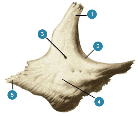
Frontal bone
Frontal bone os frontale, in an adult forms the anterior part of the cranial vault and partially its base. It consists of four parts: the frontal scales, two orbital parts and the nasal part.Frontal scales
Frontal scales, squama frontalis, convex anteriorly, has the following surfaces: external, or frontal, two temporal, or lateral, and internal, or cerebral.
Outside surface, facies externa, smooth, convex anteriorly. An elevation is not always noticeable along the median line - a metopic suture, sutura metopica) is a trace of the coalescence of the early childhood half of the frontal bone. In the anterior sections, the frontal surface of the scales passes into the orbital surface, facies orbitalis, forming on each side the supraorbital margin, margo supraorbitalis being top orbital margin, margo orbitalis... Above and parallel to the supraorbital margin, the arcuate eminence - the superciliary arch, protrudes more or less prominently, arcus superciliaris... Above each brow arch, a rounded eminence is visible - the frontal tubercle, tuber frontale... Between the protuberances of the superciliary arches and slightly above their surface of the frontal scales in the glabella region, it looks like a somewhat deepened platform - this is a glabella, glabella... The inner third of the supraorbital margin has a small supraorbital notch, incisura supraorbitalis... This notch is highly variable and can be expressed in the form of the supraorbital foramen, foramen supraorbitale... Closer to the midline, i.e. more medially, lies an equally pronounced frontal notch, incisura frontalis, (in the supraorbital notch pass the lateral branch of the supraorbital nerve and vessels, in the frontal - the medial branch of the same nerve and vessels). At the site of the specified notch, a frontal foramen may form, foramen frontale.
Laterally, the supraorbital margin turns into a blunt, triangular zygomatic process, processus zygomaticus, its serrated edge is connected to the frontal process of the zygomatic bone, forming a frontal-zygomatic suture, sutura frontozygomatica.
The temporal line is directed upward and backward from the zygomatic process, linea temporalis, it separates the frontal surface of the scales from the temporal surface. Temporal surface facies temporalis, is the anterosuperior part of the temporal fossa, fossa temporalis, where the anterior bundles of the temporal muscle begin.
Inner surface, facies interna, concave. It has weakly pronounced finger-like impressions ( impressiones digitatae, and inconsistent arterial grooves, sulci arteriosi, (as an imprint of the relief of the adjacent brain and blood vessels).
In the middle of the inner surface of the frontal scales, there is a groove of the superior sagittal sinus, sulcus sinus sagittalis superioris... Its both edges, going up and back, pass into the groove of the parietal bones of the same name, and below they join into a sharp frontal ridge, crista frontalis, (the process of the dura mater is attached to it - the sickle of the large brain). The lowest part of the crest and wing of the cock's crest of the ethmoid bone, ala cristae galli ossis ethmoidalis, form a channel - a blind hole, foramen cecum, in which there is a vein that carries blood from the nasal cavity to the superior sagittal sinus.
The upper, or posterior, edge of the frontal scales is the parietal edge, margo parietalis, thickened; its serrated edge is connected to the frontal edge of the parietal bones, forming a coronal suture, sutura coronalis... The lower sections of the scales are triangular in shape, connected to the frontal edge of the large wings of the sphenoid bone.
Each orbital part pars orbitalis, the frontal bone is part of the upper wall of the orbit. From the supraorbital edge of the frontal scales, it goes back and horizontally. It distinguishes between the lower orbital and upper cerebral surfaces.
Orbital surface, facies orbitalis facing into the cavity of the orbit, smooth and concave. In the lateral part of it, at the base of the zygomatic process, there is a shallow fossa of the lacrimal gland, fossa glandulae lacrimalis, - the location of the lacrimal gland.
In the medial part of the orbital surface, there is a weakly pronounced block fossa, fovea trochlearis, near which there is often a cartilaginous block spine, spina trochlearis, (here the cartilaginous ring is attached, which is a block of the tendon of the superior oblique muscle of the eyeball).
Superior cerebral surface facies cerebratis, the orbital part has well-defined imprints of the adjacent surface of the frontal lobes of the brain in the form of finger-like impressions, impressiones digitatae, gyrorum).
Orbital parts
The orbital parts are separated from each other by a lattice notch, incisura ethmoidalis, in which the lattice plate is located, lamina cribrosa, ethmoid bone. The notch on the sides is bounded by the edge, outward from which there are dimples covering the cells of the upper part of the ethmoid labyrinth open upward, forming their upper wall. Between the ethmoid dimples, two grooves pass in the transverse direction - anterior and posterior, which, together with the same grooves of the ethmoid labyrinth, form tubules. The latter open on the inner wall of the orbit - that two small holes: the anterior lattice hole, foramen ethmoidale anterius, (the anterior ethmoid vessels and nerve pass through it), and the posterior ethmoid foramen, foramen ethmoidale posterius, (the posterior ethmoid vessels and nerve pass through it). The edge of the ethmoid notch connects to the superior edge of the orbital plate, lamina orbitalis, ethmoid bone, forming a fronto-ethmoid suture, sutura frontoethmoidalis, and in front - with the lacrimal bone - the frontal-lacrimal suture, sutura frontolacrimalis.
The posterior edge of the orbital part, ragged and serrated, connects with the lesser wing of the sphenoid bone, forming the inner section of the sphenoid-frontal suture, sutura sphenofrontalis.
The lateral edge of the orbital part is rough, triangular in shape. It connects to the frontal edge of the greater wing of the sphenoid bone and forms the outer portion of the sphenoid-frontal suture.
Nasal part
Nose part, pars nasalis, the frontal bone in the form of an arc closes in front of the ethmoid notch. In front, in the middle of the nose, protrudes (sometimes double) obliquely downward and forward nasal spine ( spina nasalis, pointed at the end and flattened from the sides. It is surrounded in front and side by a serrated nasal edge, margo nasalis... It connects to the upper edge of the nasal bone, forming a frontal-nasal suture, sutura frontonasalis, and with the frontal process ( processus frontalis) of the upper jaw, forming a frontal-maxillary suture, sutura frontomaxillaris... The lower surface of the posterior parts of the nasal part has shallow dimples, which, as noted, cover the cells of the ethmoid labyrinths open upward.
On each side of the nasal spine there is one aperture of the frontal sinus, apertura sinus frontalis; heading up and anteriorly, it leads into the cavity of the corresponding frontal sinus.
Frontal sinus sinus frontalis, - a paired cavity lying between both plates of the frontal bone in its anteroinferior parts. Frontal sinus Carried in the air bones of the sinuses. The right sinus is separated from the left by a vertical septum of the frontal sinuses, septum sinuum frontalium... Deviating to the side, the septum determines the unequal size of the cavities of both sinuses. The boundaries vary dramatically. Sometimes the frontal sinuses reach up to the frontal tubercles, down to the supraorbital edges, posteriorly to the small wings of the sphenoid bone and to the sides - to the zygomatic processes. The frontal sinus opening connects the frontal sinus and the middle nasal passage, meatus nasi medius, nasal cavity. The sinus cavity is lined with mucous membrane.
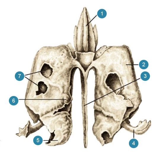
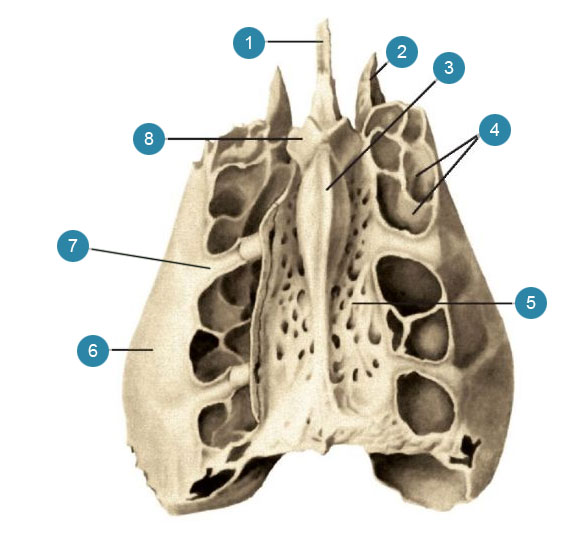
Ethmoid bone
Ethmoid bone, os ethmoidale, unpaired. Most of it lies in the upper parts of the nasal cavity, a smaller part - in the anterior parts of the base of the skull. It has the shape of an irregular cube, consists of air cells and belongs to the group of air bones, ossa pneumatica.In the ethmoid bone, an ethmoid plate extending horizontally, a perpendicular plate lying vertically, and ethmoid labyrinths located on both sides of the latter are distinguished.
Lattice plate, lamina cribrosa, is the upper wall of the nasal cavity, is located horizontally in the lattice notch of the frontal bone, forming a fronto-lattice suture, sutura frontoethmoidalis... It is perforated with 30-40 small holes, foramina fibrosae through which the nerves (fibers of the olfactory nerves) and blood vessels pass.
Perpendicular plate, lamina perpendicularis, is divided into two parts: a smaller upper one, which lies above the lattice plate, and a large lower one, located under this plate. Top part forms a cock's comb, crista galli, and is directed into the cranial cavity (a sickle of the large brain is attached to the crest - a process of the dura mater).
The border of the antero-lower edge of the cock's comb on each side is a non-permanent formation - the wing of the cock's comb, ala cristae galli... Both processes delimit the blind hole behind and above, foramen cecum, frontal bone. Bottom part a perpendicular plate of an irregular quadrangular shape, directed vertically downward into the nasal cavity, and forms the anteroposterior part of the bony septum. From above, it adjoins the nasal spine of the frontal bone, in front - to the nasal bones, behind - to the wedge-shaped crest, from below - to the vomer, and in front and below - to the cartilaginous part of the nasal septum. There is often a deviation of all or part of the perpendicular plate to the side.
Lattice maze labyrinthus ethmoidalis, - paired formation, located on both sides of the perpendicular plate, adjacent to the lower surface of the lattice plate. Consists of numerous air-bearing lattice cells, cellulae ethmoidales communicating both with each other and through a series of holes with the nasal cavity. The ethmoid cells are lined with a mucous membrane, which is a direct extension of the nasal mucosa.
The cells located in front open into the middle nasal passage, the middle and rear ones communicate with the upper nasal passage.
The lateral wall is a thin smooth orbital plate, lamina orbitalis, forming most of the inner wall of the orbit. The plate is connected at the top with the frontal bone, forming a fronto-ethmoid suture, sutura fronto-ethmoidalis, below - with upper jaw- ethmo-maxillary suture, sutura ethmoidomaxillaris, and with the orbital process of the palatine bone - palatine-ethmoid suture, sutura palato-ethmoidalis, in front - with the lacrimal bone - lacrimal-ethmoid suture and behind - with the sphenoid bone - wedge-ethmoid suture, sutura spheno-ethmoidalis... Along the upper edge of the labyrinth, there are two small grooves - the anterior and posterior ethmoid grooves, which, with the grooves of the same name of the frontal bone, form tubules that open with the anterior and posterior ethmoid openings, foramina ethmoidales anterius et posterius, (vessels and nerves of the same name pass through these holes).
The medial wall of the labyrinth is a rough, grooved plate that forms most of the lateral wall of the nasal cavity. On its surface, facing the perpendicular plate, there are two thin, slightly curved at the edges and outwardly curved processes: the upper - the upper turbinate, concha nasalis superior, and the lower one is the middle turbinate, concha nasalis media... Sometimes above the superior turbinate there is a rudimentary process in the form of a thin bony ridge - the highest turbinate, concha nasalis suprema... In the upper-posterior part of the medial wall of the labyrinth, between the upper and middle turbinates, a slit-shaped space is formed - the upper nasal passage, meatus nasi superior... The gap under the middle nasal concha is the middle nasal passage, meatus nasi medius.
From the lower anterior surface of each labyrinth, anteriorly and downwardly from the middle nasal concha, a hook-shaped process, bent backward and downward, departs, processus uncinatus... On the whole skull, it connects with the ethmoid process, processus ethmoidalis, the inferior turbinate.
One of the most large cells, having the appearance of a swelling, - a lattice bubble, bulla ethmoidalis.
Between the hooked process below and in front and a large ethmoid vesicle behind and above there is a gap - an ethmoid funnel, infundibulum ethmoidale, the upper end of which communicates with the opening of the sinus of the frontal bone. The posterior edge of the uncinate process and the lower surface of the large ethmoid vesicle form a semilunar cleft, hiatus semilunaris through which the sinus of the maxillary bone communicates with the middle nasal passage.
Coulter
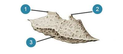
Coulter, vomer, is an unpaired, rhombus-shaped plate that forms the posterior part of the nasal septum.The coulter, excluding its posterior edge, is usually slightly bent to the side,
Top edge the coulter is thicker than others. It is separated by the coulter furrow, sulcus vomeris, into two outwardly bent processes - opener wings, alae vomeris... They adjoin the lower surface of the body of the sphenoid bone and cover its beak, forming a wedge-vomer suture, sutura sphenovomeriana... Such seams will slope to schindiles, schyndilesis... This area is the wedge-shaped part of the opener, pars cuneiformis vomeris.
The posterior edge of the bone is the choanal crest, crista choanalis vomeris, slightly pointed, separates the posterior openings of the nasal cavity - choanas, choanae.
The anterior and lower margins are rough. The lower edge is connected to the nasal crest of the upper jaw and palatine bone, and the anterior (oblique) - at the top with the perpendicular plate of the ethmoid bone, at the bottom - with the cartilage of the nasal septum.
Temporal bone
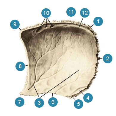
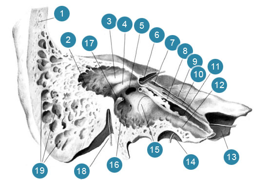
Temporal bone, os temporale, steam room, participates in the formation of the base of the skull and the lateral wall of its vault. It contains the organ of hearing and balance. It articulates with the lower jaw and is the support of the chewing apparatus.There is an external auditory opening on the outer surface of the bone, porus acusticus externus, around which three parts of the temporal bone are located; above - a scaly part, inwardly and behind - a stony part, or a pyramid, in front and below - a drum part.
Scaly part of the temporal bone
Scaly part, pars squamosa, has the shape of a plate and is located almost in the sagittal direction. External temporal surface, facies temporalis, the scaly part is slightly rough and slightly convex. In the back, it passes in vertical direction sulcus of the middle temporal artery, sulcus arteriae temporalis mediae
In the posterior inferior part of the scaly part, there is an arcuate line that continues into the inferior temporal line, linea temporalis inferior, parietal bone.
From the scaly part, above and somewhat anterior to the external auditory opening, the zygomatic process departs in a horizontal direction, processus zygomaticus... It is, as it were, a continuation of the supra-mastoid ridge, crista supramastoidea located horizontally along the lower edge of the outer surface of the scaly part. Starting with a broad root, the zygomatic process then narrows. It has an inner and outer surface and two edges - a longer top and bottom, shorter. The anterior end of the zygomatic process is serrated. The zygomatic process of the temporal bone and the temporal process, processus temporalis, the zygomatic bones are connected using the temporomandibular suture, sutura temporozygomatica, forming a zygomatic arch, arcus zygomaticus.
On the lower surface of the root of the zygomatic process is a transverse-oval mandibular fossa, fossa mandibularis... The anterior half of the fossa, up to the stony-scaly fissure, is the articular surface, facies articularis, temporomandibular joint. In front, the mandibular fossa is limited by the articular tubercle, tuberculum articulare.
The outer surface of the scaly part is involved in the formation of the temporal fossa, fossa temporalis, (here the bundles of the temporal muscle begin, m. temporalis).
Inner brain surface facies cerebralis slightly concave. There are finger-like indentations on it, impressiones digitatae as well as the arterial groove, sulcus arteriosus, (it contains the middle meningeal artery, a. meningea media).
The scaly part of the temporal bone has two free edges - the sphenoid and the parietal.
Antero-inferior wedge-shaped edge, margo sphenoidalis, wide, serrated, connects with the scaly edge of the large wing of the sphenoid bone and forms a wedge-scaly suture, sutura sphenosquamosa... Upper-posterior parietal margin, margo parietalis, pointed, longer than the previous one, connected to the scaly edge of the parietal bone.
Temporal bone pyramid
The pyramid, the rocky part - pars petrosa, the temporal bone consists of the posterolateral and anteromedial sections.
The posterolateral part of the petrous part of the temporal bone is the mastoid process, processus mastoideus, which is located posterior to the external auditory opening. It distinguishes between the outer and inner surfaces. The outer surface is convex, rough and is the site of muscle attachment. Downward, the mastoid process turns into a cone-shaped protrusion, which is easily felt through the skin,
On the inside, the process is limited by a deep mastoid notch, incisura mastoidea, (the posterior abdomen of the digastric muscle originates from it, venter posterior m. digastrici). Parallel to the notch and somewhat posteriorly is the groove of the occipital artery, sulcus arteriae occipitalis, (trace of adherence of the artery of the same name).
On the inner, cerebral, surface of the mastoid process there is a wide S-shaped sulcus of the sigmoid sinus, sulcus sinus sigmoidei, passing at the top into the groove of the parietal bone of the same name and then into the groove of the transverse sinus of the occipital bone (the venous sinus lies in it, sinus transversa). Downward, the sigmoid sinus groove continues as the eponymous groove of the occipital bone.
The posterior border of the mastoid process is the serrated occipital margin, margo occipitalis, which, connecting with the mastoid edge of the occipital bone, forms the occipito-mastoid suture, sutura occipitomastoidea... In the middle of the suture length or in the occipital margin there is a mastoid opening, foramen mastoideum, (sometimes there are several of them), which is the place of occurrence of the mastoid veins, vv. emissariae mastoidea connecting the saphenous veins of the head with the sigmoid venous sinus, as well as the mastoid branch of the occipital artery, ramus mastoideus a. occipitalis.
From above, the mastoid process is bounded by the parietal edge, which forms a parietal notch on the border with the edge of the same name of the scaly part of the temporal bone, incisura parietalis; it includes the mastoid angle of the parietal bone, forming the parieto-mastoid suture, sutura parietomastoidea.
At the place of transition of the outer surface of the mastoid process into the outer surface of the scaly part, you can see the remnants of the scaly-mastoid suture, sutura squamosomastoidea which is well pronounced on the skull of children.
On the cut of the mastoid process, the bony air cavities inside it are visible - mastoid cells, cellulae mastoideae... These cells separate from one another the bony mastoid walls ( paries mastoideus). The permanent cavity is the mastoid cave, antrum mastoideum, in the central part of the process; mastoid cells open into it, it connects to the tympanic cavity, cavitas tympanica... The mastoid cells and the mastoid cavity are lined with mucous membranes.
The anteromedial part of the stony part lies medially from the scaly part and the mastoid process. It has the shape of a trihedral pyramid, the long axis of which is directed from the outside and back to front and medially. The base of the stony part faces outwards and backwards; the top of the pyramid, apex partis petrosae, directed inward and anteriorly.
In the stony part, three surfaces are distinguished: anterior, posterior and lower, and three edges: upper, posterior and anterior.
The front surface of the pyramid, facies anterior partis petrosae, smooth and wide, facing the cranial cavity, directed obliquely from top to bottom and in front and passes into the brain surface of the scaly part. It is sometimes separated from the latter by a stony-scaly crack, fissura petrosquamosa... Almost in the middle of the front surface there is an arcuate eminence, eminentia arcuata, which is formed by the underlying anterior semicircular canal of the labyrinth. Between the elevation and the stony-scaly fissure, there is a small platform - the roof of the tympanic cavity, tegmen tympani, under which the tympanic cavity is located, cavum tympani... On the anterior surface, near the apex of the stony part, there is a small trigeminal depression, impressio trigemini, (the junction of the trigeminal node, ganglion trigeminale).
Lateral to the depression is the cleft of the canal of the greater stony nerve, hiatus canalis n. petrosi majoris, from which a narrow groove of the large stony nerve is directed medially, sulcus n. petrosi majoris... Anteriorly and somewhat laterally from the indicated opening, there is a small cleft of the canal of the small stony nerve, hiatus canalis n. petrosi minoris, from which the groove of the small stony nerve is directed, sulcus n. petrosi minoris.
The back surface of the pyramid, facies posterior partis petrosae, as well as the anterior one, faces the cranial cavity, but goes up and posteriorly, where it passes into the mastoid process. Almost in the middle of it is round shape internal auditory opening, porus acusticus internus which leads to the inner ear canal, meatus acusticus internus(the facial, intermediate, vestibular cochlear nerves pass through it, nn. facialis, intermedius, vestibulocochlearis as well as artery and vein of the labyrinth, a. et v. labirinthi). Slightly higher and lateral from the internal auditory foramen there is a well-pronounced in newborns, small depth subarc fossa, fossa subarcuata, (it includes the process of the dura mater of the brain). Even more lateral lies the slit-like outer aperture of the water supply of the vestibule, apertura externa aqueductus vestibuli opening into the water supply of the vestibule, aqueductus vestibuli... Through the aperture from the cavity inner ear the endolymphatic duct comes out.
The lower surface of the pyramid, facies inferior partis petrosae, rough and uneven, forms part of the lower surface of the base of the skull. There is a rounded or oval jugular fossa on it, fossa jugularis, (the place of attachment of the upper bulb of the internal jugular vein).
At the bottom of the fossa, a small groove is noticeable (the auricular branch of the vagus nerve passes through it). The groove leads to the opening of the mastoid canaliculus, canaliculus mastoideus, which opens in the drum-mastoid fissure, fissura tympanomastoidea.
The posterior edge of the jugular fossa is limited by the jugular notch, incisura jugularis, which is a small intracranial process, processus intrajugularis, divides into two parts - anteromedial and posterolateral. A rounded foramen lies in front of the jugular fossa; it leads to the sleepy canal, sa nalis caroticus opening at the top of the rocky part.
Between the anterior circumference of the jugular fossa and the external opening of the carotid canal, there is a small petrous dimple, fossula petrosa, (the place of attachment of the lower node of the glossopharyngeal nerve). In the depths of the dimple there is a hole - a passage into the tympanic tubule, canaliculus tympanies, (the tympanic nerve and the lower tympanic artery pass through it). The tympanic tubule leads to the middle ear, auris media, or the tympanic cavity, cavum lympani), cavitas tympanies).
Laterally from the jugular fossa, the styloid process directed downward and somewhat anteriorly protrudes, processus styloideus, from which the muscles and ligaments begin. In front of the outside of the base of the process descends the bony protrusion of the tympanic part - the sheath of the styloid process, vagina processus styloidei... There is a styloid foramen behind the base of the process, foramen stytomastoideum, which is the outlet of the facial canal, canalis facialis.
The top edge of the pyramid, marge superior partis petrosae, separates its front surface from the back. A groove of the superior stony sinus runs along the edge, sulcus sinus petrosi superioris, - the imprint of the superior petrosal venous sinus lying here and the attachment of the tentorium of the cerebellum - part of the dura mater of the brain. This groove passes posteriorly into the groove of the sigmoid sinus of the mastoid process of the temporal bone.
Rear edge of the pyramid, margo posterior partis petrosae, separates its back surface from the bottom. Along it, on the cerebral surface, there is a groove of the lower stony sinus, sulcus sinus petrosi inferioris, (trace of adherence of the inferior petrosal venous sinus). Almost in the middle of the posterior margin, near the jugular notch, there is a triangular funnel-shaped depression, in which the external aperture of the cochlear tubule lies, apertura externa canaliculi cochleae, the snail tubule ends in it, canaliculus cochleae.
The anterior edge of the stony part, located on the lateral side of its anterior surface, is shorter than the upper and posterior; it is separated from the scaly part of the temporal bone by a stony-scaly gap, fissura petrosquamosa... On it, lateral to the inner opening of the carotid canal, is the opening of the musculocutaneous canal leading to the tympanic cavity.
Channels and cavities of the petrous part of the temporal bone:
Sleepy channel, canalis caroticus, begins in the middle sections of the lower surface of the stony part with an external opening. First, the canal is directed upward, located here in front of the middle ear cavity, then, bending, follows anteriorly and medially and opens at the top of the pyramid with an internal opening (the internal carotid artery, accompanying its veins and plexus of sympathetic nerve fibers pass through the carotid canal).
Sleepy drum tubules, canaliculi caroticotympanici, are two small tubules branching from the carotid canal and leading into the tympanic cavity (through which the carotid-tympanic nerves pass).
Facial canal, canalis facialis, begins at the bottom of the internal auditory canal, meatus acusticus internus, (in the field of the facial nerve, area n. facialis). The canal runs horizontally and almost at right angles to the axis of the stony part, goes to its anterior surface, to the cleft of the canal of the greater stony nerve, hiatus canalis n. petrosi majoris... Here, turning at right angles, it forms the knee of the facial canal, geniculum canalis facialis, and passes to the posterior section of the medial wall of the tympanic cavity (accordingly, on this wall of the tympanic cavity there is a protrusion of the facial canal, prominentia canalis facialis). Further, the canal, heading posteriorly, follows along the axis of the stony part to the pyramidal elevation, eminentia pyramidalis; from here it goes vertically down and opens with a styloid opening, foramen stylomastoideum, (facial and intermediate nerves, arteries and veins pass in the canal).
Drum string duct canaliculus chordae tympani, begins on the outer wall of the facial canal, a few millimeters above the styloid opening. Heading forward and upward, the tubule enters the tympanic cavity and opens on the back wall of it (a branch of the intermediate nerve passes through the tubule - the tympanic string, chorda tympani, which, having entered the tympanic cavity through the tubule, leaves it through the stony-tympanic fissure, fissura petrotympanica).
Tympanic tubule, canaliculus tympanicus, begins on the lower surface of the stony part, deep in the stony dimple. Then it goes to the lower wall of the tympanic cavity and, piercing it, enters the tympanic cavity, passes along its medial wall and is located in the groove of the cape, sulcus promontorii... Then it follows to the upper wall of the tympanic cavity, where it opens with a cleft of the canal of the small stony nerve ( hiatus canalis n. petrosi minoris).
Muscular-tubal canal, canalis musculotubarius, is a continuation of the anteroposterior part of the tympanic cavity. The outer opening of the canal is located at the notch between the stony and scaly parts of the temporal bone, at the anterior end of the stony-scaly fissure. The canal is located laterally and slightly posterior to the horizontal part of the carotid canal, almost along the longitudinal axis of the stony part. The horizontally located septum of the musculocutaneous canal, septum canalis musculotubarii, divides the canal into the upper smaller half-cap of the muscle straining the eardrum, semicanals m. tensoris tympani, and the lower greater palukanal of the auditory tube, semicanals lubae auditivae, (in the first there is a muscle that strains the eardrum, the second connects the tympanic cavity with the pharyngeal cavity.
Mastoid tubule, canaliculus mastoideus, begins deep in the jugular fossa, passes across the lower part of the facial canal and opens into the tympanic-mastoid fissure (the ear branch of the vagus nerve passes through the tubule).
Tympanic cavity, cavum tympani... - an elongated, laterally compressed cavity lined with a mucous membrane. Three auditory ossicles lie inside the cavity: the malleus, malleus, anvil, incus, and stirrup ( stapes), which, articulating with each other, form a chain of the auditory ossicles (more about the structure of these canals, the tympanic cavity, the auditory ossicles and the labyrinth.
The tympanic part of the temporal bone
Drum part, pars tympanlca, - the smallest part of the temporal bone. It is a slightly curved annular plate and forms the anterior, lower walls and part of the posterior wall of the external auditory canal, meatus acusticus extenus... Here you can see the border drum-scaly crack, fissura tympanosquamosa, which, together with the stony-scaly fissure, separates the tympanic part from the mandibular fossa of the scaly part. The outer edge of the tympanic part, closed from above by the scales of the temporal bone, limits the external auditory opening, porus acusticus externus... At the posterior-superior outer edge of this hole there is a supraspinous spine, spina suprameatica... Under it is the supraanal dimple, foveola suprameatica... On the border of the larger, inner, and smaller, outer, parts of the external auditory canal, the tympanic groove is located, sulcus tympanicus, (the place of attachment of the tympanic membrane). At the top, it is limited by two curved protrusions: in front - a large tympanic spine, spina tympanica major, and behind - a small tympanic spine, spina tympanica minor... Between these protrusions is the drum cut ( incisura tympanica) opening into a drum groove, recessus epitympanicus.
Between the medial part of the tympanic part and the scaly part of the temporal bone, the lower process of the roof of the tympanic cavity is wedged. In front of this process is a stony-scaly slit, fissura petrosquamosa, and behind - a stony-tympanic fissure, fissura petrotympanica, (a nerve comes out of the last - a drum string and small vessels). Both furrows continue outward into the drum-scale fissure, fissura tympanosquamosa.
The lateral part of the tympanic part passes into the stony crest, the elongated part of which forms the sheath of the styloid process, vagina processus styloidei... In a newborn, the external auditory canal is still absent and the tympanic part is represented by the tympanic ring, anulus tympanicus, which then grows, forming a significant part of the external auditory canal.
On the inner surface of the tympanic spine, the spinous crest is clearly visible, at the ends of which there are anterior and posterior tympanic processes, and along it there is a groove of the malleus.
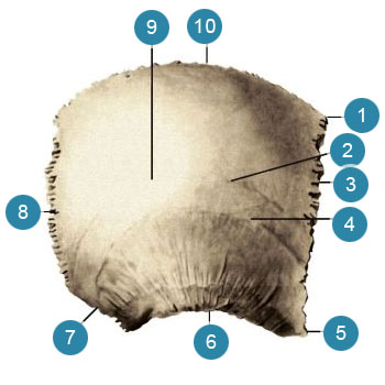
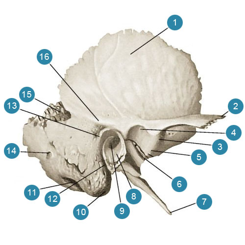
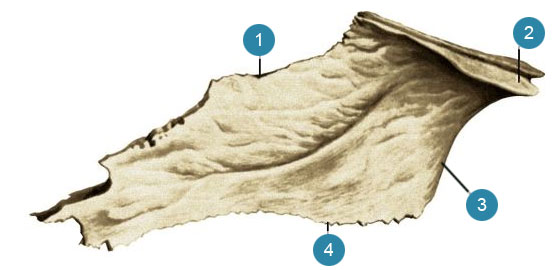
Parietal bone
Parietal bone, os parietale, steam room, forms the upper and lateral parts of the cranial vault. It has the shape of a quadrangular, outwardly convex plate, in which two surfaces are distinguished: outer and inner - four edges: upper, lower, anterior and posterior.Outside surface, facies externa, smooth and convex. The place of the greatest convexity of the bone is the parietal tubercle, tuber parietale... Below the parietal tubercle, there is an arcuate rough upper temporal line horizontally, linea temporalis superior, which starts from the anterior edge of the bone and, being a continuation of the line of the same name of the frontal bone, stretches across the entire surface of the parietal bone to its posterior inferior angle. Below this line, parallel to the lower edge of the parietal bone, there is another, more pronounced lower temporal line, linea temporalis inferior, (the first is the site of attachment of the temporal fascia, fascia temporalis, the second - the temporal muscle, m. temporalis).
Inner surface, facies interna, concave; it has weakly pronounced prints of the relief of the adjacent brain in the form of finger-like impressions, impressiones digitatae, and tree-like branching arterial grooves, sulci arteriosi, (traces of the branches of the middle meningeal artery adjacent here, a. meningea media).
An incomplete groove of the superior sagittal sinus runs along the upper edge of the inner surface of the bone, sulcus sinus sagittalis superioris... With the groove of the same name of the other parietal bone, it forms a complete groove (a process of the dura mater is attached to the edges of the groove - the sickle of the large brain, falx cerebri).
In the back of the same upper edge of the bone, there is a small parietal foramen, foramen parietale, through which the branch of the occipital artery passes to the dura mater and the parietal emissary vein. In the depths of the sagittal sinus sulcus and in the vicinity of it (especially on the parietal bones in old age) there are many small granulation dimples, foveolae granulares, (outgrowths come here - granulation of the arachnoid membrane)).
On the inner surface, at the posterior inferior angle of the parietal bone, there is a deep groove of the sigmoid sinus, sulcus sinus sigmoidei, (imprint of the sigmoid venous sinus of the dura mater). Anteriorly, this groove passes into the groove of the temporal bone of the same name, posteriorly - into the groove of the transverse sinus of the occipital bone.
Upper, sagittal, edge, margosagittalis, straight, strongly serrated, longer than the others, connected to the same edge of the other parietal bone in a sagittal suture, sutura sagittalis... Bottom scaly edge, margo squamosus, pointed, arched; its anterior section is covered by the posterior part of the upper edge of the greater wing of the sphenoid bone; farther posteriorly, the temporal bone scales are superimposed by its parietal edge; the most posterior area is connected by teeth with the mastoid process of the temporal bone. According to these three areas, three seams are formed: a scaly seam, sutura squamosa, parieto-mastoid suture, sutura parietomastoidea, and a wedge-shaped parietal suture, sutura sphenoparietalis.
Front, frontal, edge, margo frontalis, toothed; it connects with the parietal edge of the scales of the frontal bone, forming a coronary suture, sutura coronalis.
Back, occipital, edge, margo occipitalis, serrated, connects to the lambdoid edge of the occipital bone and forms a lambdoid suture, sutura lambdoidea.
According to the four edges, the parietal bone has four corners:
Anteroposterior frontal angle, angulus frontalis, approaches a straight line (limited by the coronary and sagittal sutures);
antero-inferior wedge-shaped angle, angulus sphenoidalis, acute (limited by the coronal and wedge-parietal sutures);
posterosuperior occipital angle, angulus occipitalis, blunt (limited to lambdoid and sagittal sutures).
postero-inferior mastoid angle, angulus mastoideus, more blunt than the posterior superior (limited by the lambdoid and parieto-mastoid sutures); its anterior portion fills the parietal notch, incisura parietalis, the temporal bone.
Inferior turbinate
Inferior turbinate, concha nasalis inferior, steam room, is a curved bone plate and has three processes: lacrimal and ethmoid.Maxillary process, processus maxillaris, forms an acute angle with the bone; this corner includes the lower edge of the maxillary cleft. The process is clearly visible from the side of the maxillary sinus after its opening.
Lacrimal process processus lacrimalis, connects the inferior turbinate with the lacrimal bone.
Ethmoid process, processus ethmoidalis, departs from the junction of the jaw process with the body of the bone and protrudes into the maxillary sinus. It often fuses with the hook-shaped process of the ethmoid bone.
The inferior shell by the anterior part of the upper edge is strengthened on the shell ridge of the upper jaw, crista conchalis maxillae, and the posterior part - on the shell crest of the perpendicular plate of the palatine bone, crista conchalis lamini perpendicularis os palatini... There is a longitudinal slit under the inferior concha - the inferior nasal passage, meatus nasi inferior.
Lacrimal bone
Lacrimal bone os lacrimale, steam room, is located in the anterior part of the medial wall of the orbit and has the shape of an oblong quadrangular plate. Its upper edge is connected to the orbital part of the frontal bone, forming a frontal-lacrimal suture, sutura frontolacrimalis, posterior - with the anterior edge of the orbital plate of the ethmoid bone and forms the ethmoid-lacrimal suture, sutura ethmoidolacrimalis... The lower edge of the lacrimal bone on the border with the orbital surface of the upper jaw forms a lacrimal-maxillary suture, sutura lacrimomaxillaris, and with the lacrimal process of the inferior concha - the lacrimal-concha suture, sutura lacrimoconchalis... In front, the bone connects with the frontal process of the upper jaw, forming a lacrimal-maxillary suture, sutura lacrimomaxillaris.The bone covers the anterior cells of the ethmoid bone and carries the posterior lacrimal crest on its lateral surface, crista lacrimalis posterior, which divides it into a posterior section, a larger one, and an anterior, smaller one. The ridge ends with a ledge - a lacrimal hook, hamulus lacrimalis... The latter is directed to the lacrimal groove on the frontal process of the upper jaw. The posterior section is flattened, the anterior one is concave and forms a lacrimal groove, sulcus lacrimalis... This groove, together with the lacrimal groove of the upper jaw, sulcus lacrimalis maxillae, forms a fossa of the lacrimal sac, fossa sacci lacrimalis which continues into the nasolacrimal canal, canalis nasolacrimalis... The canal opens into the lower nasal passage, meatus nasalis inferior.
Nasal bone
Nasal bone, os nasale, steam room, has the shape of a quadrangle, slightly elongated and somewhat convex anteriorly. Its upper edge is connected to the nasal part of the frontal bone, the lateral edge is connected to the anterior edge of the frontal process of the upper jaw.The anterior surface of the bone is smooth and perforated with one or more holes (a trace of the passage of blood vessels and nerves). The posterior surface is slightly concave and has a lattice groove, sulcus ethmoidalis, - trace of the anterior ethmoid nerve. With the inner, slightly serrated edges, both nasal bones form an internasal suture, sutura intenasalis, on which the longitudinal groove is located.
Both bones with their inner surfaces adjoin the nasal spine of the frontal bone and the perpendicular plate of the ethmoid bone.
Upper jaw
Upper jaw, maxilla, steam room, is located in the upper anterior part of the facial skull. Refers to the number of air bones, since it contains an extensive cavity lined with a mucous membrane - the maxillary sinus, sinus maxillaris.In the bone, a body and four processes are distinguished.
Upper jaw body, corpus maxillae, has four surfaces: orbital, anterior, nasal and infratemporal.
There are the following bone processes: frontal, zygomatic, alveolar and palatine.
Orbital surface, facies orbitalis, smooth, has the shape of a triangle, slightly inclined anteriorly, outward and downward, forms the lower wall of the orbit, orbita.
Its medial edge is connected in front with the lacrimal bone, forming the lacrimal-maxillary suture, posteriorly from the lacrimal bone - with the orbital plate of the ethmoid bone in the ethmoid-maxillary suture and further posteriorly - with the orbital process of the palatine bone in the palatine-maxillary suture.
The anterior edge of the orbital surface is smooth and forms a free infraorbital edge, margo infraorbitalis being bottom orbital edge of the orbit, margo orbitalis... Outside, it is serrated and passes into the zygomatic process. Medially, the infraorbital margin forms an upward bend, sharpens and passes into the frontal process, along which the longitudinal anterior lacrimal ridge stretches, crista lacrimalis anterior... At the place of transition to the frontal process, the inner edge of the orbital surface forms a lacrimal notch ( incisura lacrimalis), which, together with the lacrimal hook of the lacrimal bone, limits the upper opening of the nasolacrimal canal.
The posterior edge of the orbital surface, together with the lower edge of the orbital surface of the large wings of the sphenoid bone, running parallel to it, forms the lower orbital fissure, fissura orbitalis inferior... In the middle part of the lower wall of the gap there is a groove - the infraorbital groove, sulcus infraorbitalis, which, heading anteriorly, becomes deeper and gradually passes into the infraorbital canal, canalis infraorbitalis, (the infraorbital nerve, artery and veins lie in the furrow and to the pale). The canal describes an arc and opens on the anterior surface of the upper jaw body. In the lower wall of the canal, there are many small openings of the dental tubules - the so-called alveolar openings, foramina alveolaria, nerves pass through them to the group of the anterior teeth of the upper jaw.
The infratemporal surface facies infratemporalis, facing the infratemporal fossa, fossa infratemporalis, and pterygo-palatine fossa, fossa pterygopalatina, uneven, often convex, forms a tubercle of the upper jaw, tuber maxillae... On it, two or three small alveolar openings are distinguished, leading to the alveolar canals, canales alveolares through which the nerves pass to the back teeth of the upper jaw.
Front surface, fades anterior slightly curved. Below the infraorbital margin, a rather large infraorbital foramen opens on it, foramen infraorbitale, below which there is a small depression - the canine fossa, fossa canina, (here the muscle that lifts the corner of the mouth originates, m. levator anguli oris).
Below, the anterior surface without a noticeable border passes into the anterior (buccal) surface of the alveolar process, processus alveolaris, on which there is a number of protuberances - alveolar eminences, juga alveolaria.
Inwardly and anteriorly, towards the nose, the anterior surface of the upper jaw body passes into the sharp edge of the nasal notch, incisura nasalis... At the bottom, the notch ends with an anterior nasal spine, spina nasalis anterior... The nasal notches of both maxillary bones limit the pear-shaped aperture ( apertura piriformis) leading into the nasal cavity.
Nasal surface, facies nasalis, the upper jaw is more complex. In its upper-posterior corner there is a hole - a maxillary cleft, hiatus maxillaris leading to the maxillary sinus. Behind the cleft, the rough nasal surface forms a suture with the perpendicular plate of the palatine bone. Here, a large palatine groove runs vertically along the nasal surface of the upper jaw, sulcus palatinus major... It forms one of the walls of the large palatine canal, canalis palatinus major... Anterior to the maxillary cleft, there is a lacrimal groove, sulcus lacrimalis, limited in front by the posterior edge of the frontal process. The lacrimal bone is adjacent to the lacrimal groove at the top, and the lacrimal process of the inferior shell below. In this case, the lacrimal groove closes in the nasolacrimal canal, canalis nasolacrimalis... Even more anteriorly on the nasal surface there is a horizontal protrusion - the concha ridge, crista conchalis, to which the inferior turbinate is attached.
From the upper edge of the nasal surface, at the place of its transition to the front, the frontal process is straightened up, processus frontalis... It has a medial (nasal) and lateral (facial) surface. Lateral surface of the anterior lacrimal crest, crista lacrimalis anterior, divides into two sections - front and back. The posterior section passes downward into the lacrimal groove, sulcus lacrimalis... The border of her from the inside is the tear edge, margo lacrimalis, to which the lacrimal bone is adjacent, forming a lacrimal-maxillary suture with it, sutura lacrimo-maxillaris... On the medial surface from front to back, there is an ethmoid ridge, crista ethmoidalis... The upper edge of the frontal process is serrated and connects with the nasal part of the frontal bone, forming a frontal-maxillary suture, sutura frontomaxillaris... The anterior edge of the frontal process is connected to the nasal bone at the nasomaxillary suture, sutura nasomaxillaris.
Zygomatic process processus zygomaticus, departs from the outer upper corner of the body. Rough end of the zygomatic process and zygomatic bone, os zygomaticum, form a zygomatic-maxillary suture, sutura zygomaticomaxillaris.
Palatine process processus palatinus, is a horizontally located bone plate, which departs from the inside from the lower edge of the nasal surface of the upper jaw body and, together with the horizontal plate of the palatine bone, forms a bony septum between the nasal cavity and the oral cavity. The inner rough edges of the palatine processes, both maxillary bones are connected, forming the median palatine suture, sutura palatina mediana... To the right and left of the suture is the longitudinal palatine ridge, torus palatinus.
In the median palatine suture, the palatine processes form an acute marginal protrusion directed towards the nasal cavity - the so-called nasal crest, crista nosalis, which is adjacent to the lower edge of the opener and the cartilaginous septum of the nose. The posterior edge of the palatine process is in contact with the anterior edge of the horizontal part of the palatine bone, forming a transverse palatine suture with it, sutura palatina transversa... The upper surface of the palatine processes is smooth and slightly concave. The lower surface is rough, there are two palatine grooves near its posterior end, sulci palatini, which are separated from one another by small palatine spines, spinae palatinae, (vessels and nerves lie in the grooves). The right and left palatine processes at their anterior edge form an oval incisal fossa, fossa incisiva... At the bottom of the fossa there are incisal holes, foramina incisiva, (there are two of them), which open the incisor canal, canalis incisivus, also ending with incisal openings on the nasal surface of the palatine processes. The canal can be located on one of the processes, in this case, the incisal groove is located on the opposite process. The area of the incisal fossa from the palatine processes is sometimes separated by an incisal suture, sutura incisiva), in such cases, the incisor bone is formed, os incisivum.
Alveolar ridge ( processus alveolaris), the development of which is associated with the development of teeth, departs from the lower edge of the upper jaw body downward and describes an arc directed by a bulge forward and outward. The lower surface of this area is the alveolar arch, arcus alveolaris... It has holes - dental alveoli, alveoli dentales, in which the roots of the teeth are located - 8 on each side. The alveoli are separated from one another by interalveolar septa, septa interalveolaria... Some of the alveoli, in turn, are divided by inter-root septa, septa interradicularia, into smaller cells according to the number of tooth roots.
The anterior surface of the alveolar process, respectively, of the five anterior alveoli has longitudinal alveolar elevations, juga alveolaria... A part of the alveolar process with the alveoli of the two anterior incisors represents a separate incisor bone in the embryo, os incisivum, which merges early with the alveolar process of the upper jaw. Both alveolar processes are connected and form the intermaxillary suture, sutura intermaxillaris.
Palatine bone
Palatine bone os palatinum- paired bone. It is a curved plate lying in the posterior part of the nasal cavity, making up part of the bottom of this cavity - the bony palate, palatum osseum, and a side wall. It distinguishes between horizontal and perpendicular plates.Horizontal plate, lamina horizon-talis, each of the palatine bones, joining together along the midline of the bony palate, participates in the formation of the posterior part of the median palatine suture, and joining with two anterior palatine processes of the maxillary bones, forms a transverse palatine suture, sutura palatina transversa.
Upper, nasal, surface, facies nasa-lis, the horizontal plate faces the nasal cavity, and the lower palatal surface ( facies palatina) is part of the bony palate, palatum osseum, the upper wall of the oral cavity itself, cavitas oris propria.
At the posteromedial end of the horizontal plate there is a posterior nasal spine ( spina nasalis posterior, along the medial edge - the nasal crest, crista nasalis... The upper surface of each horizontal plate is slightly concave and smooth, the lower one is rough.
A thick pyramidal process extends back from the outer part of the base of the perpendicular plate, processus RU- ramidalis... It wedges into the notch between the plates of the pterygoid process of the sphenoid bone and limits the pterygoid fossa from below, fossa pterygoidea.
On the lower surface of the pyramidal process there are 1-2 holes - small palatine holes, foramina palatina mi-on r a, entrances to the small palatine canals, canales palatini minores, in which the nerves of the same name pass. Anterior to them, along the lateral edge of the horizontal plate, on its lower side, the lower edge of the great palatine sulcus forms a large palatine opening with the same edge of the sulcus on the upper jaw, foramen palatinum majus, which is located in the palatine-maxillary suture.
Perpendicular plate, lamina re r-pendicularis, the palatine bone forms a right angle with the horizontal plate. This thin bone plate is adjacent to the anterior edge of the medial surface of the pterygoid process and to the posterior part of the nasal surface of the upper jaw body. On the maxillary surface, facies ma-xillaris, there is a large palatine groove, sul-cus palatinus major, which forms a large palatine canal, canalis palatinus major opening on the bony palate with a large palatine opening, foramen palatinum majus.
On the nasal surface, facies nasalis, the perpendicular plate of the palatine bone has a shell crest, crista concha lis, - a trace of fusion with the posterior part of the nasal concha on it.
Slightly higher is the lattice comb ( crista ethmoidalis), where the middle nasal concha of the ethmoid bone has attached.
The upper edge of the perpendicular wall ends in two processes of the orbital process, processus orbitalis, and a wedge-shaped appendage c tkom, processus sphenoidalis, which are separated from one another by the wedge-palatine notch, cisura sphenopalatina... The latter, with the body of the sphenoid bone adhering here, forms a sphenoid-palatine opening, men sphenopalatinum.
Orbital process processus orbitalis, adjacent to the orbital surface of the jaw; it often has a cell connecting with the posterior i kami of the ethmoid bone.
Sphenoid process, processus sphenoi dalis, approaches the lower surface of the sphenoid bone, its shell and the wings of the vomer.
Cheekbone
Cheekbone, os zygomaticum, steam room, enters from the lateral parts of the facial skull. Three surfaces are distinguished. The lateral surface is facing outwards, facies lateralis, irregularly quadrangular shape, convex, especially in the area of the protruding tubercle.Inwardly and anteriorly directed concave orbital surface, facies orbitalis, is a part of the outer and lower walls of the orbit and converges with the lateral surface with a sharp arcuate edge, complementing the infraorbital edge below, margo infraorbitalis.
Temporal surface facies temporalis, facing towards the temporal fossa.
The frontal process departs from the upper corner of the bone body, processus frontalis... It connects with the zygomatic process of the frontal bone, forming a fronto-zygomatic suture, sutura frontozygomatica, and with a large wing of the sphenoid bone, making up the sphenoid-zygomatic suture, sutura sphenozygomatica... Along the posterior edge of the upper third of the frontal process of the zygomatic bone, there is a marginal tubercle, tuberculum marginale... On the orbital surface of the frontal process, there is often a well-defined orbital eminence, eminentia orbitalis.
Connecting with the upper jaw, the zygomatic bone forms a zygomatic-maxillary suture, sutura zygomaticomaxillaris.
On the orbital surface of the bone there is a zygomatic orbital opening, foramen zygomaticoorbitale, which leads to the tubule bifurcating inside the bone. One branch of this tubule opens on the anterior surface of the bone in the form of a zygomatic-facial foramen, foramen zygomaticofaciale, the other - on the temporal surface in the form of a zygomatic opening (nerves pass through these tubules). On the same surface, the orbital eminence is often expressed, eminentia orbitalis.
The temporal process departs from the posterior corner of the zygomatic bone, processus temporalis... It connects to the zygomatic process of the temporal bone through the temporomandibular suture, sutura temporozygomatica, forming a zygomatic arch, arcus zygomaticus.
Lower jaw
Lower jaw, mandibula, unpaired, forms the lower part of the facial skull. In the bone, a body and two processes are distinguished, called branches (go from the posterior end of the body upwards).Body, corpus, formed from two halves connecting along the midline (chin symphysis, symphysis mentalis), which grow together into one bone in the first year of life. Each half is curved outwardly with a convexity. Its height is greater than its thickness. On the body, the lower edge is distinguished - the base of the lower jaw, basis man-dibulae, and the upper - the alveolar part, pars alveolaris.
On the outer surface of the body, in its middle sections, there is a small chin protrusion ( protuberantia mentalis) outwards from which the chin tubercle immediately protrudes, tuberculum mentale... Above and outward of this tubercle lies the chin opening, foramen mentale, (the place where the vessels and nerve exit). This hole corresponds to the position of the root of the second small molar. An oblique line is directed upward from the chin opening, linea obliqua, which passes into the anterior edge of the branch of the lower jaw.
The development of the alveolar part depends on the teeth contained in it.
This part is thinned and contains alveolar eminences, juga alveolaria... At the top, it is bounded by an arched free edge- alveolar arch, arcus alveolaris... In the alveolar arch there are 16 (8 on each side) dental alveoli, alveoli dentales separated from one another by inter-alveolar septa, septa interalveolaria.
On the inner surface of the body of the lower jaw, near the midline, there is a single or bifurcated chin spine, spina mentalis, (the place of the beginning of the sublingual and sublingual lingual muscles). At its lower edge there is a depression - a digastric fossa, fossa digastrica, trail of attachment of the digastric muscle. On the lateral sections of the inner surface on each side towards the branch of the lower jaw, the maxillary-hyoid line runs obliquely, linea mylohyoidea(this is where the jaw-hypoglossal muscle and the jaw-pharyngeal part of the upper pharyngeal constrictor begin).
Above the maxillary-hyoid line, closer to the hyoid spine, is the hyoid fossa, fovea sublingualis, - the trace of the adjacent sublingual gland, and below and posterior to this line - the often poorly expressed submandibular fossa, fovea submandibularis, a trace of adherence of the submandibular gland.
Branch of the lower jaw, ramus mandibulae, is a wide bone plate that rises from the posterior end of the body of the lower jaw up and obliquely backward, forming an angle of the lower jaw with the lower edge of the body, angulus mandibulae.
On the outer surface of the branch, in the corner area, there is a rough surface - chewing tuberosity ( tuberositas masseterica) trace of attachment of the muscle of the same name. On the inner side, respectively, of the chewing tuberosity, there is less roughness - pterygoid tuberosity, tuberositas pterygoidea, trail of attachment of the medial pterygoid muscle.
In the middle of the inner surface of the branch there is an opening of the lower jaw ( foramen mandibulae) limited from the inside and in front by a small bony protrusion - the tongue of the lower jaw ( lingula mandibulae). This hole leads to the canal of the lower jaw, canalis mandibulae, in which the vessels and nerves pass. The canal lies in the thickness of the cancellous bone. On the front surface of the body of the lower jaw, it has an outlet - the chin opening, foramen mentale.
From the opening of the lower jaw down and forward, along the upper border of the pterygoid tuberosity, passes the maxillary-hyoid groove, sulcus mylohyoideus, (trace of the occurrence of the vessels and nerves of the same name). Sometimes this groove or part of it is covered by a bone plate, turning into a canal. Slightly higher and anterior to the opening of the lower jaw, the mandibular ridge is located, torus mandibularis.
At the upper end of the branch of the lower jaw there are two processes, which are separated by the notch of the lower jaw, incisura mandibulae... Anterior, coronoid, process, proces-sus coronoideus, on the inner surface often has a roughness due to the attachment of the temporal muscle. Posterior, condylar, process, processus condylaris, ends with the head of the lower jaw, caput mandibulae... The latter has an elliptical articular surface, participating together with temporal bone skull in the formation of the temporomandibular joint, articulatio temporomandibularis.
The head goes into the neck of the lower jaw, collum mandibulae, on the inner floor of the circle of which a pterygoid fossa is noticeable, fovea pterygoidea, is the place of attachment of the lateral pterygoid muscle.
Hyoid bone
Hyoid bone os hyoideum) lies under the body of the tongue, has the shape of a horseshoe and at thin people can be felt through the skin. It connects to other bones through ligaments. The hyoid bone consists of the body, corpus, and big and small horns, cornua majora et cornua minora.The body of the bone is in the form of a plate, convex anteriorly; it bears transverse and vertical ridges. The upper edge of the plate is pointed, the lower one is thickened. Side edges the bodies are connected to the large horns using articular surfaces or fibrous or hyaline cartilage.
Large horns extend from the body of the bone towards the back and outward. They are thinner and longer than the body and have small thickenings at the ends.
The small horns extend from the junction of the body of the bone with the large horns. Sometimes they remain cartilaginous. The small horns are connected to the body of the hyoid bone either by means of a joint with a loosely stretched capsule, or by means of connective tissue. Their ends are enclosed in the stylohyoid ligament, lig. stylohyoideum... This ligament sometimes contains one or more small bones.
STATE BUDGET EDUCATIONAL
ESTABLISHMENT
HIGHER PROFESSIONAL EDUCATION
"KUBAN STATE MEDICAL
UNIVERSITY "
MINISTRY OF HEALTH
RUSSIAN FEDERATION
DEPARTMENT OF NORMAL ANATOMY
Scull
Study guide
for independent work of 1st year students
medical, pediatric and dental faculties
Krasnodar
UDC 611.714 (075.8)
Compiled by:
Sever I.S. - Associate Professor, Candidate of Biological Sciences
Vologina N.I. - Associate Professor, Candidate of Medical Sciences
Vanyants A.B. - assistant
Edited by
Baybakova S.E., professor, doctor of biological sciences, head. Department of Normal Anatomy, KubSMU
Reviewers:
Skorikova L.A. - professor, head. Department of Propedeutics and Prevention of Dental Diseases, KubSMU
Abushkevich V.G. - Professor of the Department of Normal Physiology, Kuban State Medical University
The teaching aid is intended for independent work of 1st year students of the medical, pediatric and dental faculties, studying the section of human anatomy "Skull". The manual is compiled in accordance with FGOS-3 of HPE on the basis of work programs in the discipline "Anatomy" for students of the medical and pediatric faculties and in the discipline "Human Anatomy - Head and Neck Anatomy" for students of the Faculty of Dentistry.
Foreword
The training manual contains basic information on the anatomy of the skull. When compiling it, textbooks, an anatomical atlas and additional literature recommended by standard curricula were used.
The presented material is systematized. First, the characteristics of the individual bones of the skull are given. Parts of bones, surfaces, edges, processes and the main structures located on them are indicated briefly and in a certain sequence (in the text they are separated by a hyphen). Along with the Russian names for bone structures, the corresponding Latin terms are given. Then there is a description of the skull as a whole and a brief description of its formations is given: cranial fossa, orbit, nasal cavity, temporal, infratemporal, pterygo-palatine fossa. The used Latin terms are given in accordance with the accepted International Anatomical Terminology.
The methodological manual contains test questions, situational tasks and is illustrated with pictures, which are given in the form of an "Appendix".
Introduction
The section "Osteology", concerning the anatomy of the skull, is important and rather difficult for first-year students to study due to the large volume of material, the abundance of Latin terms. This methodological manual has been compiled in order to help students in the study of both individual bones of the skull and the skull as a whole in the process of independent work. The presented material is systematized for its better assimilation. The bones of the cerebral skull and facial skull are described separately, indicating their parts and the main structures located on them. When describing the formations of the skull as a whole (orbit, nasal cavity, temporal, infratemporal and pterygo-palatine fossae), attention is focused on the structure of their walls, communications between themselves and with other cranial cavities.
The teaching aid will help students to systematize the knowledge gained in practical classes and in a lecture course, and to navigate when working with natural preparations. In the process of independent work with bone preparations, along with study guide it is necessary to use the textbook and anatomical atlas.
This study guide has been designed in accordance with the basic requirements of the human anatomy curriculum.
SCULL
Scull,cranium , is the skeleton of the head and is a complex of interconnected bones . In the cranial cavity are located the brain, organs of vision, hearing and balance, smell, taste and the initial sections of the digestive and respiratory systems.
The skull is divided into two sections:
Brain skull, cranium cerebrale (neurocranium), in which the brain is located;
Facial (visceral) skull, cranium viscerale (viscerocranium), which forms the bone base of the face and the beginning of the digestive and respiratory systems.