The functions of the motor system. Musculoskeletal system
The organs of movement are a single system, where each part and organ is formed and functions in constant interaction with each other. The elements that make up the system of organs of movement are divided into two main categories: passive (bones, ligaments and joints) and active elements of the organs of movement (muscles).
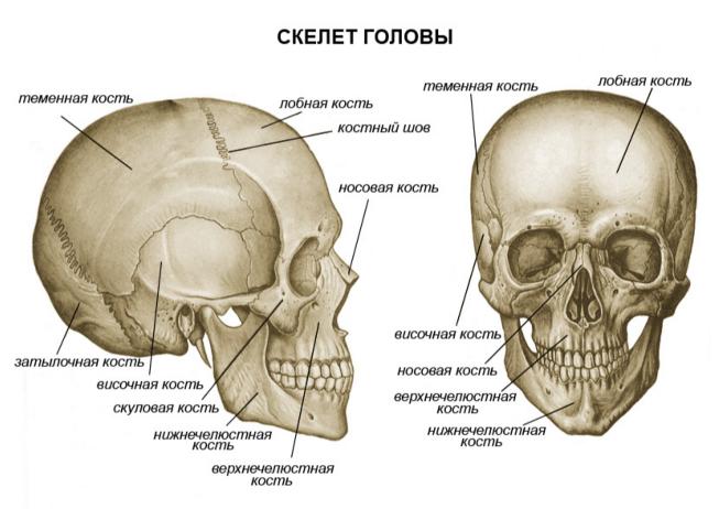
The size and shape of the human body is largely determined by the structural basis - the skeleton. The skeleton provides support and protection for the entire body and individual organs. As part of the skeleton, there is a system of movably articulated levers, set in motion by muscles, due to which various movements of the body and its parts in space are performed. Individual parts of the skeleton serve not only as a receptacle for vital organs, but also provide their protection. For example, the skull, rib cage and pelvis serve as protection for the brain, lungs, heart, intestines, etc.
Until recently, the prevailing opinion was that the role of the skeleton in the human body is limited to the function of body support and participation in movement (this was the reason for the appearance of the term "support locomotor apparatus"). Thanks to modern research the understanding of the functions of the skeleton has expanded significantly. For example, the skeleton is actively involved in metabolism, namely, in maintaining at a certain level mineral composition blood. Substances that make up the skeleton such as calcium, phosphorus, lemon acid and others, if necessary, easily enter into exchange reactions. Muscle function is also not limited to the inclusion of bones in movement and the performance of work, many muscles, surrounding body cavities, protect internal organs.
General information about the skeleton. Bone shape
The human skeleton is similar in structure to the skeleton of higher animals, but has whole line features that are associated with upright posture, movement on two limbs, high development hands and brain.
The human skeleton is a system of 206 bones, of which 85 are paired and 36 are unpaired. Bones are organs of the body. The weight of the skeleton in a man is approximately 18% of the body weight, in a woman - 16%, in a newborn - 14%. The skeleton includes bones of various sizes and shapes.
By shape, bones are divided into:
a) long (located in the skeleton of the limbs);
b) short (located in the wrist and tarsus, that is, where both great strength and mobility of the skeleton are needed);
v) wide or flat (form the walls of the cavities in which the internal organs are located - hip bone, bones of the cerebral skull);
G) mixed (have a different shape).
Bone joints
The bones are articulated different ways... According to the degree of mobility, joints are distinguished: a) motionless; b) sedentary; c) movable joints of bones, or joints.
An immobile joint is formed as a result of bone fusion, while movement may be extremely limited or completely absent. For example, the immobility of the bones of the cerebral skull is ensured by the fact that the numerous protrusions of one bone enter the corresponding depression of the other. This connection of bones is called a suture.
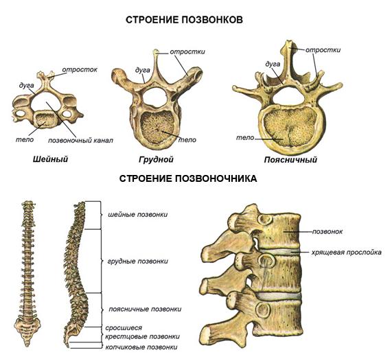
The presence of elastic cartilage pads between the bones allows for little mobility. For example, there are spacers between the individual vertebrae. During muscle contraction, the pads contract and the vertebrae draw closer together. During active movements (walking, running, jumping), the cartilage acts as a shock absorber, thereby softening sharp shocks and protecting the body from shock.
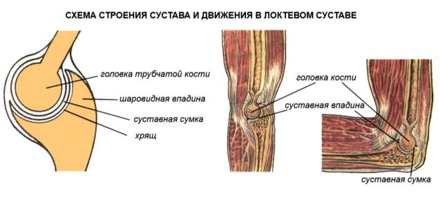
Mobile joints of bones are more common, which is provided by joints. The ends of the bones that form the joint are covered with hyaline cartilage 0.2 to 0.6 mm thick. This cartilage is very elastic, has a smooth shiny surface, therefore friction between the bones is significantly reduced, which greatly facilitates their movement.
From a very dense connective tissue, an articular bag (capsule) is formed, which surrounds the area of articulation of the bones. The strong outer (fibrous) layer of the capsule firmly connects the articulating bones. The inside of the capsule is lined with a synovial membrane. The joint cavity contains synovial fluid, which acts as a lubricant and also helps to reduce friction.
Outside, the joint is reinforced with ligaments. A number of joints are strengthened by ligaments and inside. In addition, there are special devices inside the joints that increase the articulated surfaces: lips, discs, menisci made of connective tissue and cartilage.
The joint cavity is hermetically sealed. The pressure between the articular surfaces is always negative (less than atmospheric), and therefore the external Atmosphere pressure prevents their divergence.
Joint types
According to the shape of the articular surface and along the axes of rotation, joints are distinguished:
a) with three;
b) with two;
v) with one axis of rotation.
The first group consists of spherical joints - the most mobile (for example, the joint between the scapula and humerus). The joint between the ring bone and the thigh, called the nutty joint, is a type of ball joint.
The second group consists of ellipsoidal (for example, the joint between the skull and the first cervical vertebra) and saddle joints (for example, the joint between the metacarpal bone of the first finger and the corresponding wrist bone).
The third group includes block (joints between the phalanges of the fingers), cylindrical (between the ulna and radius) and screw-shaped joints (forming the elbow joint).
Any loose body has six degrees of freedom, because it produces three translational and three rotational movements along the coordinate axes. A fixed body can only rotate. Since all body links are secured, joints with three axes of rotation are the most mobile and have three degrees of freedom. Joints with two axes of rotation are less mobile, therefore they have two degrees of freedom. Joints with one axis of rotation have one degree of freedom, which means the least mobility.
Bone structure
Each bone represents complex organ consisting of bone tissue, periosteum, bone marrow, blood and lymphatic vessels and nerves. With the exception of the connecting surfaces, the entire bone is covered with the periosteum - a thin connective tissue membrane rich in nerves and blood vessels that penetrate from it into the bone through special openings. Ligaments and muscles are attached to the periosteum. The cells that make up the inner layer of the periosteum grow and multiply, which ensures the growth of bone in thickness, and in the event of a fracture, the formation of callus.
By sawing the tubular bone along the long axis, you can see that there is a dense (or compact) bone substance on the surface, and cancellous under it (in depth). V short bones such as vertebrae, spongy substance predominates. Depending on the load on the bone, the compact substance forms a layer of different thickness. The spongy substance is formed by very thin bony beams oriented parallel to the main stress lines. This allows the bone to withstand significant loads.
The dense layer of bone has a lamellar structure and is similar to a system of cylinders inserted into each other, which also gives the bone strength and lightness. Bone cells lie between the plates of the bone substance. Bone plates make up the intercellular substance of bone tissue.
The tubular bone consists of a body (diaphysis) and two ends (pineal glands). On the pineal glands are the articular surfaces, which are covered with cartilage involved in the formation of the joint. On the surface of the bones, there are hillocks, tubercles, grooves, ridges, notches to which muscle tendons are attached, as well as holes through which vessels and nerves pass.
Bone chemistry
Dried and defatted bone has next lineup: organic matter - 30%; minerals - 60%; water - 10%.
Bone organic matter includes fibrous protein (collagen), carbohydrates, and many enzymes.
Bone minerals are represented by salts of calcium, phosphorus, magnesium and many trace elements (such as aluminum, fluorine, manganese, lead, strontium, uranium, cobalt, iron, molybdenum, etc.). The skeleton of an adult contains about 1200 g of calcium, 530 g of phosphorus, 11 g of magnesium, that is, 99% of all calcium in the human body is contained in bones.
In children, organic matter predominates in the bone tissue, therefore their skeleton is more flexible, elastic, easily deformed under prolonged and heavy load or wrong positions body. The amount of minerals in bones increases with age, which makes bones more fragile and more likely to break.
Organic and mineral substances make the bone strong, firm and elastic. The strength of the bone is also ensured by its structure, the location of the spongy bone crossbars in accordance with the direction of the forces of pressure and tension.
Bone is 30 times harder than brick, 2.5 times harder than granite. Bone is stronger than oak. It is nine times stronger than lead and is almost as strong as cast iron. In an upright position, the human femur can withstand the pressure of the load up to 1500 kg, and the tibia - up to 1800 kg.
Development skeletal system in childhood and adolescence
During the period intrauterine development in children, the skeleton is made up of cartilage tissue. Ossification points appear in 7-8 weeks. The newborn has ossified diaphysis of tubular bones. After birth, the process of ossification continues. The timing of the appearance of ossification points and the end of ossification are different for different bones. Moreover, for each bone they are relatively constant, they can be used to judge normal development skeleton in children and their age.
The skeleton of a child differs from that of an adult in its size, proportions, structure and chemical composition. The development of the skeleton in children determines the development of the body (for example, the muscles develop more slowly than the skeleton grows).
There are two pathways for bone development.
1. Primary ossification, when the bones develop directly from the embryonic connective tissue - the mesenchyme (bones of the cranial vault, facial part, partly the clavicle, etc.). First, a skeletal mesenchymal syncytium is formed. In it, cells are laid - osteoblasts, which turn into bone cells - osteocytes, and fibrils, saturated with calcium salts and turning into bone plates. Thus, bone develops from connective tissue.
2. Secondary ossification, when the bones are initially laid in the form of dense mesenchymal formations with approximate outlines of future bones, then turn into cartilaginous tissues and are replaced by bone tissues (bones of the base of the skull, trunk and extremities).
With secondary ossification, the development of bone tissue occurs by replacement both outside and inside. Outside, the formation of bone substance occurs by osteoblasts of the periosteum. Inside, ossification begins with the formation of ossification nuclei, gradually the cartilage is absorbed and replaced by bone. As the bone grows, it is absorbed from the inside by special cells - osteoclasts. The build-up of bone matter goes outside. Bone growth in length occurs due to the formation of bone substance in the cartilage located between the pineal gland and the diaphysis. These cartilages gradually move towards the pineal gland.
Many bones in human body are not laid entirely, but separate parts, which then fuse into a single bone. For example, the pelvic bone first has three parts that fuse together by the age of 14-16. Tubular bones are also laid in three main parts (ossification nuclei in the places of formation of bone protrusions are not taken into account). For example, the embryo's tibia initially consists of solid hyaline cartilage. Ossification begins in the middle around the eighth week intrauterine life... Replacement of the diaphysis with bone occurs gradually and goes first from the outside, and then from the inside. In this case, the epiphyses remain cartilaginous. The ossification nucleus in the upper pineal gland appears after birth, and in the lower - in the second year of life. In the middle part of the epiphyses, the bone first grows from the inside, then from the outside, as a result of which two layers of epiphyseal cartilage that separate the diaphysis from the epiphyses remain.
In the upper epiphysis femur the formation of bone beams occurs at the age of 4–5 years. After 7–8 years, they elongate and become uniform and compact. The thickness of the epiphyseal cartilage reaches 2–2.5 mm by the age of 17-18. By the age of 24, the growth of the upper end of the bone ends and the upper pineal gland grows together with the diaphysis. The lower pineal gland grows to diaphysis even earlier - by the age of 22. With the end of ossification of the tubular bones, their growth in length stops.
Ossification process
General ossification of tubular bones ends by the end of puberty: in women - by 17-21, in men - by 19-24 years. Due to the fact that men puberty ends later than women, they are on average taller.
From five months to one and a half years, that is, when the child gets to his feet, the main development of the lamellar bone occurs. By the age of 2.5–3, the remains of coarse-fibrous tissue are already absent, although during the second year of life, most of the bone tissue has a lamellar structure.
Decreased glandular function internal secretion(anterior pituitary gland, thyroid, parathyroid, thymus, genital) and lack of vitamins (especially vitamin D) can delay ossification. Acceleration of ossification occurs with premature puberty, enhanced function the anterior part of the adenohypophysis, thyroid gland and the adrenal cortex. The delay and acceleration of ossification is most often manifested up to 17-18 years, and the difference between the "bone" and passport ages can reach 5-10 years. Sometimes ossification occurs faster or slower on one side of the body than on the other.
With age, the chemical composition of bones changes. Children's bones contain more organic matter and less inorganic matter. As it grows, the amount of calcium, phosphorus, magnesium and other elements increases significantly, and the ratio between them changes. So, in young children, calcium is most of all retained in the bones, however, as they grow older, a shift towards greater phosphorus retention occurs. Inorganic substances in the composition of the bones of a newborn make up one-half of the weight of a bone, in an adult - four-fifths.
Change in structure and chemical composition bones and their change physical properties... In children, bones are more elastic and less brittle than in adults. Cartilage in children is also more flexible.
The age-related differences in the structure and composition of bones are especially clearly manifested in the number, location, and structure of the Haversian canals. With age, their number decreases, and the location and structure change. How older child, the more dense substance in its bones, in small children there is more spongy substance. By the age of 7, the structure of tubular bones is similar to that of an adult, but between 10–12 years of age the spongy substance of bones changes even more intensively, its structure stabilizes by 18–20 years.
How younger child, the more the periosteum is fused with the bone. The final delineation between bone and periosteum occurs by age 7. By the age of 12, the dense substance of the bone has an almost homogeneous structure, by the age of 15, single areas of resorption of the dense substance completely disappear, and by the age of 17 large osteocytes predominate in it.
From 7 to 10 years of age, the growth of the bone marrow cavity in the tubular bones sharply slows down, and it is finally formed from 11–12 to 18 years of age. The increase in the bone marrow canal occurs in parallel with the uniform growth of dense matter.
Bone marrow is located between the plates of the spongy substance and in the bone marrow canal. Due to the large number of blood vessels in the tissues, newborns have only red bone marrow - hematopoiesis occurs in it. From six months, a gradual process of replacement in the diaphysis of the tubular bones of the red bone marrow with yellow, consisting mostly of fat cells, begins. Replacement of the red brain ends by 12-15 years. In adults, red bone marrow is preserved in the epiphyses of tubular bones, in the sternum, ribs and spine and is approximately 1500 cubic meters. cm.
Healing of fractures and the formation of callus in children occurs after 21-25 days, in infants this process is even faster. Dislocations in children under 10 years of age are rare due to the great extensibility of the ligamentous apparatus.
Consists of a skeleton and muscles, it performs the following functions:
Protective (limits the cavities in which the internal organs are located);
Support function;
Provides active human movements;
Performs a hematopoietic function;
Participates in the metabolism.
The passive part of the support motor system a skeleton made up of bones, cartilage, joints and ligaments. There are more than 200 bones in the human skeleton.
Each bone is an organ made up of bone tissue.
Bone= cells with processes + intercellular substance + nerves + vessels + connective tissue membrane
Bones:
(bone properties): organic matter (flexibility and elasticity), inorganic matter (hardness).
Growth direction (source of new cells): in length (cartilage), in thickness (periosteum).
Joint of bones: movable, semi-movable, immovable
Joint- articulating bone with the glenoid cavity + articulated bone with the head + strong ligaments + articular capsule + articular fluid
Human skeleton consists of 200 bones.
Main departments:
Muscle- the active part of the musculoskeletal system, providing all the variety of movements performed in the human body. Thanks to the muscles, the body maintains balance, moves in space, breathing movements are carried out by the chest and diaphragm, swallowing, a voice is formed, eye movements are carried out, the work of internal organs, including the heart. A person has two types of muscles: smooth and striated.
Smooth muscles are found in internal organs: the walls of blood vessels, bladder, ureters, intestines. Their reduction is arbitrary.
The striated muscles provide muscle attachment to the tendons and bones of the skeleton. Skeletal muscles set in motion the bones relative to each other in the compositions, in addition, they are involved in the formation of the walls of the abdominal and chest cavities, the pelvis. Are part of the wall of the upper part of the esophagus and larynx. The movement of the apple, respiratory and swallowing movements are carried out. All skeletal muscles can be divided into two groups - flexors and extensors.
The facial muscles are the muscles of the face that are not associated with the joints.
The heart muscle is a special striated muscle, where the fibers are connected, and it contracts rapidly.
In humans, every muscle contains all types of muscle fibers; their ratio varies depending on the purpose of each muscle. For each muscle, there are blood vessels that penetrate the outer shell and break down in the muscle into a network of capillaries. Through the blood, muscle fibers are supplied with oxygen and nutrients. In addition, a nerve is connected to each muscle, which transmits signals.
Abstract in biology on the topic:
Pupil 9 "G" class
high school № 117
South-West Administrative District Moscow
Yuditsky Alexander.
Moscow 2004
Plan:
I. Introduction.
II. Skeleton.
1. The spine.
2. Chest.
3. Extremities.
4. Leg and arm.
III. Two types of muscle tissue.
1. Smooth muscles.
2.Muscles of the skeleton.
3. Nerve connections in the muscles.
4. Muscles generate heat.
5. Strength and speed of muscle contraction.
IV. Fatigue and rest.
1. Causes of fatigue.
V. Statics and dynamics human body.
1. Conditions of balance.
Vi. Everyone needs sports.
1.Training muscles.
2. Labor and sports.
3. Anyone can become an athlete.
Vii.
VIII. Conclusion.
XI.
Musculoskeletal system
The musculoskeletal system consists of the bones of the skeleton with joints, ligaments and muscles with tendons, which, along with movements, provide the supporting function of the body. Bones and joints are passively involved in movement, obeying the action of muscles, but they play a leading role in the implementation of the supporting function. A certain shape and structure of bones give them great strength, the reserve of which for compression, expansion, bending significantly exceeds the loads possible during the daily work of the musculoskeletal system. For example, the human tibia, when compressed, withstands a load of more than a ton, and is almost as strong as cast iron in tensile strength. Ligaments and cartilages also have a large margin of safety.
The skeleton is made up of interconnected bones. It provides our body with support and shape retention, as well as protects the internal organs. In an adult, the skeleton consists of approximately 200 bones. Each bone has a certain shape, size and occupies a certain position in the skeleton. Some of the bones are connected by movable joints. They are set in motion by the muscles attached to them.
Spine. The spine is the original structure that constitutes the main support of the skeleton. If it consisted of a solid bone rod, then our movements would be constrained, devoid of flexibility and would deliver the same discomfort like riding in a cart without springs on a cobblestone pavement.
The elasticity of hundreds of ligaments, cartilaginous layers and bends makes the spine strong and flexible support. Thanks to this structure of the spine, a person can bend, jump, tumble, and run. Very strong intervertebral ligaments allow the most difficult movements and at the same time create reliable protection spinal cord. It is not subjected to any mechanical stretching, pressure at the most incredible bends of the spine.
The bends of the spinal column correspond to the effect of the load on the skeletal axis. Therefore, the lower, more massive part becomes a support when moving; the upper one, with free movement, helps to maintain balance. Vertebral column could be called a vertebral spring.
The undulating curves of the spine provide elasticity. They appear with development motor abilities the child, when he begins to hold his head, stand, walk.
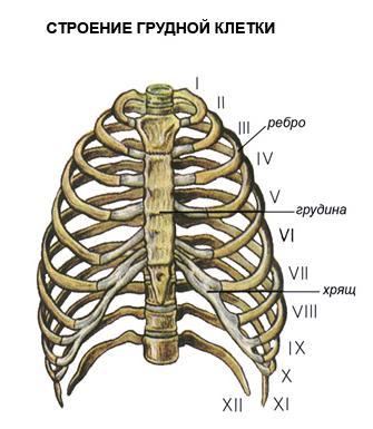
Rib cage. The rib cage is formed by the thoracic vertebrae, twelve pairs of ribs, and the flat sternum, or sternum. The ribs are flat, arched bones. Their posterior ends are movably connected to the thoracic vertebrae, and the anterior ends of the ten upper ribs are connected to the sternum using flexible cartilage. It provides mobility chest when breathing. The two lower pairs of edges are shorter than the others and end freely. The rib cage protects the heart and lungs, as well as the liver and stomach.
It is interesting to note that the ossification of the chest occurs later than other bones. By the age of twenty, the ossification of the ribs ends, and only by the age of thirty there is a complete fusion of the parts of the sternum, consisting of the handle, the body of the sternum and the xiphoid process.
The shape of the chest changes with age. In a newborn, it has, as a rule, the shape of a cone with the base facing downward. Then the chest circumference in the first three years increases faster than the length of the body. Gradually, the thorax from a cone-shaped one acquires a characteristic for a person rounded shape... Its diameter is greater than its length.
The development of the chest depends on the lifestyle of the person. Compare the athlete, swimmer, athlete with a person who does not play sports. It is easy to understand that the development of the chest, its mobility depends on the development of the muscles. Therefore, in adolescents of twelve to fifteen years old who are involved in sports, the chest circumference is seven to eight centimeters larger than that of their peers who do not go in for sports.
Improper seating of students at the desk, compression of the chest can lead to its deformation, which disrupts the development of the heart, large vessels and lungs.
Extremities. Due to the fact that the limbs are attached to a reliable support, they have mobility in all directions, and are able to withstand heavy physical exertion.
The light bones - the clavicle and shoulder blades, which lie on the upper part of the rib cage, encircle it like a belt. This is the support of the arms. The ridges and ridges on the clavicle and scapula are where the muscles attach. The greater the strength of these muscles, the more developed bone processes and irregularities. In an athlete, a loader, the longitudinal ridge of the scapula is more developed than in a watchmaker or bookkeeper. The clavicle is a bridging bridge between the bones of the trunk and arms. The scapula and collarbone provide a reliable spring support for the hand.
By the position of the shoulder blades and collarbones, one can judge the position of the hands. Anatomists helped restore broken arms ancient greek statue Venus de Milo, determining their position by the silhouettes of the shoulder blades and collarbones.
The pelvic bones are thick, wide and almost completely fused. In humans, the pelvis lives up to its name - it, like a bowl, supports the internal organs from below. This is one of the typical features of the human skeleton. The massiveness of the pelvis is proportional to the massiveness of the bones of the legs, which bear the main load when a person moves, therefore, the skeleton of the human pelvis can withstand a great load.
Leg and arm. With an upright posture, a person's hands do not carry a constant load as support, they acquire lightness and variety of action, freedom of movement. The hand can perform hundreds of thousands of different motor operations. The legs, on the other hand, bear the entire weight of the body. They are massive, have extremely strong bones and ligaments.
The head of the shoulder has no limitation in wide circular motions hands, for example when throwing a javelin. The head of the femur protrudes deeply into the depression of the pelvis, which limits movement. The ligaments of this joint are the strongest and keep the weight of the torso on the hips.
Exercise and training achieve great freedom of movement of the legs, despite their massiveness. Ballet art, gymnastics, martial arts can be a convincing example of this.
The tubular bones of the arms and legs have a huge margin of safety. It is interesting that the arrangement of the openwork beams of the Eiffel Tower corresponds to the structure of the spongy substance of the heads of the tubular bones, as if J. Eiffel designed the bones. The engineer used the same structural laws that govern the structure of the bone, giving it lightness and strength. This is the reason for the similarity between the metal structure and the living bone structure.
Elbow joint provides complex and varied hand movements in a person's working life. Only he is characterized by the ability to rotate the forearm around its axis, with a characteristic movement of unwinding or twisting.
Knee-joint guides the lower leg when walking, running, jumping. The knee ligaments in humans determine the strength of the support when the limb is straightened.
The hand begins with a group of bones in the wrist. These bones do not experience strong pressure, they perform a similar function, so they are small, monotonous, and difficult to distinguish. It is interesting to mention that the great anatomist Andrei Vesalius could, blindfolded, identify each carpal bone and say whether it belongs to the left or right hand.
The bones of the metacarpus are moderately mobile, they are located in the form of a fan and serve as a support for the fingers. Phalanges of fingers - 14. All fingers have three bones, except for the thumb - it has two bones. A person has a very mobile thumb. It can become at right angles to everyone else. Its metacarpal bone is able to oppose the rest of the bones of the hand.
Development thumb associated with labor movements of the hand. The Indians call the thumb "mother", the Javanese call the "big brother." In ancient times, captives were cut off their thumb in order to humiliate their human dignity and make them unfit for participation in battles.
The brush makes the most subtle movements. In any working position of the hand, the hand retains complete freedom movement.
The foot became more massive due to walking. The bones of the tarsus are very large and strong in comparison with the bones of the wrist. The largest of them are the talus and calcaneus. They can withstand considerable body weight. In newborns, the movements of the foot and its thumb are similar to their movements in monkeys. Strengthening the supporting role of the foot during walking led to the formation of its arch. When walking, standing, you can easily feel how the entire space between these points "hangs in the air."
A vault, as is known in mechanics, can withstand more pressure than a platform. The arch of the foot provides elasticity of the gait, eliminates pressure on the nerves and blood vessels. His education in the history of human origin is associated with upright posture and is distinctive feature a person acquired in the process of his historical development.
Two types of muscle tissue.
Smooth muscles. When we talked about muscle, we usually thought of skeletal muscle. But, besides them, in our body in the connective tissue there are smooth muscles in the form of single cells, in some places they are collected in bundles.
There are many smooth muscles in the skin, they are located at the base of the hair follicle. By contracting, these muscles lift the hair and squeeze oil out of the sebaceous gland.
In the eye, smooth annular and radial muscles are located around the pupil. They work all the time, imperceptibly for us: in bright light, the annular muscles constrict the pupil, and in the dark, the radial muscles contract and the pupil expands.
In the walls of all tubular organs - respiratory tract, vessels, digestive tract, urethra and others - there is a layer of smooth muscles. It contracts under the influence of nerve impulses. For example, reducing it in the windpipe delays the flow of air containing harmful impurities - dust, gases.
Due to the contraction and relaxation of the smooth cells of the walls of blood vessels, their lumen either narrows or expands, which contributes to the distribution of blood in the body. The smooth muscles of the esophagus contract and push a lump of food or a sip of water into the stomach.
Complex plexuses of smooth muscle cells are formed in organs with a wide cavity - in the stomach, bladder, uterus. The contraction of these cells causes compression and narrowing of the lumen of the organ. The force of each contraction of the cells is negligible, since they are very small. However, the addition of the forces of entire beams can create a tremendous force contraction. Powerful contractions create a sensation of intense pain.
Muscles of the skeleton. Skeletal muscles carry out both static activity, fixing the body in a certain position, and dynamic, ensuring the movement of the body in space and its individual parts relative to each other. Both types of muscle activity interact closely, complementing each other: static activity provides a natural backdrop for dynamic activity. As a rule, the position of the joint is changed with the help of several muscles in different directions, including the opposite action. Complex movements joints are performed by coordinated, simultaneous or sequential contraction of muscles of undirected action. Consistency (coordination) is especially necessary for the performance of motor acts in which many joints are involved (for example, skiing, swimming).
Skeletal muscles are not only the executive motor apparatus, but also a kind of sense organs. Muscle fibers and tendons contain nerve endings - receptors that send impulses to cells at various levels of the central nervous system. As a result, a closed cycle is created: impulses from various formations of the central nervous system, going along the motor nerves, cause muscle contraction, and impulses sent by muscle receptors inform the central nervous system about each element of the system. The cyclical system of links ensures the accuracy of movements and their coordination. Although the control of the movement of skeletal muscles is carried out by various sections of the central nervous system, the leading role in ensuring interaction and goal setting motor reaction belongs to the cerebral cortex. In the cerebral cortex, the motor and sensory zones of the representations form a single system, with each muscle group corresponding to a certain section of these zones. Such a relationship allows you to perform movements, referring them to factors acting on the body environment... The control of voluntary movements can be schematically represented as follows. The tasks and purpose of a motor action are formed by thinking, which determines the direction of a person's attention and efforts. Thought and emotions accumulate and direct these efforts. The mechanisms of the highest nervous activity form the interaction of psychophysiological mechanisms of motion control at various levels. On the basis of the interaction of the musculoskeletal system, the deployment and correction of motor activity are provided. Analyzers play an important role in the implementation of the motor reaction. The motor analyzer provides the dynamics and interconnection of muscle contractions, participates in the spatial and temporal organization of the motor act. The balance analyzer, or vestibular analyzer, interacts with the motor analyzer when changing the position of the body in space. Vision and hearing, actively perceiving information from the environment, participate in spatial orientation and correction of motor reactions.
The name "muscle" comes from the word "muscle", which means "mouse".
This is due to the fact that anatomists, observing the contraction of skeletal muscles, noticed that they seemed to run under the skin, like mice.
The muscle is made up of muscle plexuses. The length of the muscle plexuses in humans reaches 12 cm. Each such plexus forms a separate muscle fiber.
Numerous rod-shaped nuclei are located under the muscle fiber sheath. Along the entire length of the cell, there are several hundred thinnest filaments of the cytoplasm - myofibrils, capable of contracting. In turn, myofibrils are formed by 2.5 thousand protein filaments.
In myofibrils, light and dark discs alternate, and under a microscope the muscle fiber looks transversely striated. Let's compare the function of skeletal and smooth muscles. It turns out that the striated musculature cannot lengthen as much as the smooth one. On the other hand, skeletal muscles contract faster than the muscles of internal organs. It is not difficult, therefore, to explain why a snail or an earthworm, devoid of striated musculature, moves slowly. The swiftness of movements of a bee, lizard, eagle, horse, man is ensured by the rapidity of contraction of the striated muscles.
Muscle fiber thickness different people not the same. For those who go in for sports, muscle fibers develop well, their mass is large, which means that the contraction force is also great. The limited work of muscles leads to a significant reduction in the thickness of the fibers and muscle mass in general, and entails a decrease in the force of contraction.
There are 656 skeletal muscles in the human body. Almost all muscles are paired. The position of the muscles, their shape, the method of attachment to the bones have been studied in detail by anatomy. The location and structure of muscles is especially important for the surgeon to know. That is why the surgeon is primarily an anatomist, and anatomy and surgery are sisters. World services in the development of these sciences belong to our national science, and above all to N.I. Pirogov.
Nerve connections in the muscles. It is wrong to think that the muscle itself can contract. It would be difficult to imagine even one coordinated movement if the muscles were uncontrollable. Nerve impulses "start up" the muscle. One muscle receives an average of 20 impulses per second. For example, up to 300 muscles take part in each step, and many impulses coordinate their work.
The number of nerve endings in different muscles is not the same. There are relatively few of them in the muscles of the thigh, and the oculomotor muscles, which perform fine and precise movements all day long, are rich in motor nerve endings. The cerebral cortex is unevenly connected with individual muscle groups. For example, huge areas of the cortex are occupied by motor areas that control the muscles of the face, hand, lips, feet, and relatively insignificant areas - by the muscles of the shoulder, thigh, and lower leg. The magnitude separate zones the motor area of the cortex is proportional not to the mass of muscle tissue, but to the subtlety and complexity of the movements of the corresponding organs.
Each muscle has a double neural subordination. One nerves send impulses from the head and spinal cord... They cause muscle contraction. Others, moving away from the nodes that lie on the sides of the spinal cord, regulate their nutrition.
Nerve signals governing muscle movement and nutrition are consistent with neural regulation of muscle blood supply. It turns out a single triple nervous control.
Muscles generate heat. The striated muscles are "engines" in which chemical energy is immediately converted into mechanical energy. The muscle uses 33% of the chemical energy for movement, which is released during the breakdown of animal starch - glycogen. 67% of the energy in the form of heat is transferred by the blood to other tissues and warms the body evenly. That is why in the cold a person tries to move more, as if warming himself up at the expense of the energy generated by the muscles. Small involuntary muscle contractions cause tremors - the body increases the generation of heat.
The strength and speed of muscle contraction. The strength of a muscle depends on the number of muscle fibers, its cross-sectional area, the size of the surface of the bone to which it is attached, the angle of attachment and the frequency of nerve impulses. All these factors have been identified by special studies.
The strength of a person's muscles is determined by what kind of load he can lift. Muscles outside the body develop strength several times greater than that which manifests itself in human movements.
The working quality of a muscle is associated with its ability to suddenly change its elasticity. Muscle protein becomes very elastic when contracted. After muscle contraction, it again regains its original state. Becoming elastic, the muscle holds the load, this is where muscle strength is manifested. A human muscle for every square centimeter of section develops a force of up to 156.8 N.
One of the strongest muscles is the calf. It can lift 130 kg. Every healthy person is able to "stand on tiptoe" on one leg and even lift an additional load at the same time. This load falls mainly on the gastrocnemius muscle.
Under the influence of constant nerve impulses, the muscles of our body are always tense, or, as they say, are in a state of tone - prolonged contraction. You can check the muscle tone on yourself: close your eyes with force, and you will feel the trembling of the contracted muscles in the eye area.
It is known that any muscle can contract with different strengths. For example, in raising small stone and a pound weight, the same muscles are involved, but they expend different strength. The speed at which we can set our muscles in motion is different and depends on the training of the body. The violinist performs 10 movements per second, and the pianist up to 40.
Fatigue and rest
The causes of fatigue. Fatigue is an indicator that the body cannot work in full force... Why does muscle fatigue occur? For science, this question has long been unresolved. Various theories were created.
Some scientists have suggested that the muscle is depleted from a lack nutrients; Others said that she was “suffocated”, lack of oxygen. It has been suggested that fatigue occurs due to poisoning, or clogging, of the muscle with poisonous waste products. However, none of these theories have satisfactorily explained the causes of fatigue. As a result, it has been suggested that the cause of fatigue does not lie in the muscle. It was hypothesized about nerve fatigue. However, an outstanding Russian physiologist, one of the students of I.M.Sechenov, Professor N.E. Vvdensky proved by example that nerve conductors are practically not fatigued.
The path to solving the mystery of fatigue was discovered by the Russian physiologist I. M. Sechenov. He developed the nervous theory of fatigue. He found that the right hand, after prolonged work, restored its working capacity if, during the period of its rest, movements were made with the left hand. The nerve centers of the left hand seemed to energize the tired nerve centers right hand... It turned out that fatigue is removed more quickly when the rest of the working hand is combined with the work of the other hand than with complete rest. With these experiments, I.M.Sechenov outlined the ways of relieving fatigue and the ways of their rational organization of rest, thereby realizing his noble desire to facilitate human labor.
Human body statics and dynamics
Equilibrium conditions. Every body has a mass and a center of gravity. The plumb line through the center of gravity (line of gravity) always falls on the support. The lower the center of gravity and the wider the support, the more stable the balance. So, when standing, the center of gravity is placed approximately at the level of the second sacral vertebra. The line of gravity is between both feet, inside the support area.
The stability of the body is significantly increased if the legs are apart: the area of support increases. As the legs come closer together, the support area decreases, and, consequently, stability decreases. The stability of a person standing on one leg is even less.
Our body is very mobile, and the center of gravity is constantly shifting. For example, when you carry a bucket of water in one hand, for stability you bend over in opposite side, with the other hand stretching out almost horizontally. If you carry a heavy object on your back, then the body leans forward. In all these cases, the line of gravity approaches the edge of the support, so the balance of the body is stable. If the projection of the center of gravity of the body goes beyond the area of support, the body will fall. Its stability is ensured by a shift in the center of gravity, corresponding to a change in body position. To create a counterweight, the torso is tilted to the opposite side of the load. The line of gravity remains within the support area.
By performing various gymnastic exercises, you can determine how balance and stability are maintained if the center of gravity goes beyond the fulcrum.
For greater stability, rope-walkers take a pole in their hands, which they tilt in one direction or the other. By balancing, they move the center of gravity to a limited support.
Everyone needs sports
Muscle training. Active physical activity- one of mandatory conditions harmonious development person.
Constant exercise lengthens the muscles, developing their ability to stretch better. During training, muscle mass increases, muscles become stronger, nerve impulses cause muscle contraction of great strength.
Muscle strength and bone strength are intertwined. When playing sports, the bones become thicker, and accordingly the developed muscles have sufficient support. The entire skeleton becomes stronger and more resistant to stress and injury. Good motor load - necessary condition normal growth and the development of the body. A sedentary lifestyle is harmful to health. Lack of movement is the cause of muscle laxity and weakness. Physical exercises, labor, games develop efficiency, endurance, strength, agility and speed.
Labor and sports. Movements in work and sports are forms of muscle activity. Labor and sport are interconnected and complement each other.
Two students came to the workshop, stood at the workbench for the first time. One goes in for sports, the other does not. It is easy to see how quickly an athlete learns labor skills.
Sport develops important motor qualities- agility, speed, strength, endurance.
These qualities are also improved in work.
Labor and physical education help each other. They are conducive to mental work. When moving, the brain receives from the muscles an abundance of nerve signals that support it normal condition and develop. Overcoming fatigue during physical labor increases performance during mental work.
Anyone can become an athlete. Do I need to have any natural qualities to become an athlete? There can be only one answer: no. Diligence and systematic training ensure the achievement of high sports results. Sometimes it is advisable to consider general features physique for choosing a particular sport.
And this is not always necessary. Some athletes have achieved first-class results in sports for which they seem to have no data. Vitaly Ushakov, despite the small lung capacity before playing sports, became a first-class swimmer and gave better performance than some other athletes with "natural buoyancy".
The famous wrestler I.M.Poddubny wrote that wrestlers are not born, wrestling develops a person and he, from an ordinary boy, becomes a mighty strongman.
Desire and perseverance, training and thoughtful attitude to physical activities do miracles. Even sick, physically weak and pampered people can be great athletes. For example, the European champion in race walking A.I. Yegorov suffered from rickets in childhood, did not walk until 5 years old. Under the supervision of a doctor, he began to play sports and achieved high performance.
Great people about the benefits of exercise.
Gymnastics as a means of physical education originated in Ancient China and India, but especially developed in Ancient Greece... The nude Greeks went in for sports under the rays of the southern sun. Hence, in fact, the word "gymnastics" comes from: translated from the ancient Greek "hymnos" means "naked".
Even the great thinkers of antiquity Plato, Aristotle, Socrates noted the influence of movements on the body. They themselves were engaged in gymnastics to a ripe old age.
MV Lomonosov was the first to raise his voice in defense of the health of the Russian people. He himself was great physical strength and athletic build. Lomonosov considered it necessary "to try in every possible way to be in the movement of the body." He thought to introduce Olympic Games in Russia. The great scientist spoke about the benefits of physical activity after strenuous mental work. "Movement," he said, "can serve as a medicine."
A.I. Radishchev deeply believed that physical education you can "strengthen the body, and with it the spirit."
A. V. Suvorov introduced, and did military gymnastics himself, demanded training and tempering of the troops. "My offspring," said the great commander, "I ask you to take my example."
A.S. Pushkin's contemporaries wrote about him that he was of the strongest build, muscular, flexible, and gymnastics contributed to this.
LN Tolstoy was fond of cycling, horseback riding. At 82 years old, he made horseback walks 20 miles or more per day. He loved to mow, dig, saw. At the age of 70, Tolstoy won ice skating among young people who visited Yasnaya Polyana. He wrote: “With diligent mental work without movement and bodily labor, sheer grief. I don’t walk, don’t work my legs and arms for at least one day, in the evening I’m no longer fit: neither read, nor write, nor even listen carefully to others, my head is spinning, but there are some stars in my eyes, and the night is spent without sleep ".
Maxim Gorky was fond of rowing, swimming, playing in small towns, in winter he went skiing and skating.
I.P. Pavlov went in for sports to a ripe old age and loved physical work... For many years he led the gymnastic circle of doctors in St. Petersburg.
Conclusion
In legends, the Russian people endowed their heroes with extraordinary strength, glorified their heroic deeds in labor and in defending the Motherland from enemies. Labor and love for native land in the minds of the people are inseparable from each other.
The epics and legends reflect the features of our people - hard work, courage, mighty strength. The Arabic writer of the 11th century Abubekri wrote that the Slavs are a people so powerful that if they were not divided into many clans, no one would have been able to resist them.
The struggle with the harsh nature, external enemies has developed in them qualities worthy of admiration. Strong, freedom-loving, tempered, not afraid of either cold or heat, not spoiled by excesses and luxury - such were our ancestors, even according to the description of their enemies.
List of used literature.
1. "Reserves of the body" B. P. Nikitin, L. A. Nikitina. 1990 year
2. "A book for reading on human anatomy, physiology and hygiene." I. D. Zverev, 1983
3. "Russian power". Valentin Lavrov. 1991 year
4. "Secrets of Athleticism". Yuri Shaposhnikov. 1991 year
5. "Biology Human Grade 9". A.S.Batuev. 1997 year
6. www.referat.ru
Approximately one in twentieth - osteoarthritis, one in ten - regularly manifested, and from time to time or just a single experience more than 70% of the population. Problems with the musculoskeletal system are so common mainly due to the irresponsible attitude to this aspect, while prevention measures require almost no special effort.
What is it
The human musculoskeletal system is a systemically interconnected set of bones (forming the skeleton) and their joints, which allows a person to control (by means of impulses transmitted through the nervous system by the brain) the body, its statics and dynamics. The importance of the human musculoskeletal system can hardly be overestimated. A person whose SLM is not fulfilling its functions in best case- a disabled person or a paralytic lying in a layer.
Did you know? One of the founders of anatomy in its modern, scientific form was Leonardo da Vinci. He, along with other scientists and researchers of the Renaissance, performed autopsies to understand the structure of the human body.
Have healthy person functions of ODA are divided into mechanical and biological.
Basic mechanical functions
Mechanical functions are associated with the preservation of the structure and movement of the body in space.
Support
It consists in forming the basis for the rest of the body - muscles, tissues and organs are attached to the skeleton. Due to the skeleton and the muscles attached to it, a person can stand upright, his organs maintain a relatively static position relative to the axis of symmetry and to each other.
Protective
Bones protect the most important internal organs from mechanical damage: the head is protected by the skull, the dorsal - by the spine, the internal organs of the chest (lungs and others) are hidden behind the ribs, the genitals are closed by the bones of the pelvis. 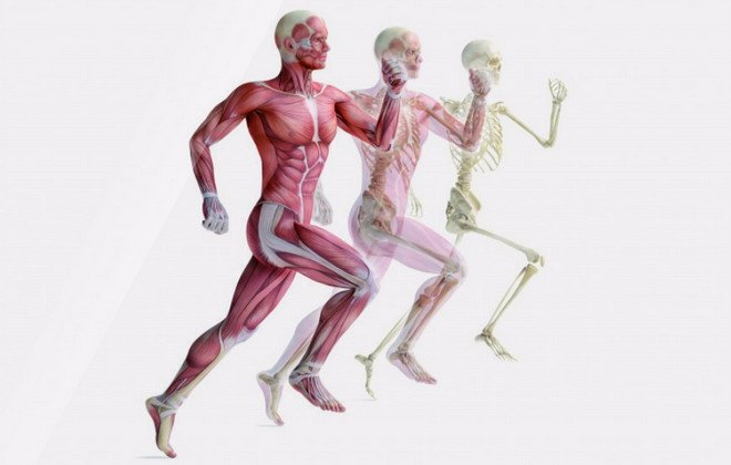 It is this protection that provides us with resistance to external influences and well trained muscles can enhance this effect.
Did you know? At the time of our birth, we have the most bones - 300. Subsequently, some grow together (and all become stronger) and their total number decreases to 206.
Motor
The most prominent function of the human musculoskeletal system. The building muscles are attached to the skeleton. Due to their contractions, various movements are performed: flexion / extension of the limbs, walking and much more.
Actually, this is one of the main differences between the representatives of the biological kingdom "Animals" - conscious and controlled movement in space.
Leaf springs
Mitigation (amortization) of movements due to the structure and position of bones and cartilage. 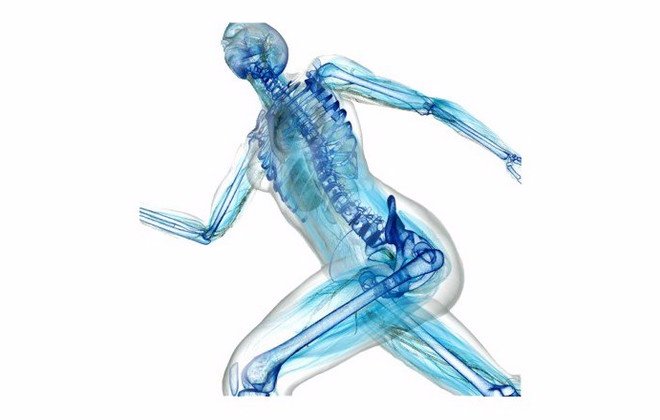 It is provided both by the shape of the bones (for example, the curvature of the foot, strong tibia - an evolutionary mechanism, most suitable for walking upright and supporting body weight with an emphasis on only one pair of limbs), and auxiliary tissues - cartilage and articular bags provide a decrease in bone friction in their places joints.
Biological functions of the system
The musculoskeletal system also has other functions that are important for life.
Hematopoietic
The process of blood formation occurs in the so-called red bone marrow, but due to its location (in the tubular bones), this function is also referred to as ODA.
In the red bone marrow, hematopoiesis (hematopoiesis) occurs - the creation of new blood cells, and partially immunopoiesis - the maturation of cells that take part in the work of the immune system.
Storing
In bones it accumulates and is stored a large number of necessary for the body substances such as, and. From there they flow to other organs, where they are included in the metabolic process. 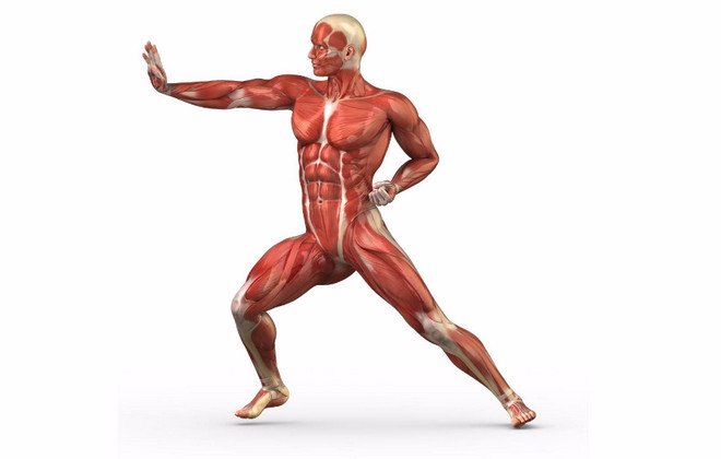 Due to these substances, the strength of bones and their resistance to external influences, as well as the rate of healing after fractures, are ensured.
Important! Calcium problems are often caused not by inadequate intake, but by rapid washout. Popular foods such as sugary sodas and oxalic acid... It is better to exclude all this from the diet.
Major problems and injuries of the ODA
Although the formation of the musculoskeletal system occurs in, its development is a process that continues throughout.
The causes of problems with ODE, as well as their consequences, can be different:- Wrong load (under or over).
- Inflammatory processes that affect bone tissue, muscle, or cartilage. The diagnosis also differs depending on the etiology and localization.
- Disorders associated with metabolism, deficiency or excess of any elements.
- Mechanical injuries (bruises, fractures) and the consequences of improper treatment.
Diseases of the musculoskeletal system
Diseases affecting our musculoskeletal system are depressing in their variety:
- Arthritis affects the joints, can flow into arthrosis.
- Infections can settle in the periarticular bursa (bursitis), muscles (myotitis), bone marrow (osteomyelitis), and large joints (periarthritis).
- The spine can bend, the ankle can lose tone.
Important! For any pain, see your doctor! On early stages ODE diseases are treated with simple and gentle methods: physical or manual therapy, therapeutic. If the disease is in a severe stage, treatment and rehabilitation will be long and difficult.
Sports injuries
Of course, with due "luck", you can fall out of the blue, and at the same time break something unexpected.
However, according to statistics, the most frequent injuries when playing sports are: muscle sprains, various injuries to the lower leg, fractures (mainly legs are affected) and ruptures (ligaments, cartilage or tendons). 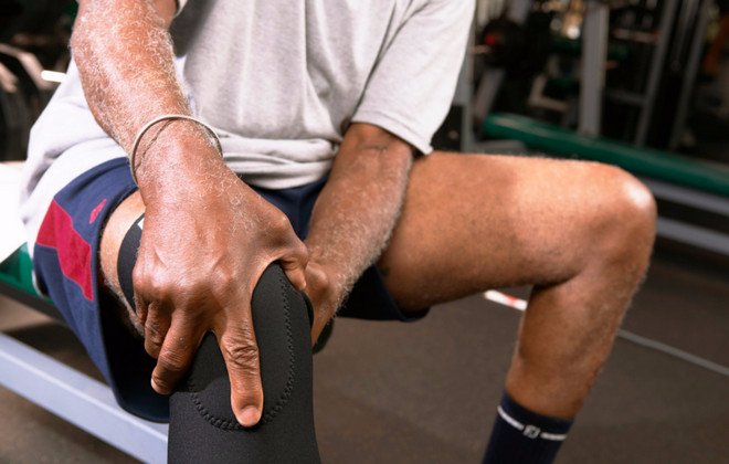
Keeping healthy: how to prevent troubles
To keep the body in good shape, and the ODA in working and healthy condition, it is important to know what measures to take to maintain normal functions of the musculoskeletal system.
Nothing supernatural is required:
- Healthy lifestyle.
- Balanced diet, rich in calcium and other minerals and trace elements.
- Regular physical activity, suitable for age and health.
- Walking in the sun (vitamin D) and fresh air.
- Maintaining optimal body weight (obesity, like dystrophy, are the enemies of ODA).
- Convenient workplace.
- Regular checkups.
As you can see, if you support the organism as a whole, everything will be in order with its systems. You don't have to go in for sports professionally. 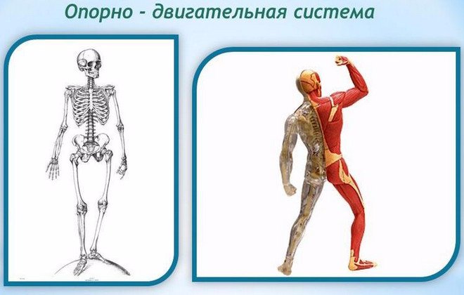 It will be enough not to neglect motor activity(in any form convenient for you, whether it is yoga, swimming or ordinary walks in the park), observe the daily regimen and maintain healthy diet nutrition. It is not so difficult. Do not be ill!