How to kiss correctly: useful tips. How to properly overshadow yourself with the Sign of the Cross
Joint destruction is practically the leader in the group of diseases in the field of degenerative dystrophy of the musculoskeletal system. In most cases, wear and tear of the joints is called osteoarthritis of the hip joints. The reasons that destroy the joints are very diverse, because most of those who are faced with such a disease are people who have crossed the age group of 40 years. Any pathology, and even such as osteoarthritis, can be cured without surgery, but subject to diagnosis at the earliest stage. The problem of many people is the lack of desire to be healthy and to visit doctors on time for examination and treatment. That is why diseases are progressing every month more and more, and the degrees are becoming more serious.
There are two types of osteoarthritis:
- primary;
- repeated.
In medical practice, destruction can occur both in one joint, and in two at the same time. It is worth noting that the manifestation of bilateral destruction of the hip joint is quite common, and with the primary lesion, the knees and vertebral discs are disturbed. If the knee joint is destroyed, treatment should be immediate.
How does osteoarthritis spread
At the initial stage of the disease, a number of abnormal pathologies can be noted in the property of the articular fluid, namely, under certain conditions, its composition acquires a viscous and thick state.

As soon as the viscosity increases, the natural process of the appearance of lubrication of the articular cartilage begins to disappear, leading to drying out and the appearance of roughness and cracks. During the development of such an anomaly, the cartilage becomes thinner and begins to weaken with daily stress on the joints, since friction during movement remains the same, but the body's indicators are much worse than in a healthy state. The length of the distance among the articulating bones of the joints becomes much less. Bones are exposed from under the cartilage, and the pressure on them begins to increase, which can lead to deformation. That is why the disease of osteoarthritis has a different terminology for deforming arthrosis of the hip joint.
There are a number of other destructions that affect joints, in addition to the process of changing joint fluids. They consist in improper blood circulation throughout the body, which can lead to a slowdown in metabolism in the articular parts by reducing the circulation of blood fluid through its capillaries and blood vessels. Gradually, every muscle in the area of \u200b\u200bthe spread of the disease begins to atrophy.
This process can be slow and very fast. Often, doctors are faced with a chronic course of the disease, accompanied by temporary severe exacerbations of pain in the articular parts. In another way, this period can be called "reactive".
With such manifestations, it is advisable to immediately contact the medical aid stations to solve the problem.
What are the reasons for the destruction of the hip joint
 There can be many reasons why the destruction of the hip joint may appear. But there are those that can be found most often.
In the presence of disturbances in the movement of blood through the joints, its outflow and inflow deteriorate. The consequence of such anomalies may be insufficient penetration of nutrients into the bone tissue, as well as the accumulation of an under-oxidized amount of products released during metabolism and activated enzymes that destroy the cartilage themselves.
- With a mechanical factor that causes constant overload of the joints, such ailments as destruction are exposed to those who are professionally related to sports. The same group of people with overload immediately includes those who suffer from overweight. Fat people already have metabolic and circulatory disorders, and with an additional effect on the joints with weight, the rapid appearance and spread of arthrosis occurs.
- A biochemical change in cartilage, impaired metabolism of the human body and the presence of hormonal problems can lead to arthrosis.
- The presence of injuries is considered a common prerequisite for the appearance of destruction of the hip joint. It is from frequent fractures and dislocations that even the youngest organism can be susceptible to such ailment.
- The presence in the body of antiseptic necrosis of the heads of the hip joints.
- Infection and inflammation of the articular regions.
- Anomaly in the spine and feet.
- Dysplasia at birth.
- Age group.
- The presence of a hereditary predisposition. The very fact of destruction is not transmitted by genes, but the presence of weakness in the development of bones, thin cartilage tissues, as a result, can lead to such consequences. Those whose relatives already have a diagnosis of osteoarthritis, it is advisable to take more careful care of their health and protect themselves from possible fractures and falls.
What are the symptoms of the disease
The process of destruction of the hip joint can have different symptoms, it is important to detect them in time and get help from specialists. It is equally important to remember that the manifestation of symptoms changes depending on the degree of development of the disease. Identification of an ailment can be in the form:
- pain in joints, hips, groin areas, knees (under exertion and in a calm state);
- stiffness and stiffness in movements;
- limping;
- shortening the sore leg;
- atrophy of the muscles of the thighs.
The main sign of the presence of osteoarthritis and the complaints that most often sound from patients is pain and discomfort. How exactly the pain manifests itself, how long it lasts and in what duration and location it can be present, directly depends on the degree of development of the disease in the body. The initial stage is the time when it is best to cure the disease and get rid of the lesions that have already begun to spread to the joints.

The initial stage of joint degradation is stretching of the retaining ligaments
But due to the fact that the initial stages of the development of the disease in most cases do not manifest themselves with any signs, people cannot diagnose the presence of certain problems in themselves, and timely treatment to doctors does not occur, which leads to disastrous consequences, in the form of destruction of bone tissue.
The presence of pain may gradually increase, the movable properties of the legs are limited, which leads to inconvenience at the first steps. As a rule, at rest, the pain subsides, but again at the initial stage. By outward signs, you can immediately find those who suffer from the disease of osteoarthritis, since a limp, rolling over to the sore side begins to be present in walking, and the step slows down significantly. Through such acquired skills of movement, people often soon encounter atrophy of the muscles of the thighs and their drying out.
With drying out and, accordingly, a decrease in the volume of muscle mass, the patient begins to be disturbed by the inconvenience in the knee joints, namely, where the tendons are attached. In knee pains, the period of development and presence is much brighter and in greater volume than in the inguinal and femoral regions. Therefore, it is often possible to face an incorrect diagnosis, which bears such a name as arthrosis of the knee joints. The consequences of misdiagnosis are very serious, since through the treatment of the knee joints, their prevention is carried out, and the femoral part remains intact with a progressive ailment.
It is important to know that pain in the thigh and groin does not always mean the presence of arthrosis, since there can be echoes of inflammation of the tendons or damaged parts in the spine. The confusion arises from the fact that patients present complaints and present a very similar picture of symptoms.
The only way out can be the choice of qualified specialists who, at the highest level, can conduct a study of the diagnosis of the hip joint. Treatment plays a very important role in health and should not be neglected.
Stages of development of the disease
 The destruction of the hip joint of the first stage is manifested by periodic pain that occurs during physical exertion. The main part of the pain is concentrated in the hip joint itself and in the thigh and knee parts. Basically, with rest, the pain disappears. The amplitude of movement has no border, namely, there are no disturbances in gait, muscle forces remain unchanged. If an X-ray examination is carried out at the first stage, then noticeable minor formations of bone tissue that do not go beyond the articular lips. All growths of bone tissue are located on the inner and outer edges of the articular surfaces of such a department as the acetabulum. The necks and heads of the femoral bones remain unchanged, and the type of joint space may be unevenly narrowed.
In the second stage, the pain intensifies, it becomes more intense. Despite such deterioration, discomfort begins to manifest itself not in the groin area, but in the thighs, in a calm and active state there is an unpleasant sensation. With a prolonged stay on the legs, lameness and collapse on the sore side appear. The normal function of the joints gradually begins to wither away. Internal rotation and hip abduction become more restricted. The muscles responsible for abduction and extension begin to weaken. X-ray examination can reveal significant bony growth on the inner and outer edges of a section such as the acetabulum, and now they begin to extend beyond the edge of the cartilaginous lips. The femoral heads begin to undergo deformation, their volumes increase and there are uneven contours. On those parts that are most congested, cyst formations are often found.
 The appearance of the femoral neck can expand and thicken. The slits of the hip joints are narrowed. The head of the hip bone is displaced upward.
At the third stage, there is a regular characteristic painful presence of discomfort, and even with complete rest and sleep. In movement, people try to use canes, since the process of movement is very limited, the muscles of the thighs, legs, buttocks atrophy. Due to the weak condition of the abductor muscle of the thigh, the pelvic part can be tilted. Thus, there may be a shortening of the limb that has anomalies. In order for the patient to reach the surface of the floor, there is a need to roll over to the side that is sick. This is how compensation, shortening of the trunk and inclination of the pelvic part occurs. The consequences of such overshooting are manifested in the movement of the center of gravity and additional loads on the joints.
X-ray examination can show a wide area that has growths in relation to the femur.
Damage diagnostics
Diagnosis is carried out through X-ray studies, which can help in identifying the degree of development of the disease and its etiology. For example, dysplastic destruction is seen due to thickening and sloping of the acetabulum and an increase in the cervico-shaft angle.
When detecting destruction of the hip joint, treatment can be different. If the development of the disease was a consequence of the presence of Perthes disease, then an abnormal shape of such a part of the femur as the proximal ends may be noticeable. With deformation of the heads and a decrease in the diaphyseal angles, the formation of coxa vara occurs.
The peculiarity of X-ray pictures of post-traumatic osteoarthritis depends on those injuries that were received earlier, and on the articular surface at the site where the bones of the hip joint have grown together.
In any treatment of bones or the whole body, it is advisable to pay maximum attention to those centers in which the examination will be carried out. Since the consequences of misdiagnosis can be dire and affect a person's life expectancy. Therefore, many began to give preference to those clinics that specialize in the study of osteoarthritis.
Arthrosis of the hip joint is a common disease among older people. Today, there are many different methods of treating this disease, due to which joint pain in patients with arthrosis is reduced, freedom of movement in the joint and the quality of life are improved.
What happens with arthrosis of the hip joint? Articular cartilage is a smooth elastic tissue that covers the surface of the femoral neck where it meets the pelvic bone. Thanks to the articular cartilage, free movement of bones in the joint is carried out, and friction is significantly reduced.
With arthrosis, there is a gradual destruction of the articular cartilage. As the destruction progresses, the cartilaginous tissue is more and more erased, over time, the head of the femur begins to be exposed and rubbing directly against the acetabulum of the pelvic bone. In the affected joint, bone growths can form - spurs, or osteophytes, accompanied by significant pain during movement.
Hip anatomy
Table of contents [Show]
Causes of arthrosis
Osteoarthritis of the hip joint can be caused by an injury to the hip at an early age. Changes in joint movement and uneven stress on the joint can ultimately lead to the destruction of the articular cartilage. An improperly spliced \u200b\u200bfemur after a fracture due to displacement of the load can also cause wear and tear of the cartilage tissue.
Cartilage injury, infection, or bleeding in the joint can lead to arthrosis of the hip joint. A fairly common reason is a person's genetic predisposition to the development of arthrosis. Aseptic necrosis of the femoral head also leads to destruction of the hip joint.
Joint arthrosis
Symptoms of arthrosis
The main and earliest symptom of arthrosis of the hip joint is the emerging pain during movement, walking, during sports, physical labor. A patient with arthrosis develops discomfort in the joint while walking, and lameness may appear.
Another symptom of this disease is stiffness in the hip joint in the morning after sleep, as well as after a long sitting in one place. The joint becomes stiff, stiff, a person needs to disperse or do exercises so that the stiffness goes away. In advanced cases, bone spurs can develop, which restrict movement in the joint and bring significant pain. In the last stages of arthrosis, pain in the joint is present even during rest and sleep.
Diagnosis of arthrosis begins with an oral questioning by the doctor of the patient, his complaints, examination. To determine the degree of destruction of the articular cartilage, an X-ray of the diseased joint is prescribed. An X-ray image can be used to determine the size of the joint space, how hard the destruction process has gone, as well as the presence of bone growths in the joint.
Magnetic resonance imaging (MRI) may be needed to determine if arthrosis is due to avascular necrosis of the femoral head. Also, the patient needs to undergo blood tests to exclude infectious and systemic diseases, such as rheumatoid arthritis, ankylosing spondylitis, systemic lupus erythematosus.
MRI diagnostics
Arthrosis treatment
It is impossible to completely cure arthrosis of the hip joint, however, with the help of various methods of treatment, it is possible to slow down and stop the destruction of the articular cartilage. Allocate non-surgical (medicines, physiotherapy, gymnastics, folk remedies) and surgical treatment.
Quite often, pain relievers are prescribed to relieve pain in the joint. Also, non-steroidal anti-inflammatory drugs such as ibuprofen and aspirin are widely used to relieve pain. Recently, new generation anti-inflammatory drugs have been used - COX-2 inhibitors. They have significantly fewer side effects on the human gastrointestinal tract and can be taken for a long time.
Chondroprotectors are widely used - drugs based on glucosamine and chondroitin sulfate. Chondroprotectors are able to stop the process of destruction of cartilage tissue, and they also have a slight anti-inflammatory effect.
In severe cases, when there is severe pain in the hip joint and there is significant inflammation, intra-articular injections of steroid anti-inflammatory drugs, such as cortisone, are used. They have powerful anti-inflammatory effects, but they have serious side effects. Repeated injections of cortisone can accelerate the destruction of articular cartilage and also increase the risk of infection of the hip joint during the injection. Therefore, cortisone injections are used in extreme cases.
Physiotherapy methods have a good therapeutic effect: magnetotherapy, laser therapy, cryotherapy, etc. These methods help relieve inflammation in the hip joint, improve blood circulation in the joint area, and also enhance recovery processes in the tissues of the joint.
Regular physiotherapy exercises, exercises to strengthen the muscles of the pelvis and hips, walking and cycling have a beneficial effect on the functional state of the hip joint.
As an additional therapy, folk remedies for the treatment of arthrosis of the hip joint are used.
In some cases, when therapeutic methods do not help, surgical methods of treatment are used. They are mainly used for grade 3 hip arthrosis.
- Arthroscopy
Currently, this operation is quite widespread. With the help of an arthroscope, the surgeon can examine the internal cavity of the joint and diagnose it. An arthroscope is a miniature camera that is inserted into the joint through a small incision and displays an image of the joint cavity on a monitor screen. With arthroscopy, the surgeon can remove free fragments of cartilage tissue inside the joint, rinse the articular cavity with saline, and perform manipulations to restore various articular ones. Thus, arthroscopy can be an effective treatment for hip arthrosis.
Joint arthroscopy
As a result of various diseases or injuries of the hip joint, the axial load on it can change, as a result of which increased pressure is exerted on some areas of the articular cartilage. This pressure leads to more rapid destruction of the articular cartilage. In such cases, an operation can be used in order to remove the load from the affected area of \u200b\u200bthe cartilage and redistribute the load to healthy areas. This can help reduce pain and slow further cartilage breakdown. This operation is called an osteotomy. During osteotomy, a part of the femur is cut into two parts, then spliced \u200b\u200bat a certain angle so that the load falls on the healthy areas of the cartilage. This operation is a temporary measure, and today it is rarely used.
Joint prosthesis
Replacing a joint with an artificial prosthesis is the most proven and long-term solution for advanced stages of arthrosis. Surgeons prefer not to put a prosthetic joint on patients under 60 years of age. This is due to the fact that young patients are usually more active and can damage and destroy the artificial joint rather quickly. With a quiet lifestyle, the prosthesis can function for about 15-20 years. Replacing a joint after the end of its life is more difficult and can cause various complications.
Interesting video on the topic:
Painful sensations in the hip joint: causes and treatment
The hip joint in the human body is very powerful. It is formed by connecting the cavity of the anonymous pelvic bone to the head and most of the neck of the femur.
The surface of this head is covered with hyaline cartilage, which makes the glide very soft.
The cause of pain can be varied - from physical injury to inflammation and infections that affect the joint itself or the tendons around it. If an injury occurs, you should see a doctor immediately to diagnose the exact problem and begin treatment.

If the pain is caused by diseases, it is possible that complications such as damage to cartilage, decrease in joint fluid and others develop. Thus, the function of the hip joint is noticeably impaired, and severe pain is constantly felt.
When the hip joint wears out, the cartilage becomes very thin, inflammation begins inside the joint, which is also accompanied by pain. If the cartilage is completely erased, the bare head rubs against the bone in the pelvic cavity, the pain becomes unbearable.
Sometimes, even due to damage to the joint, the pain may not be in it, but down the thigh to the shin. And if it hurts in the area of \u200b\u200bthe joint, the reason may lie in a spinal injury in the lumbar spine. In addition to pain, the hip does not move well and bends sideways. With serious problems, lameness develops, the legs seem to be of different lengths. After sitting for many hours in one position, the movement results in severe pain.
A very serious and dangerous problem is a fracture of the femur, especially the neck. A person becomes less active, complications develop in the form of thrombosis and infection. The older the age, the more complex the consequences become.

Fractures require surgical intervention, since conservative treatment in such cases is ineffective. It is prescribed only when the state of health does not allow the patient to undergo the operation.
Dislocations of the hip are quite rare, which can occur due to a fall from a height or a car accident, when the legs are bent at the knees and fixed. Dislocation can be posterior, obturator and suprapubic. Menisci, ligaments, and tendons in the hip joint can also be damaged.
Very rarely, but unfortunately, aseptic necrosis can occur, when the joints in the femur are destroyed due to the interruption of their blood supply. Hormonal drugs, thrombosis and other ailments can complicate the situation. Most cases were found in young men under the age of 45.
The reasons for the development of the disease can be excessive alcohol consumption, old injuries, the consequences of radiation therapy. A symptom of necrosis is a sharp pain radiating to the groin and lower back.
Often, a person cannot lean on a sore leg when walking or in a sitting position. The muscles of the thigh become thinner over time and become powerless, the person begins to limp.
If treatment is not started on time, the muscles of the lower leg may atrophy, and pain will accompany the patient even in a calm state.
Necrosis is treated with medications that restore blood flow in the head, dissolve dead areas. It is also recommended to do massages and gymnastic exercises, wear special orthopedic shoes and use a walking stick. In advanced cases, it is necessary to do an operation to replace the head.
Purulent arthritis. The most common inflammation is in the area of \u200b\u200bthe hip joint. Distinguish between primary arthritis, when microbes are introduced into the joint when injured with a sharp object. Secondary, however, occurs through the blood, when there is purulent inflammation nearby. At the same time, the patient's temperature rises, the hip hurts and swells, the skin turns red.
Treatment consists of antibiotic therapy using several drugs at once. Drainages are placed in the tissues around the hip joint to remove pus. In this case, the leg should always be at rest, for which a splint or plaster of paris is being adjusted. If the joint cavity is filled with pus, it is removed by puncture, then washed with antibiotics.
When the exacerbation phase passes, doctors prescribe physiotherapy and physiotherapy exercises. If such treatment does not give results, the installation of an endoprosthesis is required.
Tuberculous arthritis. Causes pain in childhood when tuberculosis germs enter the hip joint. The manifestation of the disease is very slow - the child becomes inactive, inactive, gets tired quickly. Then the movement of the joint is noticeably limited, it begins to become inflamed and pus accumulates. Worst of all, one leg seems to be shorter and the pain is even more intense.
Treatment consists in the application of plaster all over the leg to the chest, the use of drugs against tuberculosis. Surgical methods are carried out in connection with abscesses in the soft tissues of the thigh.
In addition, bursitis of the trochanteric bursa, infections, tumors in bones and tissues can serve as the causes of pain in the hip joint. At the very first symptoms and pain in the hip, it is imperative to consult with specialists - an orthopedist, traumatologist, neurologist, rheumatologist and physiotherapist.
Take care of your health and seek qualified medical help in a timely manner!
What is meant by the saying "joints are erased"? So that we can walk, run and jump without feeling pain, the cartilaginous layer in the joints must be absolutely healthy, because it is she who is responsible for the function of mutual sliding in the joints. The cartilaginous layer completely covers the articular surfaces of the bones and does not allow them to come into contact with each other. For a number of reasons, cartilage can break down and wear off. This process, which is not accompanied by inflammation, is called arthrosis. The most common complaint is "Erased cartilage in the knee joint" and "Erased hip joint", and now we will explain why.
Throughout life, the legs have a great load; when running or jumping, the joints take on a weight two to three times the body weight of the "owner". Everything has a limit, so by the age of 60, the cartilage in the joints becomes thinner and worn out. For some, arthrosis begins earlier, for example, women are more susceptible to the disease due to hormonal changes, athletes - due to intense exertion, and people leading an unhealthy lifestyle - “kill” joints with junk food, alcohol or drugs.
Be careful! If the skin over the joint is hot or the body temperature is elevated, the reason is not that the knee joints have worn out, this is not arthrosis. Most likely, you are faced with gout, infectious arthritis or rheumatoid arthritis.
Unfortunately, without resorting to surgery, it will not be possible to completely restore the cartilaginous surface, especially if an elderly person is faced with a problem, and not a young athlete whose body is able to fight many diseases on its own, and even more so with the help of drugs. But, despite this, it is necessary to treat arthrosis at any age: the disease does not go away by itself, but only develops, every day it will be more difficult and more difficult to move. Conservative therapy can help relieve pain, improve quality of life, and slow down the destruction of cartilage. You only need to contact an experienced traumatologist-orthopedist: he will prescribe a treatment aimed at improving joint mobility and completely eliminating pain.
In order to improve the mobility of the joint, light physical activity and procedures are recommended:
- Walking on flat terrain
- Swimming
- Massage
- Physiotherapy procedures
But prohibited:
- Jumping
- Weight lifting
- Walking stairs
- Walking on uneven terrain
- Prolonged stay in a static position
Patients who decide to undergo surgery and undergo treatment remain satisfied with the improved quality of life.
Chronic diseases of the musculoskeletal system often end sadly. Large joints suffer the most, since they have a heavy load.
Coxarthrosis is a pathology, the outcome of which is the destruction of the articular surface and limitation of its mobility. The disease affects people over 45 years of age and has a chronic course with exacerbations and remissions. A person may turn to a specialist because of symptoms of acute pain or inability to make habitual movements.
Sometimes it is too late, since pathological processes have already developed on the surface of the cartilage and deformities have begun.
The destruction of the hip joint develops for several reasons:
- Prolonged heavy loads of the joint due to sports activities or unfavorable working conditions.
- Overweight, obesity.
- Changes in the concentration of hormones in the blood and metabolic disorders.
- Inflammatory disease of the femoral neck.
- Pathologies at birth: dysplasia, congenital dislocation of the hip, which occurs during intrauterine development.
- Decreased blood flow to the joint. Tissues receive little nutrients, which leads to the accumulation of side elements that accelerate the destruction of the cartilaginous surface.
- Age over 45. Joints have their own "shelf life". Elderly people who are not involved in preventive measures have wear and tear of the hip joint.
- A sedentary lifestyle, decreased muscle strength.
- Injuries and their consequences.
- Diseases of the joints and an increase in the load on the pelvic region.
- Hereditary reason.
Changes begin to occur in the synovial fluid, due to its viscosity, the motor ability of the joint and its elasticity decrease. Lack of lubrication causes the cartilage tissue to dry out and crack. When moving, the articular surfaces wear out, and without starting therapy, bone is exposed, which leads to deformities.
The process of destruction of the hip joint can be rapid or last for years with seasonal exacerbations and attenuation.
The main symptoms of hip joint destruction are:
- Soreness. Initially, there is almost no pain, as the disease develops, unpleasant sensations grow and take an intolerable form.
- Limitation of mobility of the hip joint: there is lameness, stiffness or inability to make the usual movements. In the process of destruction of the joint, it becomes difficult for the patient to walk.
- Changes in muscle strength can contribute to irreversible atrophy of the buttock or thigh muscle.
- Scoliosis: a forced position to relieve pressure on the diseased joint.
- Shortening of the limb. It occurs due to a late visit to the doctor, as a result - the presence of destruction and deformation of bone tissue.
Diagnosis of a disease of the hip joint is made by interviewing the patient, radiography, MRI, ultrasound, general and biochemical blood tests, sometimes a puncture is required.
In the initial stages, coxarthrosis can be detected by chance when performing an overview X-ray of the pelvis. Later, on an X-ray, the changes will worsen, and more pronounced signs of changes in bone and cartilage tissue, the proliferation of osteophytes, and a narrowing of the joint space will be visible.
With the timely treatment of a person to honey. institution therapy is carried out by conservative methods.
Treatment of coxarthrosis is a long process, depending on the degree of damage and requires specialist supervision and regular examinations.
In the reactive stage, the patient is prescribed an operation, up to joint replacement.
If the symptoms of the disease are not started, then the hip joint can be treated:
- decrease in load;
- Exercise therapy. It is useful to do swimming, yoga, dancing, therapeutic walking. Correct dosage of physical activity does not bring discomfort;
- massage sessions, manual therapy, acupuncture;
- physiotherapy;
- using pain relievers, injections or gels if necessary;
- taking every six months the recommended course of chondroprotectors.
Drug therapy is aimed at eliminating symptoms and nourishing the cartilage tissue.
Non-steroidal anti-inflammatory drugs.
Medicines effectively remove inflammation and reduce pain when moving. But they have a number of side effects, act negatively on the gastrointestinal tract and are addictive.
Do not take two or three anti-inflammatory drugs at the same time.
If the medicine has stopped helping, you should consult with your doctor and change the remedy used.
Vasodilator drugs.
Medicines improve the microcirculation of the vessels of the body and relieve spasm, swelling of the affected joint. The drugs have few side effects, but uncontrolled use can cause pinpoint hemorrhages on the skin and allergic reactions.
Muscle relaxants.
These include sirdalud and midocalm. The funds should be used with great care, since they have an effect on the central nervous system and cause dizziness, nausea, or mental retardation.
Chondroprotectors.
Modern medicine has created many drugs that support the normal functioning of the joints.
Regular courses of medicines can stop the development of the disease and the destruction of the articular cartilage, and reduce inflammation.
Steroid drugs.
The funds are available in the form of injections. An intra-articular injection is done no more than 2 times a year and only if it is not possible to remove the inflammatory process by other methods.
With coxarthrosis, weight loss is an important factor. The therapeutic diet provides for the exclusion of chocolate, alcohol, flour products, spicy and overly salty foods, smoked meats.
Steam cooking or baking in an oven is allowed.
Nutrition for diseases of the hip joint:
- goat milk and fermented milk products;
- lean boiled meat or sea fish;
- jellied meat, jelly;
- cereal porridge;
- fiber-rich foods (vegetables, fruits);
- seafood;
- dried fruit compotes.
There are several methods of alternative treatment for the destruction of the hip joint.
Therapy with bee products involves the use of honey and compresses with bee venom. Apitherapy is not suitable for everyone, as allergic reactions to honey and its derivatives are not uncommon.
Salt compresses help to remove swelling and pain in the hip joint. If irritation occurs, the procedure should be canceled, and you should also not apply a bandage to wounds or pustular rashes on the skin.
Decoctions of herbs can be used as rubbing and compresses at night. Elderberry, juniper, willow, nettle relieve inflammation well. Folk remedies can be taken in a course of up to three months.
In the advanced stage of pelvic coxarthrosis, surgical restoration of the joint can be prescribed. Sometimes the destroyed part of the femur is replaced with an endoprosthesis. The operation is performed as planned, rehabilitation takes up to 6 months. The patient first walks with a cane, but over time, the joint begins to function and serves up to 15 years without replacement.
Simple rules will help prevent the development of the disease:
Physiotherapy.
Exercises are preferably carried out under the supervision of an instructor or at home after the removal of inflammatory and pain symptoms. Exercise therapy increases blood flow and reduces the risk of atrophy.
- Lying on your back, raise your leg and tighten your gluteal muscles.
- Flex and extend the knee.
- Raise straightened legs as high as possible, the upper body remains motionless.
- Sit on a chair, squeeze a fitness ball between your thighs.
Compliance with a diet and a balanced diet rich in minerals and vitamins.
Weight loss, alignment of hormonal levels and metabolism.
Timely visit to the doctor and constant examinations.
Joints require careful attention to themselves. The sooner a person pays attention to his health, the less complications the disease will bring in the future.
The human body tends to wear out over time. This also applies to cartilage tissue, which plays an important role in the structure of joints. Thinning of cartilage tissue is a natural process, and the restoration of cartilage in the knee joint causes many difficulties.
Cartilage structure
Cartilage in the human body has several functions, one of which is hinge. This is not a scientific name at all, but this word perfectly defines the importance of cartilage tissue. One can imagine the connection of two bones in the human body. If during each movement there was no special layer between them, then after a short time, they would be erased almost completely. In addition, every movement would be accompanied by unbearable pain at the junction of the bones.
The structure of the joint allows you to move without feeling pain and without erasing bones due to cartilage. The synovial fluid plays the role of natural lubricant; it covers the inner surface of the joint. And its absence has a destructive effect on the entire joint. In addition, the synovial fluid is the supplier of nutrients to the cartilage. Due to the absence or small number of capillaries in the cartilage, the blood cannot transfer nutrients here, without which the destruction of cartilage tissue is possible.
The absence of blood vessels in the cartilage is quite logical, since in the process of work the cartilage is deformed several times under the influence of external factors and in the process of movement. If there were many blood vessels in the cartilaginous tissue, then they would be constantly squeezed, which would negatively affect the entire circulatory system. Therefore, the cartilage receives nutrition from the synovial fluid.
But this fluid also has its own peculiarities. If the cartilage is completely immobilized, an example would be gypsum, then the cartilage tissue begins to thin out. This is due to the fact that the lack of movement does not allow synovial fluid to flow to those places that are in a static position. A decrease in the amount of synovial fluid leads to a decrease in cartilage tissue.
Durable and elastic at the same time
Collagen is responsible for the feature of cartilage to be strong and at the same time ideally yield to deformation. Almost half of the intercellular space of the cartilage is filled with it. Its structure allows it to remain strong and give in to stretching or twisting when necessary. And the main advantage is that having taken one form, it just as easily takes another.
The composition of the cartilage includes water, a large amount of which is in the intercellular space, which makes it possible for the cartilage to always be elastic. Also, water allows the cartilage to take on a lot of stress due to the fact that it spreads the way the body needs it. Its mechanism of excretion through the synovial fluid allows it to adapt to almost any unexpected situation.
Recovery
Damage to cartilaginous tissue is a simple matter, but what natural element is responsible for recovery?
Chondrocytes help to restore cartilage tissue. These are the cells that are responsible for the formation of the necessary connections in the cartilage. The restoration and development of new compounds occurs quickly enough, but there is a small drawback.

The fact is that the bulk of the cartilage is collagen and other compounds, and chondrocytes in the cartilage tissue are not more than 3 percent. Due to the fact that all other components significantly exceed chondrocytes quantitatively, the recovery is not always as fast as one would like.
In this regard, the work of the cartilage throughout life is maintained normally, but when injured or excessive stress, these cells will not be able to independently restore all the components of the cartilage.
What can cause cartilage thinning?
The knee joints, like the hip joints, receive a very heavy load, therefore they can be damaged due to a number of reasons. The cartilage in the knee joint becomes thinner due to constant load and, unfortunately, it is not always possible to restore it. There are several reasons for damage to the cartilage, and therefore the knee joints.
Injury. Quite common among athletes is the reason why the cartilage tissue of the joint is damaged. It often happens that during sports, a small part of the cartilage breaks off. In this case, the cartilage continues to function normally, and the restoration of the damaged area does not require additional efforts. But there are also more serious injuries, which, in addition to damage, can cause an inflammatory process, which aggravates the treatment process.
Degenerative wear. This damage can occur to literally all of us. The fact is that this group includes injuries associated with excessive stress on the joint. If deformation of the knee joint is added to everything, then this often leads to softening of the cartilage, over time, small cracks appear on the cartilage tissue and the articular cartilage becomes thinner. In this case, it is so thin that it can be 2 times thinner than the norm.

A person has pains, and the cartilaginous tissue becomes thinner and thinner. If you do not start treatment on time, then such changes will only worsen and the natural restoration of the cartilage of the knee joint will become impossible. In the last stages, the cartilage in the knee joint is completely destroyed and cannot be restored. Only surgery and a prosthesis can help restore leg performance.
Arthrosis. With this disease, for various reasons, the cartilage simply begins to wear off. At first, this disease does not cause severe pain, but it still makes itself felt. With such an ailment, it is very important to consult a doctor in time, since at the first stages of this disease, treatment can be carried out by taking pills and doing special exercises. But if the cartilage is so thin that chondrocytes will simply not be able to repair damage, you will have to resort to cardinal methods of treatment. You will most likely need a knee replacement.
How to repair cartilage on your own?
Cartilage tissue simply cannot remain without stress, therefore it is constantly damaged. But in order to normalize the process of thinning and recovery, you need to know that the body itself can produce a special hormone that is responsible for the speedy restoration of joints. It is called growth hormone. The increase in this hormone in the body allows for faster repair of damage.
In this process, everything goes along a chain: growth hormone increases the level of insulin-like growth factor in the body, which, in turn, enables chondrocytes to divide. As you know, it is chondrocytes that produce all the components of the cartilage tissue. If you start to increase the growth hormone in the body, the restoration of cartilage will occur automatically.

There are several factors to increase this hormone. Its production is significantly influenced by nutrition, this does not mean that you need a rigid diet or a switch to eating only vegetables, this, rather, will have a negative effect. Change your diet towards a healthy diet. Give up fast food and soda, this approach will not interfere with the body's work and natural production of growth hormone. You need to eat in small portions, but often, the body will not be overloaded, and nutrients will constantly be supplied. It is worth adding more protein foods, which will stimulate the production of growth hormone.
Those who like to sleep well may be delighted, because to a greater extent this hormone is produced during sleep. Healthy sleep will increase the concentration of growth hormone in the body.
Workouts
There is one option for increasing growth hormone, which is included in complex treatment for minor damage to the cartilage or when it begins to wear off. This workout is familiar to everyone. Constant exercise allows you to strengthen the muscles, and if you perform exercises to restore cartilage, then in this way two problems are solved at once. With such training, you need to remember simple rules:
- do not train to such an extent that there is not enough strength even to leave the class;
- alternate rest and workout;
- define training days and don't skip them;
- joint repair exercises should not take too long.
Doctor visits
If the doctor suspects that the hyaline cartilage of the knee joint may wear off even more, in this case, you will be prescribed complex therapy, which includes medicine and exercises.

Remember that extra weight puts additional stress on it, so you will have to lose it. To restore the cartilage in the knee joint, it is necessary to periodically perform the following exercises:
- it is necessary to lean on the back of the chair with your hands, and take the sore leg to the side and hold it in this position for a couple of seconds;
- you need to sit on a chair and swing your legs;
- do an exercise called "bicycle" in the morning;
- sit on the floor, bring your legs together and stretch out in front of you, and then reach out to your toes with your hands.
All of these exercises are considered basic, they can be changed, others are often added to them. It all depends on at what stage you have the disease and what intensity of training you need.
For severe cartilage damage
Patients do not always seek help in time for the destruction of cartilage tissue in the knee joint, because of this, drugs and exercises become literally useless in treatment.

Stem cells
One of the methods to repair damaged cartilage is to recruit stem cells. The accumulation of these cells in the joint can be stimulated or transplanted directly into the damaged area. Despite all the advantages of this method, it also has disadvantages, the main one is that not everyone is suitable for such a method of treatment, this is due to the time when the production of chondrocytes begins. The transported stem cells will produce chondrocytes in the required quantity only after a few years, during which time the damage may worsen, and treatment will not make sense. Due to all the peculiarities of this method of treatment, the patient will have to use crutches for some time when moving in order to unload the damaged knee joint as much as possible.
Transplantation
There are several methods of transplantation. One of them is autologous transplantation. This process consists in the fact that cartilage tissue is taken from the healthy area of \u200b\u200bthe joint and transplanted to the damaged area. The tissues take root well, which stimulates the process of restoring the joint performance.
Cultivation requires two operations. Due to the fact that during the first operation, intact cartilage cells are taken from the knee joint and new ones are grown with their help. During the next operation, the cells grown in the laboratory are transplanted, and the damaged areas are removed, which allows the cartilage to work again.

How to prevent destruction?
It will not be possible to completely eliminate the destruction of cartilage, since even ordinary movements can cause microtrauma, but some serious damage can be prevented.
When playing sports, do not forget about knee pads. It must be remembered that excessive stress on the joint leads to damage to the cartilage. Being overweight accelerates the thinning and damage of cartilage.
By following simple rules, you can delay damage to the cartilage tissue in the knee joint.
2017-02-18
Coxarthrosis of the hip joints
Coxarthrosis of the hip joint is a degenerative-dystrophic disease of the musculoskeletal system, which is characterized by a slow but progressive course, accompanied by destruction of the intra-articular cartilage tissue and secondary changes in the structures of the hip joints. As a result of such changes, the mobility of the joint is disturbed, and over time, if no therapeutic measures are taken, it disappears altogether. Ankylosis of the joint develops, which can cause disability and the inability to move independently.
There are other names for this disease: deforming coxarthrosis, osteoarthritis, osteoarthritis. This is the most common form of degenerative-dystrophic lesions of the locomotor system, which is of great medical and social importance. Mostly people of older and old age get sick, but pathology occurs even in adolescents and children.
What is coxarthrosis
So what happens inside the hip joint with coxarthrosis? Due to the long-term negative influence of causal or risk factors, the composition and consistency of the intra-articular fluid changes. As you know, it is this substance that nourishes the cartilage tissue, since there are no blood vessels in the cartilage itself. Synovia becomes thick, viscous, its quantity decreases, it becomes much less nutrients necessary for cartilage tissue to regenerate.
In such pathological conditions, the cartilage ceases to receive everything it needs, amenable to degenerative changes. And constant overloads and microtraumas contribute to its drying and cracking. Hyaline cartilage gradually becomes thinner, and eventually disappears altogether, exposing the articular surfaces of the bones.
The joint becomes unstable. The body, in order to somehow improve this situation, reacts with the formation of osteophytes (marginal bone growths). In severe cases, they completely "brick up" the joint space, causing deformation and complete ankylosis of the hip joint. As a rule, coxarthrosis is bilateral, but one joint may also suffer.
Causes of the disease
Depending on the causes of occurrence, coxarthrosis of the hip joint can be primary or secondary. In the first case, it is not possible to detect a specific etiological factor that led to the damage to the cartilage. In such patients, only risk factors for this disease are present. In the case of secondary coxarthrosis of the hip joints, there is a clear relationship between the primary disease and its consequence in the form of osteoarthritis.
The following diseases can lead to secondary coxarthrosis:
- congenital dysplasia and dislocation of the hip (dysplastic coxarthrosis);
- aseptic necrosis of the femoral head;
- perthes disease;
- a history of traumatic injuries in the area of \u200b\u200bthe hip joint;
- inflammatory lesions of the joint (arthritis).
Risk factors for coxarthrosis of the hip joints include:
- prolonged overload of the joint;
- frequent trauma and microtrauma of joint components;
- metabolic and endocrine disorders;
- deterioration of microcirculation in the hip joint;
- congenital or acquired diseases and deformities of the musculoskeletal system (gonarthrosis, flat feet, curvature of the spine);
- overweight;
- physical inactivity or, conversely, a very active lifestyle;
- advanced age;
- poor muscle development of the body;
- the onset of menopause in women;
- hereditary predisposition.
Symptoms of coxarthrosis of the hip joint
Signs and their severity depend on the stage of osteoarthritis. The main clinical symptoms of the disease in which coxarthrosis should be suspected can be distinguished:
- pain in the joint area, in the groin area, in the thigh and knee joint during exertion, and subsequently at rest;
- development of stiffness in the joint, stiffness of movements in it;
- lameness;
- shortening of one leg;
- crunch and clicks when moving in the hip;
- slimming the muscles of the thighs and buttocks.
Important to remember! At the initial stage of coxarthrosis, patients are concerned only with minor pain or discomfort in the articulation area. Often such signs are ignored and the disease progresses. Therefore, all alarming symptoms, albeit minor, should not be left without medical attention. If this is repeated often, then you should suspect a disease and be examined. At the first stage, the pathology lends itself well to conservative treatment.
The degree of the disease
In the clinical course of the disease, three degrees are distinguished:
- Arthrosis of the 1st degree. At the same time, pain or discomfort occurs in the area of \u200b\u200bthe hip joint after an unusual physical overload, at the end of the working day. Normal physical activity is usually not accompanied by pain. There is no limitation of movement in the joint, the muscles of the thigh are not affected. As a rule, pathology at this stage is not diagnosed, since rarely does anyone go to a doctor with such minor complaints. However, stage 1 treatment is most successful.
- Arthrosis of the 2nd degree. In this case, the pain becomes intense and occurs even after normal exertion, and sometimes at rest. Painful sensations are localized not only in the area of \u200b\u200bthe joint, but are given to the groin. The range of motion in the hip decreases (rotation and abduction are limited). When moving, there is a characteristic crunch and clicks. The thigh muscles begin to gradually atrophy. The patient may develop lameness.
- Arthrosis of the 3rd degree. In this case, the pain becomes constant, disturbs even at night. Pain medications stop working. All movements in the hip are sharply limited. Patients cannot move without additional support. Often there is such a symptom as shortening of the limb on the sore side. The muscles of the thigh and buttocks are atrophied.
4 degrees of coxarthrosis does not exist, but the 4th radiological stage of the disease is distinguished. This is a complete ankylosis of the hip joint. Such arthrosis can be treated only with the help of an operation.
Diagnostics
The diagnosis is based on history data (identification of risk factors or causative diseases), clinical data and additional examination methods.
To clarify the diagnosis and carry out differential diagnostics will help:
- x-ray of the hip joints;
- Ultrasound of joints;
- MRI or CT.
As a rule, clinical and X-ray examination is sufficient to establish an accurate diagnosis. On radiographs, specific changes typical for each stage of the disease are found: narrowing of the joint space, the presence of osteophytes, displacement and deformation of the femoral head, subchondral cysts and periarticular osteosclerosis, osteoporotic changes in bones. Depending on the presence of these pathological changes and their severity, the degree and stage of the disease is established.
Treatment
Treatment of coxarthrosis depends entirely on the stage of the pathology. If at the beginning a positive effect can be achieved with the help of therapeutic physical exercises, medicines and other conservative measures, then with a far-reaching process, only an operation will help to get back on its feet.
The main goals of the treatment program:
- eliminate pain and discomfort in the area of \u200b\u200bthe sore joint;
- adjust the nutrition of the intra-articular cartilage and start the process of its restoration;
- eliminate the deficiency of intra-articular fluid;
- activate microcirculation in the tissues of the joint;
- eliminate the increased load on the hip joint;
- strengthen the muscles that surround, protect and support the joint;
- prevent deformation and increase mobility in the hip joint.
All this can be achieved only with the help of an integrated approach, which should include not only therapeutic measures, but also lifestyle changes in order to get rid of the risk factors for coxarthrosis.
Nutrition for coxarthrosis of the hip joint
The diet for coxarthrosis of the hip joint is not specific. Nutrition for coxarthrosis should be complete and balanced, enriched with vitamins and microelements necessary for the restoration of intra-articular cartilage. A low-calorie diet is essential if you are overweight. Very often, the normalization of the mass index leads to the termination of the progression of pathological changes and the restoration of cartilage.
Exercise therapy for coxarthrosis of the hip joint
Therapeutic exercises for coxarthrosis of the hip joint is one of the main methods of treatment and prevention. Not a single patient will be able to achieve long-term improvement in the condition without performing special exercises for coxarthrosis of the hip joint. Charging should be selected depending on the stage of the pathological process, general health, the presence of concomitant diseases, the age of the patient. It is great if the therapeutic and prophylactic gymnastic complex is selected by a physiotherapist, because there are also contraindicated sports for coxarthrosis, for example, running.
Drug therapy
Treatment with medicines is used in all cases of coxarthrosis. This is an affordable method that does not require much effort and allows you to quickly relieve a person of pain. But it is important to understand and remember that the use of drugs can only temporarily eliminate pain. This is by no means the most important method of treatment, it allows you to get rid of pain so that the patient can proceed with other therapeutic measures.
Medicines from the following groups are used:
- non-steroidal anti-inflammatory drugs and analgesics;
- chondroprotectors;
- glucocorticoid hormones;
- drugs that improve microcirculation;
- muscle relaxants.
These medicines are used in the form of intravenous, intramuscular and intraarticular injections, tablets, ointments, gels for topical use. If pain is severe, your doctor may prescribe a hip block. In this case, a local anesthetic and a glucocorticoid anti-inflammatory drug are injected into the soft tissues around the joint. This procedure effectively relieves pain for several weeks.
In the joint cavity, drugs can also be injected - liquid transplants of synovial fluid (Siinvisk, Giastat, Fermatron, etc.). They are based on hyaluronic acid, which compensates for the deficiency of synovia, promotes the regeneration of cartilage tissue and reduces its injury.
Manual methods of exposure
Must be included in the complex of treatment. They allow you to improve blood circulation, improve the elasticity of muscles, strengthen them, and reduce the load on the joint structures.
Most often used:
- massage and self-massage;
- manual therapy;
- post-isometric relaxation;
- traction of the spinal column.
Physiotherapy
Physiotherapeutic methods of treatment for coxarthrosis have a secondary role. This is due to the deep bedding of the joint and the massive layer of soft tissue above it. However, laser therapy, magnetotherapy, UHF, electrophoresis, and shock wave therapy are often prescribed. Some patients feel relief from such treatment.
A large number of people with coxarthrosis resort not only to traditional, but also to alternative methods of therapy, for example, to treatment with folk remedies, hirudotherapy, apitherapy, herbal medicine. Such techniques can be used, but not as a main therapy, but only as an addition. It is also important to get expert advice on this matter, since the chosen method may be contraindicated for you.
Surgery
Surgery for arthrosis of the hip joint is prescribed for the 3rd degree of the disease, when it is no longer possible to eliminate pain and stiffness of the joint by conservative methods. Organ-preserving surgeries can be performed, but hip arthroplasty is more common and effective. Sometimes only surgical treatment can return the patient to the ability to move freely without pain.
The prognosis and consequences of coxarthrosis depend on how timely the diagnosis was made, as well as on the patient's discipline and the implementation of all medical recommendations. To avoid surgery for coxarthrosis, in no case should you be lazy. Gymnastics should be done daily and for life. Already after 3-4 months of regular training, the result will not be long in coming.
Types and methods of treatment for hip fracture
In medicine, when they talk about a fracture of the hip joint, they mean a violation of the integrity of the neck of the femur or the acetabulum of the pelvis with concomitant damage to the arteries, veins, joints and nerves. The hip joint is like a hinge; it is involved in walking, running, and bending of the body.
Due to its anatomical features and functions, a fracture of the hip joint is the most severe limb injury in the practice of a traumatologist. Consider the types of fractures in this area, features of first aid, subsequent treatment and other aspects.
- Hip anatomy
- How does a fracture occur?
- Types of hip fractures
- Causes of injury and risk groups
- Symptoms of a hip fracture
- Diagnosis of a hip fracture
- Features of fracture treatment
- Rehabilitation after hip surgery
- Let's summarize the information on the injury
Hip anatomy
The hip joint has a spherical shape and is designed in such a way that it has three axes of movement:
- frontal - for flexion and extension of the legs;
- sagittal - to move the leg to the side and return to its original position;
- vertical - for rotating the pelvis and tilting the body.
This multifunctionality of the hip joint is provided by its special structure. If you do not go into the anatomical subtleties, then we can say that the head of the femur is located in the acetabulum of the pelvis. The articular capsule in this place is arranged in such a way that on the one hand it is attached along the circumference of the acetabulum, and on the other - to the femur below its neck.
It turns out that the head of the femur is located inside the joint capsule and works like a hinge. The supply of nutrients to the hip joint is carried out using the medial and lateral arteries, which bend around the femur. Innervation is carried out using the branches of the sciatic, femoral and obturator nerves.
How does a fracture occur?
The complex anatomical structure allows the hip joint, in addition to movement in three planes, to withstand significant physical exertion. However, this is exactly the case when visible strength can turn into weakness.
The thin femoral neck and bones near the acetabulum break in various incidents. Due to the rupture of the arteries passing nearby, the supply of tissues with nutrients is disrupted, bleeding occurs, and the bones do not grow together well. Infringement or damage to the nearby nerve branches provokes the appearance of severe pain, up to painful shock. The attribution of a fracture of the hip joint to severe injuries is explained by the combination of these factors.
The most difficult to treat fracture of the hip joint in the elderly. In a person aged 50 years and older, the musculoskeletal system loses its former strength and bones can break even if they fall during icy conditions. Simultaneously with the violation of the structure of bone tissue, the functioning of other organs worsens, which complicates the choice of treatment tactics.
Types of hip fractures
In traumatology, it is customary to classify fractures of the pelvic bones in the acetabulum by severity: simple and complex (see table).
In this case, fractures are almost always accompanied by dislocation of the hip joint.
As for the femur, its fracture can be of the following types:
- major - head fracture;
- subcapital - the fracture segment runs along the base of the head;
- transcervical - localization in the area of \u200b\u200bthe femoral neck;
- basic cervical - there is a fracture of the femoral neck and, at the same time, her body.
The task of the traumatologist is to determine the type of fracture based on X-rays. Based on the nature of the injury, a decision will be made on the method of surgical treatment.
Causes of injury and risk groups
A fracture of the hip joint occurs when the received physical activity is higher than the maximum allowable. In most cases, the injury occurs with a sharp and strong blow to the thigh area:
- during an accident;
- due to a fall from a height;
- with strong point strikes.
In this case, the likelihood of a fracture depends on the state of health of the victim. We have already written that the described injury often occurs in older people due to a violation of the structure of the bone tissue. In addition to this problem, a number of others can be distinguished:
- postmenopausal period (from about 50 years old);
- the presence of osteoporosis - a chronic decrease in bone density;
- congenital malformations of bone tissue.
People involved in active and extreme sports have a risk of getting a fracture of the hip joint louse in about 1.5-2 times.
Symptoms of a hip fracture
A person who has received a fracture of the hip joint is somehow not up to finding out the symptoms. But in life there are situations when loved ones or just random people get into trouble. What are the signs that you can recognize an injury and what to do before an ambulance arrives?
Symptoms to watch out for.
- Groin pain. In a calm state, it is weak, and when you try to move, it sharply increases. Similar to hip hernia pain.
- The foot of the injured limb, as a rule, is in contact with the outer part of the horizontal surface.
- It is impossible to turn the foot or knee inward (the victim experiences severe pain).
- There is a strong pulsation of the blood vessels, which can be felt when palpating the thigh.
- An injured person can bend their knees, but the foot, without changing position, glides along the surface.
- Due to the peculiarities of the structure of the joint, the hematoma may not appear immediately, but 2-3 days after the incident.
It is not necessary to check all the signs on the victim. In most cases, a person unconsciously tries to make the usual movements of the injured limb. You just need to observe the reaction, the position of the legs and feet, and draw conclusions.
How to provide first aid
Keep the patient at rest until the doctor arrives. If possible, put it on your back, fix it with straps in the area of \u200b\u200b\\ u200b \\ u200bthe waist, hip and knee joints.
While performing these actions, listen carefully to the reaction of the victim. In most cases, the pain of a fractured hip joint is so severe that the person cannot speak. In this case, watch your lips: if you aggravate the condition by your actions, the victim's lips will turn pale. You can give the pain reliever, but remember to tell the emergency doctor the name.
Diagnosis of a hip fracture
When diagnosing an injury, the doctor pays attention to the symptoms described above. This allows the healthcare professional to make a preliminary diagnosis. To confirm the initial conclusion, the victim is sent for an x-ray. It is from the results of the X-ray that the final diagnosis is made.
In some cases, computed tomography may be required. With this method, the doctor can understand if there are any pathologies in the area of \u200b\u200binjury that may interfere with therapeutic measures.
If, according to the results of an X-ray, the need for an operation becomes obvious, the doctor prescribes a referral for other examination methods:
- Ultrasound - if there is a suspicion of damage to organs in the pelvic cavity;
- MRI - if the ultrasound result cannot be accurately interpreted.
Additionally, a general analysis of blood and urine is taken, allergic tests for anesthetics are taken.
Features of fracture treatment
Treatment tactics are selected based on the nature of the fracture, damaged bones, individual characteristics of the patient and other factors. Consider the options for medical care for a fracture of the femoral neck.
Conservative fracture treatment
Conservative treatment is well suited for young patients. Its essence lies in the imposition of a hip plaster cast with the abduction of the injured leg by 300 and subsequent fixation. After 3 months, the doctor allows you to move on crutches in such a way as to exclude physical exertion on the damaged bones.
It is possible to step on a leg with a fracture of the femoral neck only after six months. For other damages, the period may be increased or decreased. With a favorable course, a return to the usual way of life is possible after 7-8 months.
The imposition of a plaster cast on the thigh area is practically not used in the treatment of fractures in the elderly. At this age, this tactic provokes complications, so traumatologists prescribe skeletal traction. With this method, the injured limb is taken to the side by 300 and a load weighing up to 10 kg is attached. The patient has to stay in this position for 710 days, then he is allowed to rise on his elbows.
Fracture surgery
Due to the peculiarities in the anatomical structure of the hip joint, conservative treatment is associated with the risk of complications. According to medical statistics, 25% of patients develop post-traumatic aseptic necrosis of the femoral head. What it is?
The blood supply to the middle part of the femur is carried out with the help of arteries, which begin just in the area of \u200b\u200bthe hip joint. When a fracture occurs, the blood vessels are damaged and the delivery of nutrients to the bones is interrupted. Under these conditions, the fusion of the broken bone is carried out due to the layer of connective tissue located inside the hip bone. Such recovery is, roughly speaking, unreliable. The desired result can only be achieved by surgery.
There are two tactics of surgical treatment for a fracture of the femoral neck - open and closed.
An open operation is carried out in the following sequence:
- the surgeon dissects the soft tissues and the hip joint;
- exposes the fracture site;
- under visual control, punches the pin and secures the damaged areas of the bone.
Open surgery for a fracture of the hip joint often causes complications such as coxarthrosis. With this pathology, the articular cartilage is affected, deformation and dystrophy of the bones develop. Therefore, open surgery is rarely performed and only if there are contraindications to other surgical techniques.
Closed surgery in this case is preferable - it is carried out under X-ray control and excludes damage to the joint capsule. Its course depends on the nature of the fracture. The surgeon performs the operation in approximately the following sequence:
- conducts general or local anesthesia;
- together with assistants takes the injured leg to the side by 300;
- with the help of an X-ray, it is convinced of the correct position of the broken sections;
- in the subtrochanteric region, it cuts tissue to the bone;
- from this point, under X-ray control, inserts the pin;
- after making sure of the achieved result, he sews up the wound and applies a plaster cast.
In a closed operation, breathing exercises are prescribed the next day. The patient is then allowed to stand up on their elbows and sit up in bed. After 4 weeks, you can already walk on crutches, after six months, physical stress on the injured leg is allowed. You can return to your usual way of life a year after the operation.
Hip arthroplasty
Medical advances in the field of traumatology make it possible to completely replace the damaged hip joint with an artificial analogue. Depending on the model, endoprostheses can serve for 20 years, providing a person with the ability to move normally. Endoprosthetics has a number of contraindications and is used only if, for some reason, other treatment tactics are impossible.
The sequence of the operation is as follows:
- performing spinal anesthesia or general anesthesia;
- treatment of the operating field, dissection of soft tissues to the bone;
- opening the capsule of the hip joint, removing the head of the femur and removing it;
- bone formation in accordance with the endoprosthesis model;
- fixing the prosthesis to the bone using special cement;
- cleaning the acetabulum from the remnants of cartilage tissue;
- installation and fixation of the endoprosthesis cup;
- suturing of soft tissues, installation of drainage.
The operation lasts up to 3.5 hours, after which the patient is transferred to further treatment and observation. If the patient follows the doctor's recommendations, the risk of postoperative complications decreases and a person can move independently on the third day.
Rehabilitation after hip surgery
The rehabilitation program for a fracture of the hip joint is compiled by the attending physician individually. Particular attention should be paid to the recommended physical activity and restrictions. For a faster recovery and reduce the likelihood of complications, the following measures are taken:
- a course of antibiotic therapy - to prevent the development of infections;
- breathing exercises - to improve blood circulation;
- physiotherapy exercises under the supervision of a medical specialist;
- manual therapy;
- physiotherapy.
Special attention is paid to the diet, which is selected individually. During the recovery period, the patient should receive a sufficient amount of protein, vitamins and minerals with food. Fried, smoked and pickled dishes, as well as coffee, strong tea and alcohol are excluded from the diet. This measure allows you to protect organs from unnecessary stress and allows the body to throw all its strength into the restoration of bones and joints.
Let's summarize the information on the injury
A hip fracture is a serious injury that requires proper treatment and care. Even if the recommendations are followed, recovery after surgery takes from several months to a year. At the same time, it is important to adhere to the instructions, regularly attend the prescribed procedures, and monitor the diet. This approach allows you to heal the fracture and return to a fulfilling life in a shorter time.
The destruction of the hip joint is initially asymptomatic, so the patient does not go to the doctor. Only after a few months, and sometimes years, the first signs of a degenerative-dystrophic process appear. Puffiness and morning stiffness of movements appear, intensifying. At this stage of destruction of the hip joint, irreversible pathological changes in cartilage, bones, connective tissue structures occur.

Shattered femoral head.
The earlier the patient turns to an orthopedist, the higher the likelihood of successful conservative treatment with the help of pharmacological drugs, daily, physiotherapy procedures. Arthrodesis or arthrodesis is indicated for patients with moderate to high severity of the hip joint.
Why are the hip joints destroyed?
Most often, the hip joints begin to collapse in old age, when the regeneration processes slow down, and the rate of destructive reactions, on the contrary, increases. Infectious, non-inflammatory pathologies, previous trauma, genetic predisposition and even can provoke thinning of bone, cartilaginous, soft tissues.

Only a doctor can diagnose pathological destruction of the hip joint.
Over time, joints are damaged with, or, especially in the absence of adequate adequate treatment. The risk of destruction increases significantly in the presence of the following diseases:
- occurring against the background of a violation of the blood supply to the femoral head with nutrients;
- fracture of the femoral neck or pelvic bones;

This is what a hip joint looks like when it has arthritis.
Moreover, these pathologies predispose to the development of a formidable recurrent disease, accompanied by the gradual irreversible destruction of the hip joint. His treatment is very difficult and lengthy, and the prognosis for full recovery is often poor.
Joint fluid with coxarthrosis is thick, viscous. The change in consistency leads to the drying out of the surface of the hyaline cartilage, loss of its smoothness and strength. The appearance of cracks on it becomes the cause of constant injury to the cartilage during movement, their thinning, aggravation of pathological changes in the hip joint. Then the bones begin to deform, trying to "adapt" to the increase in pressure on them. As a result, the metabolism worsens, provoking the progression of destruction, and subsequently - the atrophy of the leg muscles.

Stages of development of coxarthrosis of the hip joint.
Factors contributing to joint destruction are high physical activity, a sedentary lifestyle, pathologies of the spine (kyphosis, scoliosis), hormonal disorders, and circulatory disorders.
Clinical picture
The main symptoms of destruction of the hip joint are at first rare, and as the pathology progresses, they are strong, constant. They can radiate, ankle, spine. At an appointment with an orthopedist, patients complain of stiffness in movements. Moreover, at the initial stage of coxarthrosis, the restriction of mobility is short-term, disappearing after 1-2 hours. The person "walks", and during the day, stiffness while moving does not bother him. But at the third stage of degenerative-dystrophic pathology, the patient uses a cane or crutches for walking. Often he is not able to move around the room without assistance. Full or partial loss of mobility is accompanied by shortening of the leg and atrophy of the femoral muscles.

The dynamics of destruction.
| Stages of destruction of the hip joint | Typical symptoms and radiological signs |
| The first | Pain occurs only after serious physical exertion - lifting weights, intense sports training, long walking. Discomfort is localized directly in the joint and does not radiate to adjacent areas of the body. Gait is not changed, muscle weakness is not observed. X-ray images show bony growths on the inner and outer edges of the acetabulum |
| The second | The severity of clinical manifestations increases, the pain becomes constant, intense, arises even during the rest period, radiating to the groin, knees, thighs. The person begins to limp, trying to avoid pain when walking. The range of motion is reduced. In the morning hours, there is swelling of the skin over the joint. The images clearly show a decrease in the size of the joint space, the formation of large bony growths, an increase in the head of the femoral bone, deformation and its displacement upward, uneven edges |
| Third | The pains that arise are sharp, piercing, and often do not subside during the day and night. A person can hardly move independently due to atrophy of the muscles of the thigh, lower leg, buttocks. The leg is shortened, therefore, when walking, the patient tilts the body towards the injured limb to maintain balance. The shift in the center of gravity leads to an even greater increase in the load on the joint. There are multiple, enlarged femoral head, significant joint space narrowing |
Pharmacological preparations
Most of the drugs used in the treatment of coxarthrosis are prescribed to patients to eliminate symptoms. For pathology, drugs are used in the form of tablets and (or) ointments. And to reduce the severity of severe pain syndrome in coxarthrosis, or are used to conduct para-articular, periarticular, intra-articular blockades.
Elimination of pain
The drugs of the first choice for eliminating painful sensations arising from the destruction of the hip joint are non-steroidal anti-inflammatory drugs (). These are Piroxicam, Ketoprofen, Ibuprofen. NSAIDs are not intended for long-term use, since their active ingredients negatively affect the liver, kidneys, gastrointestinal tract, and also inhibit the ability of cartilage tissue to regenerate.

Often, together with tablets, NSAID ointments or gels are prescribed:
- Ketonal;
- Finalgel;

This method of treatment allows you to reduce the pharmacological load, minimize the likelihood of damage to internal organs. The analgesic effect also occurs when using - Finalgon, Kapsikam, Apizartron, Nayatoksa. Their components have a pronounced local irritating and distracting effect, stimulate the acceleration of blood circulation.
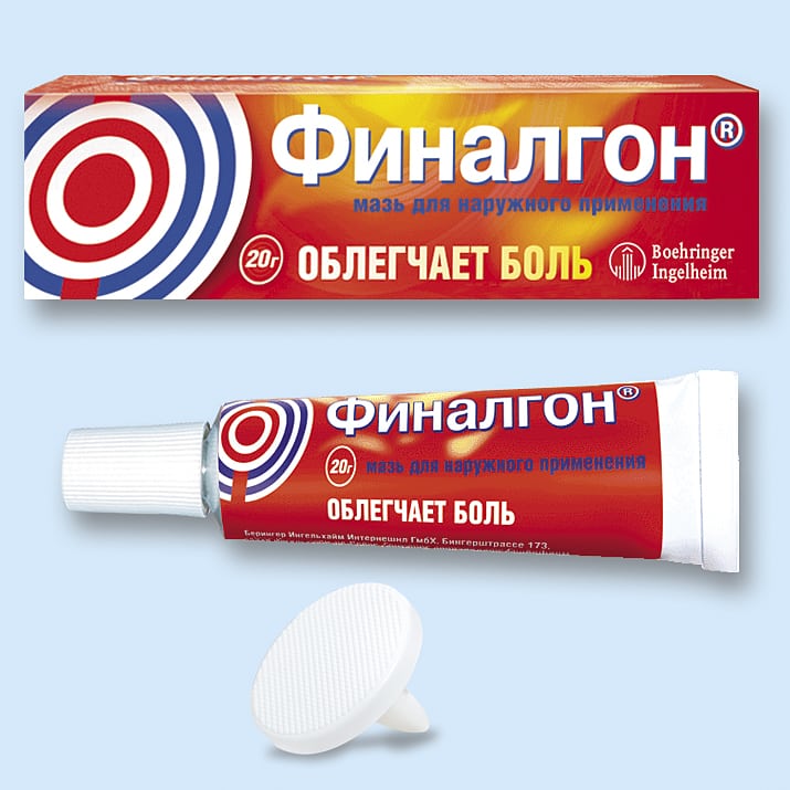
And Menovazin's solution and Espol cream relax the skeletal muscles, eliminating painful muscle spasms.
Improving the blood supply to the joint with nutrients
The use of drugs that accelerate blood circulation in it helps to stop further destruction of the hip joint. These are Stugeron, Cinnarizine, Trental, Pentoxifylline, Xanthinol nicotinate. The course intake of medicines helps to eliminate the deficiency of nutrients and bioactive substances, prevents spasms of small blood vessels.

If necessary, the therapeutic regimens include muscle relaxants (Mydocalm, Sirdalud, Tizanidine) - drugs that relax striated muscles, reducing the severity of pain.

In the treatment of pathologies accompanied by the destruction of the hip joints, magnetotherapy, laser therapy, and UHF therapy are necessarily used. Since the hip joint is a deep joint, these manipulations do not always give the desired clinical effect. Most often, electrophoresis is used, during which drug molecules are delivered to the joint under the action of an electric current. As part of complex therapy, patients are recommended:
- hardware traction, underwater traction.

Underwater traction.
The procedures stimulate muscle strengthening, increase the mobility of the hip joint, and, in combination with drug treatment, reduce the load on the femoral head and increase the distance between it and the acetabulum.
With coxarthrosis of 3 degrees of severity, conservative therapy is not carried out. Patients are immediately offered - endoprosthetics (installation of an artificial hip joint).

Installed hip prosthesis. The pink head is ceramic, wear-resistant.
Usually, a bipolar endoprosthesis is used, which immediately replaces the acetabulum and the head of the femur. If the joint is not too severely damaged, a unipolar prosthesis is implanted that simulates the femoral head. After a thorough examination of the patient, the operation is performed under general anesthesia. In patients, a short-term intake of antibacterial drugs is indicated.
High quality ensures complete restoration of all functions of the hip joint, and its service life varies from 10 to 20 years.
With the development of civilization, mankind has learned to cope with many diseases, but we have not yet been able to defeat joint diseases. Moreover: joint diseases continue to get younger.
One of the most common joint diseases is arthrosis. About 10-15% of the world's population suffers from arthrosis, and today they are not only old people, as several decades ago, but also quite young people.
Deforming arthrosis of the hip joint is the most severe, as it can lead to complete immobilization of this joint.
Of course, considerable experience has been accumulated in the treatment of arthrosis in Russian clinics. However, not all patients with arthrosis of the hip joint can be helped here. For this reason, many seek help from foreign clinics.
10-15% of the world's population suffers from arthrosis.
Before making a decision about treatment abroad, it is worth "getting to know" your disease, the causes that cause it and the methods of treatment.
Leading clinics abroad
Why does deforming arthrosis of the hip joint develop?
Osteoarthritis in essence is the destruction of the cartilage tissue of the joint, in this case, the hip. Degenerative-dystrophic changes occur in the joint, it is slowly destroyed.
Reasons for the development of arthrosis of the hip joint
The main reason for the development of the disease is the loss of elasticity of the cartilage in the joint, which can be caused by:
- Metabolic disorders in the joint,
- Hormonal disorders
- Deterioration of blood supply to the joint,
- Hereditary factors
- Excessive stress on the joint
- Old age,
- Flat feet and scoliosis,
- Certain inflammatory conditions such as arthritis.
As a result of these reasons, the resistance of the joint to stress worsens, or the stress is excessive for the joint (overweight, hard work, scoliosis), and this causes its destruction.
Leading specialists of clinics abroad
Signs and symptoms of arthrosis of the hip joint
- Pain with stress on the joint,
- Decreased joint mobility
- Feeling of muscle tension in the hip joint,
- Crunch when making movements
- Swelling and redness of the skin around the joint
- Joint deformity.
The last symptom is typical for one of the types of arthrosis - deforming arthrosis of the hip joint. When the cartilage is severely destroyed, the bones thicken. Thus, the body, as it were, compensates for the resulting destruction. Also, osteophytes - bone processes can develop on the articulating parts of the joint. They cause severe pain.

- Arthrosis of the hip joint 1 degree can only occasionally be accompanied by minor pain when the load on the hip joint. Pathological processes in the joint are just beginning, therefore, preventive procedures and treatment of 1st degree hip arthrosis will be most effective.
- Arthrosis of the hip joint 2nd degree - this is already the beginning of the destruction of the joint, therefore, all the symptoms of the disease appear. Treatment of arthrosis of the hip joint of the 2nd degree is carried out without surgery.
- Arthrosis of the hip joint, grade 3 means damage not only to the joint itself, but also to disruption of the muscles and ligaments. At the end of the third stage of the disease, arthrosis of the hip joint can lead to fusion of bone surfaces or closure of the joint, that is, it completely ceases to "work".
How to treat arthrosis of the hip joint?
Arthrosis treatment abroad begins with a complete diagnosis, which is based on the use of modern equipment. This helps in prescribing the correct treatment. It should be aimed at eliminating degenerative changes and restoring lost functions. Significant results can only be achieved with the help of comprehensive treatment.
Comprehensive treatment gives the most effective results.
Treatments for deforming arthrosis of the hip joint include:
- drug treatment,
- arthroscopy,
- treatment with folk remedies,
- physiotherapy,
- spa treatment,
- physiotherapy exercises.
In the treatment of grade 3 hip arthrosis, joint replacement or complete replacement is performed.
Medication - how to treat arthrosis of the hip joint
Medical treatment of arthrosis of the hip joint is carried out using the following drugs:
- Non-steroidal anti-inflammatory drugs,
- Chondroprotectors (drugs that restore cartilage tissue),
- Preparations for improving microcirculation,
- Muscle relaxants,
- Hormones.
Arthroscopy
This procedure is carried out both for the purpose of diagnosis and treatment. Endoscopic intervention can solve many joint problems. This technology is less traumatic and effective enough.
An arthroscopic procedure for diagnostic purposes can cost about $ 2,000-3,000, if it is performed for a therapeutic purpose, then it is twice as expensive.
 Partial replacement of the affected joint consists in the introduction of a metal-ceramic implant into it. These implants can last over 30 years. This procedure, together with rehabilitation, will take about two weeks to complete. But a complete replacement of the joint will require a longer period of treatment and rehabilitation: about one and a half months.
Despite the fact that many clinics in the USA, European and Asian countries have mastered operations on hip joints, traditionally most patients go to clinics in Germany for endoprosthetics or joint replacement. Some of them perform 5,000 partial or complete hip replacements per year.
How much does a hip replacement surgery cost?
Not less than $ 10,000, but more often $ 15,000 - $ 18,000.
In Germany - an average of 17,000 euros.
Joint replacement is performed at the third stage of arthrosis. But many patients seek help at an earlier stage of the disease, when “heavy artillery” can be dispensed with.
Prevention and treatment of arthrosis of the hip joint with folk remedies
Traditional methods as prevention and treatment offer therapeutic exercises and proper nutrition for arthrosis of the hip joint.
- Physiotherapy... Joints collapse from excessive stress, but if there is no stress on them, then they collapse even faster. Therefore, it is impossible to cure any joint disease without a set of exercises. As a rule, exercise therapy for arthrosis of the hip joint is exercises specially designed to improve the mobility of the joint and strengthen the muscles that ensure its work.
- Proper nutrition... Proper nutrition should help the body repair cartilage tissue, help cleanse it and maintain optimal weight. The diet for arthrosis of the hip joint does not have particularly strict prohibitions. It is highly recommended to include jellied meat and jelly as often as possible in the diet. These products contain natural collagens that act as chondroprotectors. By the way, their regular use by healthy people will reduce the risk of developing arthrosis.
Other treatments
A good effect is given by physiotherapy procedures, which many medical institutions are able to provide in sufficient volumes. Spa treatment with mud can be performed in the same Germany or the Dead Sea in Israel. The course of treatment of arthrosis of the hip joint, including treatment with folk remedies, is the specialization of Chinese doctors.
Exercises for arthrosis of the hip joint
The proposed therapeutic exercises for arthrosis of the hip joints can improve the quality of life of people with this type of osteoarthritis. The following range of exercises to maintain joint mobility can be performed to prevent and treat arthrosis of the hip joint at home. Each exercise must be repeated at least 5 times.

Performing the exercises of this complex for arthrosis of the hip joint, remember to be careful: movements should not cause pain or discomfort.
If this happens, you should stop exercising, and next time reduce the range of motion and exercise intensity. If possible, then it is worth consulting a rehabilitation doctor.
If you have a problem with a hip joint, you may have already been diagnosed, or you are just about to see a doctor, do not delay. Today, many doctors in different parts of the world know how to cure arthrosis of the hip joint and are ready to help in solving your problems.
See the section for more information.