Treatment of severe diaper rash in newborns. Everything about diaper rash in newborn babies: causes of appearance, effective methods of prevention and treatment. Prevention of irritation
1 pregnancy screening is the most exciting first trimester event. With bated breath, the expectant mother is waiting for news about her child - the woman does not know anything about him yet! How does the baby develop? Is everything all right with him? The survey will not leave any of these questions unanswered.
The concept of "screening" literally translated from a foreign language means "sifting" and accurately reflects the specifics of the procedure. It is as if expectant mothers are "sifted" when the indicators of the intrauterine state of their children are determined. Screening is carried out at 11-13 weeks of gestation. This is a mandatory procedure for all expectant mothers. Such an early period will not prevent accurate equipment and experienced specialists from detecting abnormalities in the development of the fetus, caused by genetics or chromosomal damage.
Features of the first screening examination
Of paramount importance is a comprehensive examination for pregnant women of the following high-risk groups:
- the mom-to-be celebrated her 35th birthday;
- in the patient's family, there were cases of the birth of children with Down syndrome and other anomalies on the basis of genetics;
- women who had spontaneous abortion or had sick (with genetic abnormalities) children;
- victims of infectious diseases that overtook women some time after conception.
The examination includes two procedures: ultrasound and biochemical screening, during which markers specific to the "interesting" position are found in the blood. These research methods can tell the doctor a lot: whether the growth rate of the baby corresponds to the gestational age and whether there are any pathologies in its development. The test results are interpreted taking into account the age and body weight of the expectant mother, as well as the presence of chronic diseases and bad habits.
When decoding the data obtained as a result of the examination, the specialist pays attention not only to absolute indicators, but also to deviations corresponding to the time of screening during pregnancy. All doctors agree that the results of a clinical blood test cannot be taken as the ultimate truth and on this basis a final diagnosis cannot be made. Whatever the results of the study, there is always the opportunity to undergo additional examination.
How pregnancy screening is done
Preparation for the procedure begins within the walls of the antenatal clinic, where the expectant mother can ask the gynecologist any questions that interest her. The specialist will talk to the woman about the importance of the first screening and explain how to prepare for it.
The laboratory, as a rule, conducts ultrasound and biochemical blood tests on the same day. The examination is completely painless, the most unpleasant moment can be considered only the procedure for taking blood.
Screening is done early in the morning, before breakfast. 1 - 2 days before the procedure, the expectant mother should pull herself together and give up her favorite foods, including chocolate, seafood, meat and all fatty foods. A forced diet is considered the key to accurate screening results.
The doctor will most likely advise the woman to refrain from sexual intercourse, otherwise the overall picture of the tests may be distorted.
Before going to the laboratory, the pregnant woman must weigh herself. The laboratory assistant will enter the data on the patient's body weight into a special form immediately before the ultrasound procedure and blood sampling for analysis.
It is better not to drink water before screening, but if the desire is overwhelming, then no more than 100 ml.

Screening during pregnancy: the purpose of ultrasound
At the initial stage of gestation, an ultrasound machine is used to determine the main parameters of a growing baby. The laboratory assistant examines the internal organs of the child, pays attention to the location of his arms and legs. In addition, the procedure makes it possible to see the position of the placenta and assess its structure. By the features of the baby's physique during this period of pregnancy, it can be judged whether the fetus is forming correctly. If a deviation in the development of a child from generally accepted norms is detected on time, the doctor will have time to conduct an additional examination, think over and develop tactics for further pregnancy management.
During the procedure, a specialist studies the following specific features of a developing child:
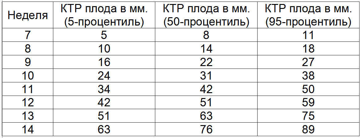
Coccyx-parietal distance (CTE)
- 11 weeks - from 34 to 50 mm;
- 12 weeks - from 42 to 59 mm;
- 13 weeks - 51 to 75 mm.
Condition of the nasal bone
At 11 weeks of gestation, it should be clearly distinguishable, but its size is still difficult to determine. Screening at 12 weeks of pregnancy shows that the nasal bone has grown - now its size ranges from 2 to 2.5 mm or more. At 10 - 12 weeks of gestation, the bone is clearly visible in 98% of healthy babies.
Collar thickness (TVP)
In other words, these are folds in the neck.
- 11 weeks - from 0.8 to 2.2 mm;
- 12-13 weeks - from 0.8-2.7 mm.
Heart rate (HR)
- 11 weeks - from 155 to 178 beats per minute;
- 12 weeks - from 150 to 174 beats per minute;
- 13 weeks - 147 to 171 beats per minute
Biparietal size
This is the distance between the outer and inner edges of both parietal bones.
- 11 weeks - from 13 to 21 mm;
- 12 weeks - from 18 to 24 mm;
- 13 weeks - 20 to 28 mm.
The appearance of the maxillary bone
A poorly expressed maxillary bone at 11 to 13 weeks may indicate a high risk of developing Down syndrome in a baby.

Based on these indicators, the doctor can draw a conclusion about the characteristics of the child's development. So the presence of a number of specific signs on the monitor of the ultrasound machine can inform the specialist about the possible pathologies of the baby's development. Let's note what they pay attention to in the first place:
- if the nasal bone is still not identified in the fetus within the established screening times during pregnancy or its size is significantly behind the norm, and the facial contours are inexpressive and smoothed, the specialist will suspect Down syndrome;
- completely invisible or very small nasal bone, the presence of an umbilical hernia, one umbilical artery instead of two, and a low heart rate indicates Edwards syndrome;
- visualized umbilical cord hernia, developmental disorder of many body systems and high heart rate are most likely indicative of the development of Patau syndrome.
Screening during pregnancy: blood test rates
As a result of a biochemical blood test, the indicators of free beta-hCG and protein-A (PAPP-A) are of the greatest value to the doctor.
In a pregnant woman, the amount of human chorionic gonadotropin in the blood is increased. An increase in the concentration of a specific substance occurs at a rapid pace in the first weeks after fertilization of the egg. It is on this basis that one can judge whether a pregnancy has taken place or not already 6 to 7 days after intercourse.
Starting from the 12th week of gestation, the hCG levels in the body begin to gradually decrease, and its level remains practically unchanged throughout the second trimester.
The norms of free hCG during pregnancy are:
- 17.4 to 130.4 ng / ml at 11 weeks of gestation;
- 13.4 to 128.5 ng / ml at 12 weeks of gestation;
- from 14.2 to 114.7 ng / ml at 13 weeks of gestation.
A free hCG level higher than normal may indicate the following conditions:
- multiple pregnancy;
- Down syndrome;
- severe toxicosis.
Significantly underestimated indicators of this substance indicate such probable pathologies:
- undeveloped pregnancy;
- delayed development of the fetus;
- the likelihood of spontaneous miscarriage;
- Edwards syndrome.
The standards for the PAPP-A substance are:
- from 0.46 to 3.73 mU / ml at 11 weeks;
- from 0.79 to 4.76 mU / ml at 12 weeks;
- from 1.03 to 6.01 mU / ml at 13 weeks.
- pathology of the development of the neural tube of the fetus;
- frozen pregnancy;
- the likelihood of spontaneous abortion;
- the risk of premature birth;
- Rh-conflict between mother and child;
- Edwards syndrome;
- Down syndrome;
- Smith-Opitz syndrome;
- Cornelia de Lange syndrome.

Biochemical analysis of the first screening during pregnancy can detect fetal pathologies in the form of Down, Patau and Edwards syndromes in 68% of cases. Taking into account the ultrasound data, the reliability of the blood test results is confirmed in 90% of cases. However, experts always remember that, for some reason, the data obtained may be distorted.
For example, the results of a biochemical blood test depend on many factors, among which the following have the greatest influence on the blood composition:
- multiple pregnancy;
- pregnancy that was obtained using the IVF procedure;
- large body weight of the patient (the more it is, the higher the indicators of specific substances in the blood);
- a course of treatment with drugs, the active substance of which is progesterone;
- diabetes mellitus in a pregnant woman;
- high risk of spontaneous abortion;
- incorrectly established gestational age;
- depressed state of the expectant mother due to psychological trauma.
If the findings from the first pregnancy screening are not too abnormal, doctors will try to allay their concerns at a second screening. This is a mandatory procedure for the second trimester of pregnancy.

Medical practice knows cases when, despite the unfavorable results of the biochemical blood test of the first and second screenings, absolutely healthy children were born in women. Note that this "miracle" has to do only with a blood test, since doctors almost never doubt the reliability of the results obtained during the ultrasound procedure.
Remember that unfavorable results from the first screening are not yet a reason to panic, as the test rates may vary depending on your health condition. You have the right to an additional examination of your own free will. You need to start with a consultation with a geneticist, who will discuss with you the possibility of conducting additional research methods. This is amniocentesis, cordocentesis, or chorionic villus biopsy. These procedures will answer your innermost question with amazing precision. A kind of bonus that these events will bring you will be the absolutely truthful news about who you are wearing under your heart - a future protector or a princess. This will be revealed by counting the chromosomes. In any case, you need to keep your hands on and hope for the best!
The hopes and concerns of the first pregnancy screening. Video
Some time ago, pregnant women did not know about such a procedure as prenatal or perinatal ... Now all expectant mothers are undergoing such an examination.
What is pregnancy screening, why is it done and why are its results so important? Answers to these and other questions of concern to many pregnant women about perinatal screening we have tried to give in this material.
In order to exclude in the future any misunderstanding of the information presented, before proceeding directly to the consideration of the above indicated topics, it is worth defining some medical terms.
Prenatal screening Is a special kind of a really standard procedure like screening. This comprehensive examination consists of Ultrasound diagnostics and laboratory research, in this particular case biochemistry of maternal serum. Early detection of some genetic abnormalities - this is the main task of such an analysis during pregnancy as screening.
Prenatal or perinatal means prenatal, and by the term screening in medicine, a number of studies of a large stratum of the population are meant, which are carried out in order to form a so-called "risk group" susceptible to certain diseases.
Can be universal or selective screening
.
It means that screening tests do not only to pregnant women, but also to other categories of people, for example, children of the same age to establish the diseases characteristic of a given period of life.
With help genetic screening doctors can find out not only about the problems in the development of the baby, but also react in time to complications during the course of which a woman may not even suspect.
Often, expectant mothers, hearing that they have to go through this procedure several times, begin to panic and worry in advance. However, there is nothing to be afraid of, you just need to ask the gynecologist in advance why you need screening for pregnant women, when and, most importantly, how this procedure is done.
So, let's start with what is standard screening carried out three times during the entire pregnancy, i.e. in every trimester ... Recall that trimester Is a period of three months.
What it is 1 trimester screening ? First, let's answer a common question about how many weeks it is first trimester of pregnancy ... In gynecology, there are only two ways to reliably establish the period during pregnancy - calendar and obstetric.
The first is based on the day of conception, and the second depends on menstrual cycle preceding fertilization ... So I trimester - this is the period that, according to the calendar method, begins the first week from conception and ends with the fourteenth week.
According to the second method, I trimester
Is 12 obstetric weeks. Moreover, in this case, the period is counted from the beginning of the last menstruation. Recently screening
not prescribed to pregnant women. 
However, now many expectant mothers are themselves interested in undergoing such a survey.
In addition, the Ministry of Health strongly recommends that research be ordered for all expectant mothers, without exception.
True, this is done voluntarily, since no one can force a woman to undergo any kind of analysis.
It is worth noting that there are categories of women who are simply obliged, for one reason or another, to pass screening, eg:
- pregnant women from thirty-five years old and on;
- expectant mothers with a history of information about the presence of a threat spontaneous ;
- women who in the first trimester suffered infectious diseases ;
- pregnant women who, for health reasons, are forced to take drugs prohibited for their situation in the early stages;
- women who have had various genetic abnormalities or abnormalities in fetal development ;
- women who have already given birth to children with any deviations or developmental disabilities ;
- women who have been diagnosed frozen or regressive pregnancy (cessation of fetal development);
- suffering from narcotic or women;
- pregnant women in whose family or in the family of the father of the unborn child cases were recorded hereditary genetic abnormalities .
How long do they do prenatal screening for the 1st trimester ? For the first screening during pregnancy, the period is set in the interval from 11 weeks to 13 obstetric weeks of pregnancy and 6 days. Earlier, above the indicated period, it makes no sense to conduct this survey, since its results will be uninformative and completely useless.
The first ultrasound scan at the 12th week of pregnancy is not done by chance for a woman. Since it is at this time that embryonic and begins fetal or fetal the period of development of the future person.
This means that the embryo turns into a fetus, i.e. there are obvious changes that speak of the development of a full-fledged living human body. As we said earlier, screening tests Is a complex of measures, which consists of ultrasound diagnostics and blood biochemistry of a woman.
It is important to understand that holding screening ultrasound in the 1st trimester during pregnancy plays the same important role as laboratory blood tests. Indeed, in order for geneticists to draw the correct conclusions based on the results of the examination, they need to study both the ultrasound results and the patient's blood biochemistry.
How many weeks the first screening is carried out, we talked, now let's move on to deciphering the results of a comprehensive study. It is really important to consider in more detail the norms established by doctors for the results of the first screening during pregnancy. Of course, only a specialist in this area with the necessary knowledge and, most importantly, experience can give a qualified assessment of the results of the analysis.
We believe that it is advisable for any pregnant woman to know at least general information about the main indicators prenatal screening and their normative values. Indeed, it is common for most expectant mothers to be overly suspicious of everything related to the health of their future child. Therefore, they will be much more comfortable if they know in advance what to expect from the research.
Decoding of screening of the 1st trimester by ultrasound, norms and possible deviations
All women know that during pregnancy they have to undergo an ultrasound examination (hereinafter referred to as ultrasound) more than once, which helps the doctor monitor the intrauterine development of the unborn child. In order to screening ultrasound gave reliable results, you need to prepare in advance for this procedure.
We are sure that the vast majority of pregnant women know how to do this procedure. However, it will not be superfluous to repeat that there are two types of research - transvaginal and transabdominal ... In the first case, the device sensor is inserted directly into the vagina, and in the second it contacts the surface of the anterior abdominal wall.

There are no special preparation rules for the transvaginal type of ultrasound.
If you have a transabdominal examination, then before the procedure (about 4 hours before the ultrasound), you should not go to the toilet "on a small", and in half an hour it is recommended to drink up to 600 ml of plain water.
The thing is that the examination must be carried out necessarily on the filled with liquid bladder .
In order for the doctor to obtain a reliable result Ultrasound screening, the following conditions must be met:
- examination period - from 11 to 13 obstetric week;
- the position of the fetus should allow the specialist to carry out the necessary manipulations, otherwise the mother will have to "influence" the baby to roll over;
- coccygeal-parietal size (hereinafter referred to as CTE) should not be less than 45 mm.
What is CTE during pregnancy on ultrasound
When conducting an ultrasound scan, a specialist necessarily examines various parameters or sizes of the fetus. This information allows you to determine how well the baby is formed, and whether he is developing correctly. The rates of these indicators depend on the duration of pregnancy.
If the value of one or another parameter obtained as a result of ultrasound deviates from the norm up or down, then this is considered a signal of the presence of some pathologies. Coccyx-parietal size - This is one of the most important initial indicators of the correct intrauterine development of the fetus.
The CTE value is compared with the weight of the fetus and the gestational age. This indicator is determined by measuring the distance from the bone of the crown of the child to his coccyx. As a general rule, the higher the CTE, the longer the pregnancy.
When this indicator slightly exceeds or, on the contrary, is slightly less than the norm, then there is no reason for panic. This only speaks about the peculiarities of the development of this particular child.
If the CTE value deviates from the standards in a large direction, then this signals the development of a large fetus, i.e. Presumably, the weight of the child at birth will exceed the average rate of 3-3.5 kg. In cases where the CTE is significantly less than the standard values, this may be a sign that:
- pregnancy does not develop as it should, in such cases, the doctor should carefully check the fetal heartbeat. If he died in the womb, then the woman needs urgent medical attention ( scraping of the uterine cavity ) in order to prevent a possible threat to health ( development of infertility ) and life ( infection, bleeding );
- the body of a pregnant woman produces an insufficient amount, as a rule, which can lead to a spontaneous miscarriage. In such cases, the doctor prescribes an additional examination for the patient and prescribes medications containing hormones ( , Dufston );
- mother is sick infectious diseases , including venereal;
- the fetus has genetic abnormalities. In such situations, doctors prescribe additional studies along with, which is part of the first screening test.
It is also worth emphasizing that there are often cases when a low CTE indicates an incorrectly established gestational age. This refers to a variant of the norm. All a woman needs in such a situation is to undergo a second ultrasound examination after a while (usually after 7-10 days).
Fetal BPD (biparietal size)
What is BPD on ultrasound during pregnancy? When conducting an ultrasound examination of the fetus in the first trimester, doctors are interested in all the possible characteristics of the unborn child. Since their study gives specialists maximum information about how the intrauterine development of a little man takes place and whether everything is in order with his health.
What is it BPD of the fetus ? First, let's decipher the medical abbreviation. BPR - it biparietal fetal head size , i.e. distance between walls parietal bones of the skull , in a simple way, the size of the head. This indicator is considered one of the main ones for determining the normal development of a child.
It is important to note that BPD shows not only how well and correctly the baby is developing, but also helps doctors prepare for the upcoming delivery. Since if the size of the head of the unborn child deviates from the norm in a large direction, then he simply will not be able to pass through the birth canal of the mother. In such cases, a planned cesarean section is prescribed.

When BPD deviates from the established norms, this may indicate:
- about the presence of such pathologies incompatible with life in the fetus as cerebral hernia or tumor ;
- about a sufficiently large size of the unborn child, if other basic parameters of the fetus are ahead of the established developmental standards by several weeks;
- about abrupt development, which after a while will return to normal, provided that other basic parameters of the fetus fit into the norms;
- about fetal development brain caused by the presence of infectious diseases in the mother.
The deviation of this indicator to the lower side indicates that the baby's brain is developing incorrectly.
Collar thickness (TVP)
Fetal TBP - what it is? Collar space by fetus or size cervical fold - this is a place (more precisely, an oblong formation), located between the neck and the upper skin membrane of the baby's body, in which fluid accumulates. The study of this value is carried out when screening the first trimester of pregnancy, since it is at this time that it is possible to measure TVP for the first time, and then analyze it.
Starting from the 14th week of pregnancy, this formation gradually decreases in size and by the 16th week it practically disappears from view. For TVP, certain norms have also been established, which are in direct proportion to the duration of pregnancy.
For example, the norm collar space thickness at 12 weeks should not exceed the range from 0.8 to 2.2 mm. Collar space thickness at 13 weeks should be in the range of 0.7 to 2.5 mm.
It is important to note that for this indicator, experts set averaged minimum values, the deviation from which indicates a thinning of the collar space, which, like the expansion of the TVP, is considered an anomaly.
![]()
If this indicator does not correspond to the TVP norms indicated in the above table at 12 weeks and at other periods of pregnancy, then such a result most likely indicates the presence of the following chromosomal abnormalities:
- trisomy 13 , a disease known as Patau syndrome, characterized by the presence of an additional 13 chromosome in human cells;
- trisomy on chromosome 21, known to everyone as Down syndrome , a human genetic disorder in which karyotype (i.e. a complete set of chromosomes) is represented by chromosome 47 instead of 46;
- monosomy on the X chromosome , a genomic disease named after the scientists who discovered it Shereshevsky-Turner syndrome, it is characterized by such anomalies of physical development as short stature, as well as sexual infantilism (immaturity);
- trisomy on chromosome 18 Is a chromosomal disease. For Edwards syndrome (the second name of this disease) is characterized by a plurality of malformations incompatible with life.
Trisomy Is an option aneuploidy , i.e. changes karyotype , in which there is an additional third in the human cell chromosome instead of normal diploid set.
Monosomy Is an option aneuploidy (chromosomal abnormality) , in which there are no chromosomes in the chromosome set.
What are the norms for trisomy 13, 18, 21 installed during pregnancy? It so happens that in the process of cell division, a failure occurs. This phenomenon has received the name in science aneuploidy. Trisomy - This is one of the varieties of aneuploidy, in which, instead of a pair of chromosomes, an extra third chromosome is present in the cell.
In other words, the child inherits from his parents an additional 13, 18 or 21 chromosome, which in turn entails genetic abnormalities that impede normal physical and mental development. Down Syndrome statistically, this is the most common disease caused by the presence of chromosome 21.
Children born with Edwards syndromes the same as in the case of Patau syndrome , usually do not live up to a year, unlike those who are unlucky to be born with Down syndrome ... Such people can live to a ripe old age. However, such a life can rather be called existence, especially in the countries of the post-Soviet space, where these people are considered outcasts and they try to avoid and ignore them.
In order to exclude such anomalies, pregnant women, especially those at risk, must undergo a mandatory screening examination. The researchers argue that the development of genetic abnormalities is in direct proportion to the age of the expectant mother. The younger the woman, the less likely it is that her child will have any abnormalities.
To establish trisomy in the first trimester of pregnancy, a study is carried out collar space of the fetus using ultrasound. In the future, pregnant women periodically take a blood test, in which for geneticists the most important indicators are the level alpha-fetoprotein (AFP), inhibin-A, chorionic gonadotropin (hCG) and estriol .
As mentioned earlier, the risk of having a genetic disorder in a child depends primarily on the age of the mother. However, there are cases when trisomy is recorded in young women. Therefore, doctors at screening study all possible signs of anomalies. It is believed that an experienced ultrasound specialist can identify problems already during the first screening examination.
Signs of Down syndrome, as well as Edwards and Patau
Trisomy 13 is characterized by a sharp decrease in the level PAPP-A (PAPP pregnancy-associated protein (protein) A-plasma ). It is also a marker of this genetic disorder. The same parameters play an important role in determining whether the fetus has Edwards syndrome .
When there is no risk of trisomy 18, normal values PAPP-A and b-hCG (free beta subunit of hCG)
are recorded in a biochemical blood test. If these values deviate from the standards established for each specific gestational age, then, most likely, genetic malformations will be found in the child. 
It is important to note that in the case when, during the first screening, a specialist records signs indicating a risk trisomies , the woman is referred for further examination and consultation with geneticists. To make the final diagnosis, the expectant mother will have to go through procedures such as:
- chorionic biopsy , i.e. obtaining a sample of chorionic tissue to diagnose anomalies;
- amniocentesis- it puncture of the amniotic membrane to get a sample amniotic fluid for the purpose of their further study in the laboratory;
- placentocentesis (placental biopsy) , given invasive diagnostic method experts take a sample placental tissue using a special puncture needle, which is pierced anterior abdominal wall ;
- cordocentesis , a method for diagnosing genetic abnormalities during pregnancy, in which the umbilical cord blood of the fetus is analyzed.
Unfortunately, if a pregnant woman has undergone any of the above studies and bioscreening and ultrasound the diagnosis of the presence of genetic abnormalities in the fetus was confirmed, doctors will offer to terminate the pregnancy. In addition, unlike standard screening studies, the data invasive examination methods can provoke a number of severe complications up to spontaneous miscarriage, so doctors resort to them in a fairly rare number of cases.
Nasal bone - This is a slightly elongated, quadrangular, convex in front of the paired bone of the human face. At the first ultrasound screening, the specialist determines the length of the baby's nose bone. It is believed that in the presence of genetic abnormalities, this bone does not develop correctly, i.e. its ossification occurs later.

Therefore, if the nasal bone is absent or its size is too small during the first screening, then this indicates the possible presence of various anomalies. It is important to emphasize that the length of the nasal bone is measured at 13 weeks or 12 weeks. At 11 weeks of screening, the specialist checks only for its presence.
It is worth emphasizing that if the size of the nasal bone does not correspond to the established norms, but if other basic indicators correspond, there is really no reason for concern. This state of affairs may be due to the individual characteristics of the development of this particular child.
Heart rate (HR)
A parameter such as Heart rate plays an important role not only in the early stages, but throughout pregnancy. Constantly measure and monitor fetal heart rate it is necessary already only in order to notice deviations in time and, if necessary, save the life of the baby.
The interesting thing is that though myocardium (heart muscle) begins to decrease already in the third week after conception, you can hear the heartbeat only from the sixth obstetric week. It is believed that at the initial stage of fetal development, the rhythm of its heartbeats should correspond to the mother's pulse (on average, it is 83 beats per minute).

However, already in the first month of intrauterine life, the number of baby's heartbeats will gradually increase (by about 3 beats per minute every day) and by the ninth week of pregnancy it will reach 175 beats per minute. Determine the fetal heart rate using ultrasound.
When conducting the first ultrasound, specialists pay attention not only to the heart rate, but also watch how the baby's heart develops. For this, the so-called four-chamber cut , i.e. method of instrumental diagnostics of heart defects.
It is important to emphasize that a deviation from the standards of such an indicator as heart rate indicates the presence of malformations of the heart ... Therefore, doctors carefully study the structure on a cut atria and fetal heart ventricles ... If any deviations are found, specialists refer the pregnant woman for additional research, for example, to echocardiography (ECG) with dopplerography.
Starting from the twentieth week, the gynecologist of the antenatal clinic will listen to the baby's heart using a special tube at each scheduled visit to the pregnant woman. A procedure such as auscultation of the heart does not apply at an earlier date due to its ineffectiveness, since the doctor just can't hear the heartbeat.
However, as the baby develops, his heart will be heard more clearly each time. Auscultation helps the gynecologist determine the position of the fetus in the womb. For example, if the heart is better heard at the level of the mother's navel, then the child is in a transverse position, if the navel is on the left or below, then the fetus is in cephalic presentation , and if above the navel, then in pelvic .
From 32 weeks of gestation, they use cardiotocography (abbreviated CTE ). When carrying out the above types of examinations, a specialist can record in the fetus:
- bradycardia , i.e. abnormally low heart rate , which is usually temporary. This deviation may be a symptom of the mother's autoimmune diseases, anemia, as well as cord clamping when the unborn baby is not getting enough oxygen. Bradycardia can be caused by congenital heart defects in order to exclude or confirm this diagnosis, a woman is sent for additional examinations without fail;
- , i.e. high heart rate. Experts rarely record such a deviation. However, if the heart rate is much higher than that provided for by the norms, then this indicates the mother or hypoxia , development intrauterine infections, anemia and genetic abnormalities at the fetus. In addition, the medications a woman takes can affect heart rate.
In addition to the above characteristics, during the first screening ultrasound examination, specialists also analyze the data:
- about symmetry cerebral hemispheres fetus;
- about the size of the circumference of his head;
- about the distance from the occipital to the frontal bone;
- the length of the bones of the shoulders, thighs and forearms;
- about the structure of the heart;
- about the location and thickness of the chorion (placenta or "child's place");
- the amount of water (amniotic fluid);
- about the condition of the pharynx cervix mothers;
- the number of vessels in the umbilical cord;
- about absence or presence hypertonicity of the uterus .
As a result of an ultrasound scan, in addition to the genetic abnormalities already discussed above ( monosomy or Shereshevsky-Turner syndrome, trisomy on chromosome 13, 18 and 21 , namely Down, Patau and Edwards syndromes ) the following pathologies in development can be identified:
- neural tube , For example, malformation of the spine (meningomyelocele and meningocele) or cranial hernia (encephalocele) ;
- Cornet de Lange syndrome , an anomaly in which multiple malformations are recorded, entailing both physical abnormalities and mental retardation;
- triploidy , a genetic developmental defect in which a failure occurs in the chromosome set, as a rule, the fetus does not survive in the presence of such a pathology;
- omphalocele , embryonic or umbilical hernia, pathology of the anterior abdominal wall, in which some organs (liver, intestines and others) develop in the hernial sac outside the abdominal cavity;
- Smith-Opitz syndrome , a genetic abnormality that affects the processes, which subsequently leads to the development of many severe pathologies, for example, or mental retardation.
Biochemical screening of the 1st trimester
Let's talk in more detail about the second stage of a comprehensive screening examination of pregnant women. What it is biochemical screening of the 1st trimester, and what are the standards established for its main indicators? Actually, biochemical screening Is nothing more than biochemical analysis blood of the future mother.
This study is carried out only after an ultrasound scan. This is due to the fact that, thanks to an ultrasound examination, the doctor establishes the exact duration of pregnancy, on which the normative values of the main indicators of blood biochemistry directly depend. So, remember that you need to go to a biochemical screening only with the results of an ultrasound scan.
How to prepare for your first pregnancy screening
We talked about how they do it, and most importantly, when they do a screening ultrasound, now it is worth paying attention to preparing for the delivery of a biochemical analysis. As with any other blood test, you need to prepare for this test well in advance.
If you want to get a reliable result of biochemical screening, then you will have to exactly follow the following recommendations:
- blood for biochemical screening is given strictly on an empty stomach, doctors do not even recommend drinking plain water, not to mention any food;
- a few days before the screening, you should change your usual diet and begin to adhere to a sparing diet, in which you should not eat too fatty and spicy foods (so as not to increase the level), as well as seafood, nuts, chocolate, citrus fruits and other allergenic foods, even if you have not previously had an allergic reaction to anything.
Strict adherence to these recommendations will allow you to get a reliable result of biochemical screening. Believe me, it is better to be patient for a while and give up your favorite delicacies, so that later you do not worry about the results of the analysis. After all, any deviation from the established norms, doctors will interpret as a pathology in the development of the baby.
Quite often, in all kinds of forums devoted to pregnancy and childbirth, women talk about how the results of the first screening, expected with such excitement, turned out to be bad, and they had to do all the procedures again. Fortunately, the pregnant women ended up receiving good news about the health of their babies, since the adjusted results indicated that there was no developmental disability. 
The whole point was that the expectant mothers did not prepare properly for the screening, which ultimately led to the receipt of inaccurate data.
Imagine how many nerves were wasted and bitter tears were shed while women were waiting for new test results.
Such colossal stress does not pass without leaving a trace for the health of any person, and even more so for a pregnant woman.
Biochemical screening of the 1st trimester, interpretation of results
When conducting the first biochemical screening analysis, the main role in diagnosing any abnormalities in fetal development is played by such indicators as free β-subunit of human chorionic gonadotropin (Further HCG ), as well as PAPP-A (Plasma Protein A Associated with Pregnancy) ... Let's consider each of them in detail.
PAPP-A - what is it?
As mentioned above, PAPP-A - This is an indicator of a biochemical blood test of a pregnant woman, which helps specialists to establish at an early stage the presence of genetic pathologies in the development of the fetus. The full name of this value sounds like pregnancy associated plasma protein A , which literally means - pregnancy-associated plasma protein A .
It is the protein (protein) A produced by the placenta during pregnancy that is responsible for the harmonious development of the unborn child. Therefore, an indicator such as the level of PAPP-A, calculated at 12 or 13 weeks during pregnancy, is considered a characteristic marker for determining genetic abnormalities.
It is mandatory to pass the analysis to check the PAPP-A level should:
- pregnant women over the age of 35;
- women who have previously given birth to children with genetic developmental disabilities;
- mothers-to-be with family members with genetic developmental disabilities;
- women who have had diseases such as , or shortly before pregnancy;
- pregnant women who have had complications or miscarriages before.
Standard values of such an indicator as PAPP-A depend on the duration of pregnancy. For example, the rate of PAPP-A at 12 weeks is from 0.79 to 4.76 mU / ml, and at 13 weeks - from 1.03 to 6.01 mU / ml. In cases where, as a result of the test, this indicator deviates from the norm, the doctor prescribes additional studies.
If the analysis reveals a low level of PAPP-A, then this may indicate the presence chromosomal abnormalities in the development of the child, for example, Down syndrome also this signals the risk of spontaneous miscarriage and regressive pregnancy ... When this indicator is increased, then this is most likely the result of the fact that the doctor could not calculate the correct gestational age.
That is why blood biochemistry is taken only after an ultrasound scan. However high PAPP-A may indicate the likelihood of developing genetic abnormalities in the development of the fetus. Therefore, in case of any deviation from the norm, the doctor will send the woman for additional examination.
Scientists gave this name to this hormone for a reason, since it is thanks to it that one can reliably find out about pregnancy already 6-8 days after fertilization has occurred egg cells. It is noteworthy that HCG begins to develop chorionic already in the first hours of pregnancy.
Moreover, its level is growing rapidly and already by 11-12 weeks of pregnancy exceeds the initial values by a factor of thousands. Then is gradually losing ground, and its indicators remain unchanged (starting from the second trimester) until childbirth. All pregnancy test strips contain hCG.

If the level human chorionic gonadotropin increased, then this may indicate:
- about the presence of the fetus Down syndrome ;
- O multiple pregnancy ;
- about the development of the mother;
When the hCG level is below the prescribed standards, it says:
- about the possible Edwards syndrome at the fetus;
- about the risk miscarriage ;
- O placental insufficiency .
After a pregnant woman has undergone an ultrasound scan and blood biochemistry, a specialist must decipher the results of the examination, as well as calculate the possible risks of developing genetic abnormalities or other pathologies using a special computer program PRISCA (Prisca).
The screening summary sheet will contain the following information:
- about age risk developmental anomalies (depending on the age of the pregnant woman, possible deviations change);
- about the values of the biochemical parameters of a woman's blood test;
- about the risk of possible diseases;
- MoM coefficient .
In order to calculate as reliably as possible the possible risks of developing certain abnormalities in the fetus, experts calculate the so-called MoM (multiple of median) coefficient. To do this, all the screening data obtained is entered into a program that plots the deviation of each indicator of the analysis of a particular woman from the average norm established for most pregnant women.
MoM is considered normal if it does not go beyond the range of values from 0.5 to 2.5. At the second stage, this coefficient is adjusted taking into account age, race, presence of diseases (for example, diabetes ), bad habits (for example, smoking), the number of previous pregnancies, ECO and other important factors.
At the final stage, the specialist makes a final conclusion. Remember, only a doctor can correctly interpret the screening results. In the video below, the doctor explains all the key points related to the first screening.
Screening cost for 1 trimester
The question of how much this study costs and where it is best to take it, worries many women. The thing is that not every state polyclinic can do such a specific examination for free. Based on the reviews left on the forums, many expectant mothers do not trust free medicine at all.
Therefore, you can often come across the question of where to do screening in Moscow or other cities. If we talk about private institutions, then in a fairly well-known and well-proven laboratory INVITRO, biochemical screening can be done for 1600 rubles.
True, this cost does not include ultrasound, which the specialist will definitely ask to present before the biochemical analysis. Therefore, you will have to separately undergo an ultrasound examination in another place, and then go to the laboratory to donate blood. Moreover, this must be done on the same day.
Second screening during pregnancy, when to do and what is included in the study
According to the recommendations of the World Health Organization (hereinafter referred to as WHO), every woman is obliged to undergo three screenings throughout the entire period of pregnancy. Although nowadays gynecologists refer all pregnant women for this examination, there are those who, for whatever reason, miss the screening.
However, for some categories of women, such research should be mandatory. This applies primarily to those who have previously given birth to children with genetic or developmental defects. In addition, it is imperative to undergo screening:
- women over the age of 35, since the risk of developing various pathologies in the fetus depends on the age of the mother;
- women who took drugs or other illegal drugs for pregnant women in the first trimester;
- women who have previously suffered two or more miscarriages;
- women who suffer from one of the following inherited diseases to a child - diabetes mellitus, diseases of the musculoskeletal system and cardiovascular system, as well as oncopathology;
- women who are at risk of spontaneous miscarriage.
In addition, future mothers should definitely undergo screening if they or their spouses were exposed to radiation before conception, and also transferred immediately before or during pregnancy. bacterial and infectious diseases ... As with the first screening for the second time, the expectant mother must also do an ultrasound scan and take a biochemical blood test, which is often called a triple test.
Timing of the second screening during pregnancy
So, let's answer the question of how many weeks do the second screening
during pregnancy. As we have already determined, the first study is carried out in the early stages of pregnancy, namely in the period from 11 to 13 weeks of the first trimester. The next screening study is carried out during the so-called "golden" period of pregnancy, i.e. in the second trimester, which starts at 14 weeks and ends at 27 weeks. 
The second trimester is called golden, because it was during this period of time that all the initial ailments associated with pregnancy ( nausea, weakness, and others) retreat, and a woman can fully rejoice in her new state, since she feels a powerful surge of strength.
A woman should visit her gynecologist every two weeks so that she can monitor the progress of her pregnancy.
The doctor gives the expectant mother recommendations regarding her interesting situation, and also informs the woman about what examinations and how long she should undergo. Typically, a pregnant woman takes a urine test and a general blood test before each visit to the gynecologist, and the second screening takes place from 16 to 20 weeks of pregnancy.
Ultrasound screening of the 2nd trimester - what is it?
During the second screening first, they undergo an ultrasound scan to determine the exact duration of pregnancy, so that later specialists can correctly interpret the results of a biochemical blood test. On the Ultrasound the doctor studies the development and size of the internal organs of the fetus: the length of the bones, the volume of the chest, head and abdomen, the development of the cerebellum, lungs, brain, spine, heart, bladder, intestines, stomach, eyes, nose, as well as the symmetry of the structure of the face.
In general, everything that is visualized with the help of ultrasound examination is analyzed. In addition to studying the main characteristics of the baby's development, experts check:
- how the placenta is located;
- the thickness of the placenta and the degree of its maturity;
- the number of vessels in the umbilical cord;
- condition of the walls, appendages and cervix;
- the quantity and quality of amniotic fluid.
Norms for ultrasound screening of the 2nd trimester of pregnancy:
Decoding the triple test (biochemical blood test)
In the second trimester, experts pay special attention to three markers of genetic abnormalities, such as:
- chorionic gonadotropin - it is produced by the fetal chorion;
- alpha-fetoprotein ( Further AFP ) - it plasma protein (protein), originally generated corpus luteum, and then produced liver and gastrointestinal tract of the fetus ;
- free estriol ( further hormone E3 ) Is a hormone that is produced in placenta , as well as fetal liver.
In some cases, they also study the level inhibin (hormone, produced follicles) ... Certain standards are set for each week of pregnancy. It is considered optimal to conduct a triple test at 17 weeks of gestation.
When the level of hCG at the second screening is overestimated, then this may indicate:
- about multiple pregnancy ;
- O diabetes mellitus from the mother;
- about the risk of development Down syndrome if the other two indicators are below normal.
If hCG, on the contrary, is lowered, then this says:
- about the risk Edwards syndrome ;
- O frozen pregnancy;
- O placental insufficiency .
When the AFP level is high, then there is a risk of:
- the presence of developmental abnormalities kidney ;
- defects neural tube ;
- developmental disabilities abdominal wall ;
- damage brain ;
- lack of water ;
- fetal death;
- spontaneous miscarriage;
- emergence rhesus conflict .
Decreased AFP can be a signal:
- Edwards syndrome ;
- diabetes mellitus mothers;
- low location placenta .
At a low level, there is a high risk:
- development anemia at the fetus;
- adrenal and placental insufficiency;
- spontaneous miscarriage ;
- availability Down syndrome ;
- development intrauterine infection ;
- delays in the physical development of the fetus.
It should be noted that the level hormone E3 some medications (for example), as well as improper and unbalanced nutrition of the mother, have an effect. When E3 is elevated, doctors diagnose diseases kidney or multiple pregnancy, and also predict premature birth, when the level of estriol rises sharply.
After the expectant mother goes through two stages of screening, doctors analyze the information received using a special computer program and calculate the same MoM coefficient as in the first study. The conclusion will indicate the risks for a particular type of deviation.
Values are indicated as a fraction, for example 1: 1500 (i.e. one case per 1500 pregnancies). It is considered the norm if the risk is less than 1: 380. Then the conclusion will indicate that the risk is below the cut-off threshold. If the risk is higher than 1: 380, then the woman will be referred for additional consultation with geneticists or offered to undergo invasive diagnostics.
It should be noted that in cases where at the first screening the biochemical analysis corresponded to the norms (indicators were calculated HCG and PAPP-A ), then the second and third time it is enough for a woman to do only an ultrasound.
The last screening examination of the expectant mother takes place in third trimester ... Many people wonder what they are looking at at the third screening and when this study should be done.
As a rule, if the pregnant woman was not diagnosed with any abnormalities in the development of the fetus or during pregnancy at the first or second examination, then she only needs to undergo an ultrasound examination, which will allow the specialist to draw final conclusions about the condition and development of the fetus, as well as its position in the womb. 
Determination of the position of the fetus ( cephalic or breech presentation ) is considered an important preparatory stage before childbirth.
In order for the delivery to be successful, and the woman can give birth on her own without surgery, the child must be in a cephalic presentation.
Otherwise, doctors are planning a cesarean section.
The third screening includes procedures such as:
- Ultrasound , which all pregnant women go through;
- dopplerography Is a technique that focuses mainly on the state of blood vessels placenta ;
- cardiotocography - a study that allows you to more accurately determine the heart rate of a child in the womb;
- blood biochemistry , during which attention is focused on such markers of genetic and other abnormalities as the level HCG, ɑ-fetoprotein and PAPP-A .
Timing of the third screening during pregnancy
It is worth noting that only the doctor decides at how many weeks 3 a woman should undergo screening, based on the individual characteristics of this particular pregnancy. However, it is considered optimal when the expectant mother undergoes a planned ultrasound scan at 32 weeks, and then immediately takes a biochemical blood test (if any), and also undergoes other necessary procedures.
However, for medical reasons, carry out dopplerography or CTG the fetus can be from 28 weeks of gestation. Third trimester starts at 28 weeks and ends with childbirth at 40-43 weeks. The last screening ultrasound is usually prescribed at 32-34 weeks.
Decoding ultrasound
How long does it take for a pregnant woman to undergo the third screening ultrasound, now we will talk in more detail about decoding the study. When conducting an ultrasound scan in the third trimester, the doctor pays special attention to:
- for development and structure of cardio-vascular system a child to exclude possible developmental pathologies, for example;
- on the right development brain , organs of the abdominal cavity, spine and genitourinary system;
- on located in the cranial cavity vein of Galen which plays an important role in the proper functioning of the brain in order to exclude aneurysm ;
- on the structure and development of the child's face.
In addition, ultrasound allows the specialist to assess the condition amniotic fluid appendages and uterus mother, as well as check and the thickness of the placenta ... In order to exclude hypoxia and pathology in the development of the nervous and cardiovascular system , as well as to reveal the features of blood flow in vessels of the uterus and the child, as well as in the umbilical cord, spend dopplerography .
As a rule, this procedure is carried out only according to indications simultaneously with an ultrasound scan. In order to exclude fetal hypoxia and define Heart rate, spend CTG ... This type of research focuses exclusively on the baby's heart, so cardiotocography prescribed in cases where the doctor has concerns about the condition cardiovascular child's systems.
Ultrasound in the third trimester of pregnancy allows you to determine not only the presentation of the child, but also the maturity of his lungs, on which the readiness for birth depends. In some cases, hospitalization for early delivery may be required to keep the baby and mother alive.
| Indicator | Average rate for 32-34 weeks of pregnancy |
| Placenta thickness | from 25 to 43 mm |
| Amniotic (amniotic) index | 80-280 mm |
| Placental maturity | 1-2 degree of ripening |
| Uterine tone | missing |
| Uterine pharynx | closed, length not less than 3 cm |
| Fetal growth | on average 45 cm |
| Fetal weight | on average 2 kg |
| Fetal belly girth | 266 - 285 mm |
| BPR | 85-89 mm |
| Fetal thigh length | 62-66 mm |
| Fetal chest girth | 309-323 mm |
| Fetal forearm size | 46-55 mm |
| Fetal shin bone size | 52-57 mm |
| Fetal Shoulder Length | 55-59 mm |
Based on the results of a biochemical blood test MoM factor should not deviate from the range from 0.5 to 2.5. The risk value for all possible deviations should correspond to 1: 380.
Education: Graduated from Vitebsk State Medical University with a degree in Surgery. At the university, he headed the Council of the Student Scientific Society. Further training in 2010 - in the specialty "Oncology" and in 2011 - in the specialty "Mammology, visual forms of oncology".
Experience: Work in the general medical network for 3 years as a surgeon (Vitebsk emergency hospital, Liozno CRH) and part-time as a regional oncologist and traumatologist. Work as a pharmaceutical representative throughout the year at the Rubicon company.
He presented 3 rationalization proposals on the topic "Optimization of antibiotic therapy depending on the species composition of microflora", 2 works won prizes in the republican competition-review of student research papers (1 and 3 categories).
In the first trimester of her pregnancy, every woman undergoes such a painless procedure as screening. For many, this is a new concept. Therefore, it becomes necessary to consider in more detail the issue of screening and identifying the main pathologies based on its results.

Screening consists of a blood test and. Before screening, a specialist takes into account the main characteristics of a pregnant woman (height, addiction to bad habits, possible diseases), which may affect the test results.
Thanks to an ultrasound scan, a specialist examines how the child's physique develops, and are there any obvious deviations... If there is a suspicion of a pathology, then the future woman in labor is sent for a detailed diagnosis with subsequent treatment.
 the expectant mother can find out how well the fetus is developing, and whether he has any genetic ailments. If the fetus develops with signs of Down's disease, then this will be determined by the thickness of the collar space. Pathologies are detected according to special indicators with blood test:
- : if the figure is below normal, then, most likely, the fetus will have Edwards syndrome, if higher - Down syndrome.
- Indicator PAPP-A(plasma protein) : a value less than the norm indicates that in the future the child is prone to diseases.
Ultrasound at the first screening aims to identify the following points:
- location of the fetus to exclude the likelihood of an ectopic pregnancy;
- confirmation of single or multiple pregnancy;
- tracking the heartbeat and viability of the embryo at the tenth week of development;
- calculation of CTE (coccygeal-parietal size);
- anatomical examination of the fetus (both external defects and pathologies of internal organs are determined);
- examination of the collar space, the thickness of which, with normal development, should correspond to two centimeters. If thickening is noted, then the likelihood of a defect is high;
- examination of the state of the placenta and the exclusion of its dysfunction.

It is this comprehensive study that helps identify genetic chromosomal abnormalities of the fetus... If a serious diagnosis is confirmed that cripples the life of an unborn child, then the pregnant may offer artificial termination of pregnancy.
Only according to the data of complex analyzes, the doctor determines the exact condition of the fetus. If the probability of pathology is confirmed, the woman is sent for special tests - biopsy and amniocentesis... Based on the results of which, they make final conclusions and decide on the further fate of the fetus.
When is the first pregnancy screening done?
How many weeks of pregnancy are screened? A routinely pregnant woman can be screened for 10-13 weeks... But most often, based on the individual characteristics of the pregnant woman, the leading doctor himself determines the exact time when the first screening should be done.
 When the expectant mother is registered, a set of studies that make it possible to track the condition of the pregnant woman and her fetus are mandatory. This can also include screening, which makes it possible for a woman in labor to be calm about the genetic and chromosomal development of the baby. But, if the woman herself refuses this procedure, then a doctor cannot force her to do the first screening.
First of all, gynecologists are guided by the interest of the pregnant woman. Since every mother should worry about the state of the fetus from the moment of conception. The danger of refusing to screening may lie in the fact that in the early period the fetus may have a serious brain disease, which will either lead to death before birth or to severe disability in the future. Even if the pregnancy is planned and both parents are completely healthy, and there are no people with genetic ailments among the relatives, then the leading doctor is still highly recommends to undergo a screening procedure.
There are risk groups who are required to undergo screening in the first trimester. They are not allowed to ignore the first screening. This group includes:

- women in labor over the age of 35;
- young individuals who decide to become mothers before adulthood;
- girls, among whose relatives there are people with Down's disease or other ailments of a genetic nature, also if such representatives are among the husband's relatives;
- if previous pregnancies were with fetal abnormalities or;
- pregnant women who have abused alcohol or drugs before conception;
- if the child's father is a distant relative of the woman in labor;
- pregnant women who have previously had cases of stillbirth;
- girls who use drugs for the rapid termination of pregnancy shortly before conception;
- if the previous child was born with a genetic disorder.
The gynecologist must prescribe screening for pregnant women who had viral diseases in the first trimester. This is explained by the fact that contraindicated groups of antiviral drugs that affect the health of the fetus are used for treatment.
How to prepare for your first pregnancy screening
 Preparatory activities are carried out in the antenatal clinic with the participation of a leading gynecologist. A conversation should take place between the pregnant woman and the doctor, during which the patient will find out all the questions of interest about the tests. There are also some nuances about the first screening:
- Comprehensive analyzes should take place in one day, while it is recommended to take them in the same laboratory. The woman in labor should be calm, and understand that all procedures will not bring pain, if you do not take into account the collection of venous blood.
- Before donating blood it is recommended refrain from intimacy and eating as this may affect the results.
- Before screening, the pregnant woman should be weighed, as accurate height and weight data will be required at the time of the procedure.
- Refrain from drinking liquids immediately before the procedure. With strong thirst, no more than one hundred milliliters of liquid is allowed.
- The results of the studies and conclusions about the presence of pathologies after decoding are reported by the doctor.
First screening rates for pregnancy
If the pregnant woman knows the indicators of the norm, then it will be easy for her to decipher the results of the first screening on her own. Thus, the expectant mother will already be aware of the likelihood of the risk of pathologies. For this, the norms approved by specialists are provided.
Important is the parameters of the protein (PAPP-A), which is responsible for the normal functioning of the placenta and hCG.

These indicators are the norm and do not predict the development of defects.
Norms of indicators of the first ultrasound screening during pregnancy
The first thing a doctor pays attention to is heart rate(at this time, it should be within 150-175 strokes) and CTE(not less than 45 mm.) for this period.

According to the first screening, the symmetry of the cerebral hemispheres is determined, as well as general indicators of how the internal organs of the fetus are developing. But, the main task of research is to provide data that confirm the likelihood of chromosomal pathology. When carrying a baby, it is very important exclude in the future such deviations and diseases:

- Triplodia (with normal fetal development, a double set of chromosomes is noted).
- Pathological changes in the neural tube.
- Possible umbilical hernia.
- The likelihood of developing Down syndrome.
- Disposition to Patau syndrome.
- Signs of de Lange's syndrome.
- The fetus develops with Edwards syndrome.
Therefore, in order to start timely treatment or stimulate fetal development, it is recommended that all future women in labor go for screening after the tenth week. Deciphering 1 screening during pregnancy should be dealt with by a specialist, because incorrect interpretation of the parameters will only lead to unwanted panic and anxiety.

Factors influencing the results of the first screening
Sometimes the results of complex studies may be inaccurate, and factors such as these accompany this:
- overweight pregnant woman, obesity stage;
- if conception did not occur naturally, but with the help of IVF, then the protein levels in the blood will be underestimated;
- if the pregnancy is multiple (in this case it will be difficult to determine the indicator of the norm);
- experiences and stressful situations on the eve of the test of the expectant mother;
- amneocentosis can also affect results;
- if a pregnant woman has a diagnosis.
Such cases distort the screening results and do not give a complete clinical picture of the condition of the unborn baby.
 As you know, there can be various factors that significantly affect the results of research. Shouldn't be ruled out probability of medical error... So, false results that can be easily confused with indicators of a genetic disease are present:
- in pregnant women with diabetes mellitus;
- also, a deviation from the norm of hCG can be when a woman is pregnant with twins;
- late first screening (sooner or later);
First Screening Video
We invite you to watch the video about the first screening, where you, we hope, will find answers to your remaining questions.
In order to exclude possible unpleasant pregnancy outcomes, in the early stages of each pregnant woman it is strongly recommended to be examined through screening. It is thanks to this method that most syndromes are detected. How was your first screening?
The skin of a newborn is very fragile and thin. An infection can easily penetrate it; when overheated, moisture quickly evaporates from it. Therefore, children's skin requires proper care and treatment, otherwise it will be impossible to avoid problems. Diaper rash is the most common of them.
Diaper rash is a non-infectious skin lesion in places of skin folds. They arise due to external and internal factors. Diaper rash can become infected and become inflamed and is medically referred to as interhydrous dermatitis. As a rule, it is inherent in infants with a sedentary lifestyle, more often in overweight children.
Where do they manifest
Localized lesions in such places:
- Groin. Diaper rash in this area can appear in both the girl and the boy. The reason for this is the constant friction of the skin and the appearance of foci of oozing.
- Buttocks. The problem often arises from moisture under a disposable diaper. It can be solved by paying special attention to the hygiene of the baby.
- Neck. This phenomenon is rarely observed. It occurs in large children with a large number of folds on the body.
- Armpits. Friction of the skin is the cause of the problem.
Skin damage
Defined 3 degrees of damage:

The baby's skin is itchy and sore, so he behaves anxious and naughty.
Kinds
- The most common type of diaper rash is contact dermatitis. At the same time, weeping rashes of red color or dry rough crusts appear on the skin. Such dermatitis is a consequence of the use of detergents, poor-quality clothing, diapers.
- Another type is impetigo. This problem appears more often in the groin, because it is constantly wet there and the skin folds rub against each other. The diaper cannot breathe properly and the skin will melt. Added to this problem is the constant contact of the baby's skin with urine and feces, which is an ideal environment for the development of inflammation.
- Diaper rash can also be caused by food allergies. A rash appears around the anus, which doctors call an allergic ring. A nursing mother should strictly monitor her diet and introduce complementary foods to the child in accordance with all the rules.
Reasons for the appearance
There are many reasons for this problem:

Diagnostics
Diaper rash in newborns: how to treat and how to diagnose the problem, any mother should know. A pediatrician or a dermatologist can diagnose "skin rash".
 If you do not know how to treat diaper rash in a newborn, consult a pediatrician!
 This is done after a visual examination of the child's skin. In difficult situations and a serious course, a deeper microscopic analysis becomes necessary, which detects fungi and bacteria. If it is understood that the nature of diaper rash is allergic, then an allergist consultation is necessary.
Diaper rash and redness treatment
How to treat diaper rash in newborns depends on the degree of skin damage. I degree does not require special treatment and the use of medications. It is enough just more thorough hygiene, regular diaper changes and air baths.
It is also recommended to use a skin protectant. To relieve redness, you can use both a regular baby cream and ointments containing panthenol, cetrimide and benzalkonium. Also shown are trays with herbal decoctions.
At the II degree, the same measures are carried out, and the skin is treated with drying agents:

With pustular lesions, the foci are treated with brilliant green or fucorcin.
If diaper rash is of an allergic nature, then anti-inflammatory drugs are used, as well as ointments based on steroid hormones.
Grade III lesion is the most difficult to treat. In this case, lotions with thianine solution and silver nitrate are used. After eliminating wetness, you can use zinc or antibacterial ointments, as well as synthomycin emulsion.
What not to do
Diaper rash in newborns: what to treat at home is something that mothers often think about. Among the many folk remedies for diaper rash, there are those that only worsen the course of the problem.
Ways not to use include:

- First of all, you should contact your local pediatrician to establish the correct diagnosis.
- The baby's skin should always be clean and dry. We need to contribute to this in every possible way.
- Air baths will help oxygenate your baby's skin. One has only to leave the baby to lie down completely naked for 15-20 minutes. The effect of the procedure will be significant.
- When the problem worsens, the diaper should be changed at least 8 times a day.
- Anything that comes into contact with the baby's skin should be thoroughly rinsed out after washing and dried in the open air.
Diaper rash in newborns: how to treat, advises the famous doctor E.O. Komarovsky. He strongly recommends doing everything so that such a problem simply does not arise.
To do this, it is necessary to ensure the ideal air parameters in the room in which the child is located:

In this case, the child should not be cold. You can wear warm clothes on it, but it should be made from natural materials. If skin irritation occurs, it is best to use special preparations. But prevention is much more effective.
Healing baths
To reduce skin inflammation, you can use bathing in herbal decoctions. The main condition is the absence of allergies to a particular herb.
Relieve inflammation and heal the skin:
- chamomile;
- succession;
- calendula.
A bath of oak bark is prepared as follows: 4 tablespoons. bark is poured with a liter of boiling water and left to brew for 30 minutes. The strained broth is added to the baby bath. The water temperature should be no more than 36 degrees. Bathing lasts about 7 minutes.
Some pediatricians may recommend that the mother bathe her baby in a manganese solution. But in the presence of other effective means, manganese has lost its relevance, since careless use can cause skin burns. This option is suitable for children with grass allergies.
The bath is prepared in this way: several crystals must be dissolved in 50 ml. water. This strong solution should be gradually poured into the bath, achieving a slightly pink color. Care must be taken to ensure that the crystals do not get into the water, otherwise they can burn the skin.
An effective bath is a string or chamomile. You need to take 3 tbsp. herbs per 10 liters of water. The required amount of grass must be boiled in a liter of water and the strained solution is poured into a bath.
Diaper rash talker
Talkers, which can only be bought at the pharmacy, are used to dry out irritations. The most well-known talker is cindole, which is composed of zinc, talc and glycerin. This agent denatures protein, creates a barrier on the skin that protects against external irritants. 
The suspension should be applied to clean and dry skin. Shake well before use. You can treat lesions with cotton pads. The chatterbox is left for several hours, after which it is thoroughly washed off with running warm water.
Serious diaper rash is treated with chatterbox masks, which are left overnight, covered with a gauze bandage. The agent is used on the area under the diaper as a prophylaxis.
Lotions
This procedure is effective. For weeping irritations, dressings with a 2% solution of tannin, silver nitrate and rivanol should be applied for 2-4 days. This formulation can be obtained from a pharmacy with a doctor's prescription. After the elimination of the wetting process, zinc paste and various emulsions can be used.
In the form of lotions, you can use the following broth: 3 tbsp. dried eucalyptus, you need to pour 1 glass of boiling water and heat the liquid in a water bath for 5 minutes. A cotton swab soaked in broth must be applied to the affected areas.
Talc
The powder can be used when the child is at least 2 months old. Earlier it is better to use a cream. Talc absorbs excess moisture, dries the skin, relieves itching and inflammation. The tool can be used as a prophylaxis. 
When buying talcum powder, you need to choose a homogeneous product without lumps and odor.
Powder is of this type:
- liquid talcum powder on contact with the skin turns into a powder that absorbs moisture;
- the powder reduces friction on the skin and fights excess moisture.
For a hygroscopic effect, starch is included in the powder. Zinc heals wounds, and the presence of herbs soothes the skin and is ideal for the area under the diaper. 
You cannot use the powder together with a fatty cream, because these funds have the opposite effect: the cream moisturizes, and talcum dries. The powder should only be applied to clean and dry skin.
Iodine solution
To prepare the solution, add a drop of iodine to one glass of water. In this solution, you need to moisten a cotton pad and wipe all the folds of the baby's skin. 
After that, you need to dry the folds with a soft towel and do not put any clothes or diapers on the baby for about an hour. With this solution, you can dry out inflammation and get rid of diaper rash.
Starch
Starching on baby skin is a bad idea.
It clogs in the pores and forms lumps on the skin. In addition, it is a favorable environment for the development of bacteria, which aggravates the situation many times over.
For some, such treatment is the only effective method. But do not use starch under the diaper and those places that are not in the air. Severe diaper rash and wet spots should never be sprinkled with starch.
Streptocide

This drug has antimicrobial effect. It is indicated in cases of abscesses and severe diaper rash.
Streptocide powder is used as a dusting powder, but after applying it, the skin must be moisturized with a greasy cream.
The second and third degrees of diaper rash are treated with silver nitrate and after that, the affected area is powdered with streptocide.
This remedy is prohibited for use in diseases of the kidneys, liver., urinary system, thyroid and allergies.
Oils
Since ancient times, skin inflammations were treated with vegetable oil boiled in an enamel bowl.
Various oils can be used:
- fir;
- olive;
- sunflower.
It should be applied in a thin layer twice a day. You can also use pharmacy oils, for example, the drug "Vitaon baby", which consists of essential oils. It has antimicrobial effect, relieves inflammation and heals wounds. The product relieves itching and activates the immune system of skin cells.
Ointments for diaper rash in newborns
Ointments are an excellent remedy for diaper rash. They soften the skin and accelerate its regeneration.
The most effective drugs are:

Diaper rash cream
Creams are a fairly effective remedy for diaper rash.
The most famous drugs are:


Diaper: pros and cons
While disposable diapers have saved many parents the hassle, there are also opponents. There is an opinion that the use of diapers can bend the legs of the child, and tight swaddling straightens them. This fact has not been scientifically confirmed and is a myth.
The harm of diapers regarding reproductive function is one of the common opinions. In fact, diapers do not in any way affect the boy's health, and as for the girl, if diapers are used incorrectly, cystitis can actually occur. Lack of hygiene may well lead to diseases of the genitourinary system. 
The famous pediatrician E.O. Komarovsky speaks out about this hygiene item very positively. The doctor also confirms the lack of his connection with the development of infertility and other myths. With the correct use of quality diapers, there can be no health problems.
What happens if diaper rash is not treated?
Diaper rash in newborns: how to treat and what are the consequences of neglected lesions - any mother should know.
There is no need to ignore diaper rash and hope that they will go away by themselves. In fact, this problem carries some danger for the baby. In addition to constant itching and pain, due to which the child cannot sleep well, there is a risk that an infection may join the affected areas.
Skin folds are ideal breeding grounds for bacteria, especially in humid environments. If you do not solve the problem immediately, then abscesses and cracks may appear. The spread of infection is very dangerous for the child's body. Therefore, you need to react instantly.
Prevention of diaper rash
To prevent skin problems in a newborn, there are several rules:
- you need to strictly observe hygiene and regularly wash the child, and then thoroughly dry the skin;
- you should regularly give the child the opportunity to take air baths without clothes (at least 15 minutes a day);
- oilcloths for protecting furniture should be fabric-based;
- washing of children's linen should be carried out only with proven products that do not cause allergies;
- the first complementary foods should be introduced gradually, according to generally accepted rules, in order to exclude food allergies.

With the right approach, diaper rash in newborns can be avoided altogether, but when they occur, the child's mother has enough funds and options than to treat the baby.
Article formatting: Vladimir the Great
Video: What to do with diaper rash in a newborn
How to treat diaper rash in newborns:
Children's skin is delicate and vulnerable, so parents need to know what to use so that there are no problems with it. Many funds are allowed to be used both to treat irritation that has already arisen and to prevent its appearance.
The choice of the best remedy for diaper rash in newborns depends on the cause of its appearance, the place of localization and the degree of neglect. Diaper rash (intertrigo) is most often caused by prolonged contact with moisture, rubbing with clothing, or overheating.
In places of natural folds on the skin (behind the ears, on the neck, on the bends of the arms and legs, in the groin or buttocks), redness and a rash appear. The child becomes moody, does not sleep well, the place may ache and itch. When the first signs of a problem are found, measures must be taken, otherwise weeping wounds develop. The risk of a secondary infection increases.
Together with the use of any means, it is necessary to take other measures:
- regularly carry out water procedures, you can add decoctions of medicinal herbs to the water;
- after the bath, the skin is soaked dry and the baby is left without clothes for a while;
- the diaper needs to be changed in a timely manner;
- clothes should be suitable for the temperature of the air, should not be wrapped too much, sweating worsens irritation on the skin.
How can diaper rash be cured? For irritation, a remedy in the form of oils, ointment, cream, powder can be chosen. Recipes from traditional medicine are popular.
It is useful to combine any drugs with medicinal baths.
- Potassium permanganate will help dry weeping rashes, disinfect the surface and speed up healing. A few drops of potassium permanganate are dissolved in a glass of water, after which the solution is added to the bathing water.
- Oak bark has astringent, antiseptic properties. The chopped grass is poured with boiling water and infused for 30 minutes. After that, the broth is added to the bath.
- Chamomile, yarrow, a series will effectively introduce the fight against diaper rash in a child.
Any herbal ingredients must be used with caution. There may be an allergic response.
Dealing with the problem
Protective agents will help prevent skin irritation and inflammation. It is recommended to use them before each diaper change.
The Bubchen series includes not only oils and creams, but also powder. From birth, you can apply oil to your skin. The cream will create a film on the skin that protects against adverse factors. The composition may include chamomile or calendula, zinc oxide, panthenol and other components that restore the skin.

For diaper rash, Sanosan ointment is prescribed. It is based on natural olive oil and milk protein that protect the delicate skin of babies. Prevents the appearance of diaper rash, but it is possible to treat a mild stage of damage. The skin softens, calms down, discomfort disappears. All components are hypoallergenic, there are no dyes in the composition.

Bepanten cream is often chosen in order to prevent damage. For therapeutic purposes, a remedy is prescribed in the form of an ointment. Apply to the skin daily, with light, circular movements. Helps to cope with weeping wounds, dries and prevents bacteria from entering.

From diaper rash in newborns, the Desitin remedy helps. The ointment contains zinc oxide as the main anti-inflammatory component. The active substance with additional components creates a film on the problem area that does not allow harmful bacteria to pass through. Excess moisture is absorbed, the surface is dried and disinfected. You need to apply three times a day.

Mustela cream heals, dries, softens and disinfects the surface. When applied to the skin, the composition is evenly distributed and quickly penetrates into the focus of inflammation, has no odor. The red and inflamed area quickly begins to shrink.

Pantestin has antimicrobial, wound healing, disinfecting properties due to miramistin and panthenol. Thanks to the composition, local defenses increase, and inflammation disappears.

Weleda includes plants such as calendula, chamomile. They help reduce inflammation, irritation and have an antiseptic effect. The area dries up and heals quickly. The hypoallergenic formula is suitable even for babies.

All these funds relieve itching, reduce the area of inflammation, heal small wounds on the body of the baby. They will help prevent the development of a more serious stage when an infection joins. The formulations must be applied to a dry, clean body, in a thin layer, and then left to completely absorb.
Help oils
Vegetable oil safely and effectively removes inflammation and irritation in any part of the baby's body. The oil is applied to the washed and dried folds.
For the delicate skin of babies, you can choose olive or fir oil for diaper rash. The selected type of oil is boiled for 15 minutes in a water bath and cooled. Smear the problem area with a small amount of the product. The procedure is carried out three times a day.
Sea buckthorn oil can help with diaper rash. It heals wounds and abrasions on the skin, relieves pain and eliminates the inflammatory focus.
If a problem occurs, the oil must be applied after water procedures. The effect becomes noticeable the very next day. Sea buckthorn oil should be used with caution, as it can cause an allergic reaction and irritation will only intensify.
Caring parents describe the positive properties of sea buckthorn oil: “When my daughter was three months old, I bought diapers, which caused redness and irritation in the groin. The treatment began with air baths and sea buckthorn oil. The redness passed quickly. Now I smear the folds as a prophylaxis. "
Often caring mothers for newborns choose Vaseline oil. It softens irritated skin, disinfects and relieves inflammation. The composition is applied in a thin layer to the affected area. As a preventive measure, the folds are smeared with oil after bathing before each diaper change.
Oils do not cause side reactions, gently care for the skin, soften the surface without harm to health.
The emergence of difficulties
In case of attachment of a fungal or bacterial infection against the background of severe inflammation and redness, pustules appear. The child's condition is deteriorating. In this case, only antibacterial or antifungal agents can help.
Good remedy Sudocrem fights irritation, itching and inflammation. The composition disinfects the surface, destroys bacteria and fungus. Apply directly to the problem area no more than once a day.
Synthomycin ointment treats inflammation of the skin caused by bacteria. After the first application, the child's condition improves, the place stops itching and hurting.
Candide ointment is used if a fungal infection has joined the symptoms of diaper rash. Not recommended for open wounds.
It is convenient to use Baneocin in the form of a powder. Treat the affected area on the neck or groin area. The inflammation quickly passes, the weeping wounds dry out and shrink. The use of the drug in the form of an ointment is allowed.
Diaper rash can be treated with Zinc Ointment. It has antibacterial and wound healing properties. The inflamed area on the child's skin dries up, stops itching, hurting and causing discomfort. It is recommended to use it no more than twice a day.
Salicylic ointment disinfects the surface, reduces the area of inflammation, heals wounds and absorbs excess fluid. In children, it is allowed to use only 1% ointment.
The formulations are applied in a thin layer directly to the affected area and only after examination by a doctor. It is imperative to observe the dosage and duration of treatment.


