The structure of the wall of the heart. The structure of the walls of the heart The layers from which the wall of the heart is formed
Anatomical and physiological features of the cardiovascular system
The structure of the heart
The circulatory system consists of the heart - the central organ of blood circulation, the rhythmic contraction of which determines this movement, and blood vessels. The vessels through which blood from the heart enters the organs are called arteries, and the vessels that bring blood to the heart are called veins (Fig. 3).
The heart is a hollow muscular organ weighing 240-330 grams, cone-shaped. It is located in the chest cavity between the lungs, in the lower mediastinum.
In the chest cavity, the heart occupies an oblique position and faces its wide part - the base, up, back and right, and narrow - tip, forward, down and left; 2/3 it is located in the left half of the chest cavity.
Figure 3 - Heart; lengthwise cut.
1 - superior vena cava; 2 - right atrium; 3 - right atrioventricular valve; 4 - right ventricle; 5 - interventricular septum; 6 - left ventricle; 7 - papillary muscles; 8 - tendon chords; 9 - left atrioventricular valve; 10 - left atrium; 11 - pulmonary veins; 12 - aortic arch.
The boundaries of the heart are variable and depend on age, gender, human constitution and body position. The length of the heart in adults is 8.7-14.0 cm, the largest transverse size of the heart is 5-8 cm, anteroposterior - 6-8 cm on the surface of the heart are noticeable interventricular sulci: anterior and posterior, covering the heart in front and behind, and transverse coronal sulcus, arranged in a ring. Along these furrows are the own arteries and veins of the heart. These grooves correspond to partitions dividing the heart into 4 sections: longitudinal intercostal and interventricular septa divide the organ into two isolated halves - right and left heart; a transverse partition divides each of these halves into an upper chamber - atrium and bottom- ventricle.
The atria take blood from the veins and push it into the ventricles, the ventricles eject the blood into the arteries; right - through the aorta, from which numerous arteries depart to the organs and walls of the body. Each atrium communicates with the corresponding ventricle and atrioventricular arteries. The right side of the heart contains venous blood, while the left side contains arterial blood.
Right atrium - is a cavity with a volume of 100-185 ml., resembles a cube in shape, is located at the base of the heart on the right and behind the aorta and pulmonary trunk. It serves as the confluence of the hollow veins and the veins of the heart itself. Its upper part is atrial appendage.
In the wall of the ear, the heart muscle forms muscular protrusions, located approximately in parallel, which are called comb muscles. In the area of confluence of the inferior vena cava there is a small valve, which is its damper. On the inner wall of the right atrium there is oval fossa(in the fetus, this is an opening through which blood passes from the right atrium into the left atrium, since the fetus does not have a small circle of blood circulation). Below and behind the edge of the oval fossa is the confluence coronary sinus, which collects most of the blood from the wall of the heart itself. The sinus opening is closed by the coronary sinus valve. The passage between the right atrium and the right ventricle is called the right atrioventricular orifice. During systole, the right ventricle closes. right atrioventricular(tricuspid) valve that separates the cavity of the right ventricle from the right atrium and does not allow blood to flow back into the right atrium. During ventricular diastole, the valve opens towards the ventricle.
Right ventricle it is separated from the left ventricle by the interventricular septum, most of which is muscular, and the smaller one, located in the uppermost section, closer to the atria, is membranous. Above in the wall of the stomach two holes: behind - the right atrioventricular, and in front - the opening of the pulmonary trunk. The elongated funnel-shaped section of the ventricle in this place is called arterial cone. Directly above the opening of the pulmonary trunk, consisting of anterior, left and right semilunar dampers, located in a circle, with a convex surface into the cavity of the right ventricle, and with a concave and free edge - into the lumen of the pulmonary trunk. On the free edge, each of the flaps has a thickening - a knot, which contributes to a more dense closing of the semilunar flaps when they are closed. When the muscles of the ventricle contract, the semilunar valves are pressed against the wall of the pulmonary trunk by blood flow and do not prevent the passage of blood from the ventricle; during relaxation, when the pressure in the cavity of the ventricle decreases, the return flow of blood fills the pockets between the wall of the pulmonary trunk and each of the semilunar valves and closes (opens) the valves, their edges close and do not allow blood to pass to the heart.
The right atrioventricular orifice is closed by the right atrioventricular valve, having anterior, posterior and medial cusps. The latter fill the triangular tendon plates. On the inner surface of the right ventricle, fleshy trabeculae and cone-shaped nipple muscles, from which to the edges and surfaces of the valves go tendon chords. During atrial contraction, the valve leaflets are pressed by the blood flow against the walls of the ventricle and do not prevent its passage into the cavity of the latter. When the muscles of the ventricle contract, the free edges of the valves close and are held in this position by the tendon chords and contraction of the papillary muscles, preventing blood from flowing back into the atrium.
Left atrium limited from the right intercardiac septum; It has left ear. In the posterior section of the upper wall, 4 pulmonary veins open into it, devoid of valves, through which arterial blood flows from their lungs. Communicates with the left ventricle through the left atrioventricular orifice.
left ventricle in the anterior upper section aortic opening. At the exit of the aorta from the left ventricle aortic valve, consisting of right, left and rear semilunar flaps. The atrioventricular orifice contains the left atrioventricular valve- (bicuspid mitral). Consisting of front and rear wings of a triangular shape. On the inner surface of the left ventricle there are fleshy trabeculae and 2 papillary muscles, from which thick tendinous chords originate, attached to the mitral valve leaflets.
The structure of the wall of the heart
The wall of the heart consists of three layers. The inner one is called endocardium, average - myocardium, outdoor - epicardium
Endocardium - lines all the cavities of the heart, tightly fused with the underlying muscle layer. From the side of the cavities of the heart, it is lined with endothelium. The endocardium forms the atrioventricular valves, as well as the valves of the aorta and pulmonary trunk.
Myocardium - is the thickest and functionally most powerful part of the heart wall. It is formed by cardiac striated muscle tissue and consists of cardiac myocytes (cardiomyocytes) interconnected by a large number of jumpers (intercalary discs), with the help of which they are connected into muscle complexes or fibers that form a narrow-loop network. It provides a complete rhythmic contraction of the atria and ventricles.
The muscle layer of the walls of the atria is thin due to a small load and consists of surface layer, common to both atria, and deep, separate for each of them. In the walls of the ventricles, it is the most significant in thickness; outer longitudinal, average roundabout and interior longitudinal layer. The outer fibers in the region of the apex of the heart pass into the inner longitudinal fibers, and between them are the circular muscle fibers of the middle layer. The muscular layer of the left ventricle is the thickest.
The muscle fibers of the atria and ventricles begin from the fibrous rings located around the right and left atrioventricular openings, which completely separate the atrial myocardium from the ventricular myocardium.
fibrous rings form a kind of skeleton of the heart, which also includes thin connective tissue rings around the openings of the aorta and pulmonary trunk and the right and left fibrous triangles adjacent to them.
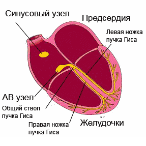
The composition of the cardiac striated muscle tissue includes typical contractile muscle cells - cardiomyocytes and atypical cardiac myocytes, which form the so-called conducting system- consisting of nodes and bundles, providing automatism of heart contractions, as well as coordination of the contractile function of the myocardium of the atria and ventricles of the heart. The centers of the conduction system of the heart are 2 nodes: 1) sinoatrial node (Kiss-Flex node), it is called the pacemaker of the heart. Located in the wall of the right atrium between the opening of the superior vena cava and the right ear and the giving branch to the atrial myocardium.
2) atrioventricular node(Ashoff-Tavara node) is located in the septum between the atrium and the ventricles. From this node departs atrioventricular bundle(bundle of His), which connects the atrial myocardium with the ventricular myocardium. In the interventricular septum, this bundle divides into right and left legs to the myocardium of the right and left ventricles. The heart receives innervation from the vagus and sympathetic nerves.
In recent years, endocrine cardiomyocytes have been described in the myocardium of the right atrium, secreting a number of hormones (cardiopatrin, cardiodilatin), which regulate the blood supply to the heart muscle.
epicardium is part of the fibro-serous membrane pericardium, covering the heart. In the pericardium, 2 layers are distinguished: the fibrous pericardium, formed by dense fibrous connective tissue, and the serous pericardium, also consisting of fibrous tissue with elastic fibers. It adheres tightly to the myocardium. In the region of the sulci of the heart, in which its blood vessels pass, under the epicardium is often possible from the surrounding organs, and the serous fluid between its plates reduces friction during heart contractions.
blood supply of the heart occurs through the coronary arteries, which are branches (right and left) of the outgoing part of the aorta, extending from it at the level of its valves. The right branch goes not only to the right, but also backwards, descending along the posterior interventricular sulcus of the heart, the left branch goes to the left and anteriorly, along the anterior interventricular sulcus. Most of the veins of the heart are collected in the coronary sinus, which flows into the right atrium and is located in the coronary sulcus. In addition, individual small veins of the heart itself flow directly into the right atrium.
The pulmonary trunk at the place of its exit from the right ventricle is located in front of the aorta. Between the pulmonary artery and the lower surface of the aortic arches is the arterial ligament, which is an overgrown ductus arteriosus (botalla) functioning during the prenatal period of life.
The structure of the heart of any organism has many characteristic nuances. In the process of phylogenesis, that is, the evolution of living organisms to more complex ones, the heart of birds, animals and humans acquires four chambers instead of two chambers in fish and three chambers in amphibians. Such a complex structure is best suited for the separation of arterial and venous blood flows. In addition, the anatomy of the human heart implies many tiny details, each of which performs its strictly defined functions.
Heart as an organ
So, the heart is nothing more than a hollow organ, consisting of specific muscle tissue, which performs the motor function. The heart is located in the chest behind the sternum, more on the left, and its longitudinal axis is directed anteriorly, to the left and down. In front, the heart borders on the lungs, almost completely covered by them, leaving only a small part directly adjacent to the chest from the inside. The boundaries of this part are otherwise called absolute cardiac dullness, and they can be determined by tapping the chest wall ().
In people with a normal constitution, the heart has a semi-horizontal position in the chest cavity, in people with an asthenic constitution (thin and tall) it is almost vertical, and in hypersthenics (dense, stocky, with large muscle mass) it is almost horizontal.

heart position
The back wall of the heart is adjacent to the esophagus and to large main vessels (to the thoracic aorta, to the inferior vena cava). The lower part of the heart is located on the diaphragm.

external structure of the heart
Age features
The human heart begins to form in the third week of the intrauterine period and continues throughout the entire period of gestation, passing through stages from a single-chamber cavity to a four-chamber heart.

development of the heart in utero
The formation of four chambers (two atria and two ventricles) occurs already in the first two months of pregnancy. The smallest structures are fully formed by childbirth. It is in the first two months that the heart of the embryo is most vulnerable to the negative influence of certain factors on the expectant mother.
The heart of the fetus is involved in the blood flow through his body, but differs in the circles of blood circulation - the fetus does not yet have its own breathing with the lungs, but it “breathes” through the placental blood. There are some openings in the fetal heart that allow the pulmonary blood flow to be "switched off" from circulation prior to delivery. During childbirth, accompanied by the first cry of the newborn, and, therefore, at the time of increased intrathoracic pressure and pressure in the heart of the child, these openings are closed. But this does not always happen, and they may remain in a child, for example, (not to be confused with such a defect as an atrial septal defect). An open window is not a heart defect, and subsequently, as the child grows, it overgrows.

hemodynamics in the heart before and after birth
The heart of a newborn child has a rounded shape, and its dimensions are 3-4 cm in length and 3-3.5 cm in width. In the first year of a child's life, the heart increases significantly in size, and more in length than in width. The mass of the heart of a newborn child is about 25-30 grams.
As the baby grows and develops, the heart also grows, sometimes significantly outpacing the development of the body itself according to age. By the age of 15, the mass of the heart increases by almost ten times, and its volume increases by more than five times. The heart grows most intensively up to five years, and then during puberty.
In an adult, the heart is about 11-14 cm long and 8-10 cm wide. Many rightly believe that the size of the heart of each person corresponds to the size of his clenched fist. The mass of the heart in women is about 200 grams, and in men - about 300-350 grams.
After 25 years, changes begin in the connective tissue of the heart, which forms the heart valves. Their elasticity is no longer the same as in childhood and adolescence, and the edges may become uneven. As a person grows up, and then aging, changes occur in all structures of the heart, as well as in the vessels that feed it (in the coronary arteries). These changes can lead to the development of numerous cardiac diseases.
Anatomical and functional features of the heart
Anatomically, the heart is an organ divided by partitions and valves into four chambers. The “upper” two are called the atria (atrium), and the “lower” two are called the ventricles (ventriculum). Between the right and left atria is the interatrial septum, and between the ventricles is the interventricular septum. Normally, these partitions do not have holes in them. If there are holes, this leads to mixing of arterial and venous blood, and, accordingly, to hypoxia of many organs and tissues. Such holes are called septal defects and refer to.

basic structure of the chambers of the heart
The boundaries between the upper and lower chambers are atrioventricular openings - the left, covered by the leaflets of the mitral valve, and the right, covered by the leaflets of the tricuspid valve. The integrity of the septa and the proper functioning of the valvular leaflets prevent mixing of blood flows in the heart, and promote a clear unidirectional flow of blood.
The atria and ventricles are different - the atria are smaller than the ventricles and have thinner walls. So, the wall of the atria is about only three millimeters, the wall of the right ventricle is about 0.5 cm, and the left one is about 1.5 cm.
The atria have small protrusions - ears. They have a slight suction function for better pumping of blood into the atrial cavity. The mouth of the vena cava flows into the right atrium near its ear, and the pulmonary veins in the amount of four (rarely five) flow into the left atrium. From the ventricles depart the pulmonary artery (more often called the pulmonary trunk) on the right and the aortic bulb on the left.

structure of the heart and its vessels
From the inside, the upper and lower chambers of the heart also differ and have their own characteristics. The surface of the atria is smoother than that of the ventricles. From the valve ring between the atrium and the ventricle, thin connective tissue valves originate - bicuspid (mitral) on the left and tricuspid (tricuspid) on the right. The other edge of the leaflet faces the inside of the ventricles. But in order for them not to hang freely, they are, as it were, supported by thin tendon threads called chords. They are like springs, stretch when the valve flaps close and contract when the flaps open. Chords originate from the papillary muscles from the wall of the ventricles - three in the right and two in the left ventricle. That is why the ventricular cavity has an uneven and bumpy inner surface.
The functions of the atria and ventricles also differ. Due to the fact that the atria need to push blood into the ventricles, and not into larger and longer vessels, they have less resistance to muscle tissue to overcome, so the atria are smaller in size and their walls are thinner than those of the ventricles. The ventricles push blood into the aorta (left) and into the pulmonary artery (right). Conventionally, the heart is divided into right and left halves. The right half serves for the flow of exclusively venous blood, and the left half for arterial blood. Schematically, "right heart" is indicated in blue, and "left heart" in red. Normally, these streams never mix.

hemodynamics in the heart
One cardiac cycle lasts about 1 second and is carried out as follows. At the moment of filling with blood, the walls of the atria relax - atrial diastole occurs. The valves of the hollow veins and pulmonary veins are open. The tricuspid and mitral valves are closed. Then the atrial walls tighten and push blood into the ventricles, the tricuspid and mitral valves open. At this point, there is systole (contraction) of the atria and diastole (relaxation) of the ventricles. After the ventricles have taken in blood, the tricuspid and mitral valves close, and the aortic and pulmonary valves open. Then the ventricles contract (ventricular systole), and the atria fill with blood again. There comes a general diastole of the heart.

cardiac cycle
The main function of the heart is reduced to pumping, that is, to pushing a certain blood volume into the aorta with such pressure and speed that the blood is delivered to the most distant organs and to the smallest cells of the body. Moreover, arterial blood with a high content of oxygen and nutrients is pushed into the aorta, which enters the left half of the heart from the vessels of the lungs (flows to the heart through the pulmonary veins).
Venous blood, with a low content of oxygen and other substances, is collected from all cells and organs from the vena cava system, and flows into the right half of the heart from the superior and inferior vena cava. Further, venous blood is pushed out of the right ventricle into the pulmonary artery, and then into the pulmonary vessels in order to carry out gas exchange in the alveoli of the lungs and to enrich it with oxygen. In the lungs, arterial blood collects in the pulmonary venules and veins, and again flows into the left half of the heart (into the left atrium). And so regularly the heart pumps blood around the body at a frequency of 60-80 beats per minute. These processes are denoted by the concept "Circulation of blood". There are two of them - small and large:
- small circle includes the flow of venous blood from the right atrium through the tricuspid valve into the right ventricle - then into the pulmonary artery - then into the arteries of the lungs - oxygenation of blood in the pulmonary alveoli - the flow of arterial blood into the smallest veins of the lungs - into the pulmonary veins - into the left atrium.
- big circle includes the flow of arterial blood from the left atrium through the mitral valve to the left ventricle - through the aorta into the arterial bed of all organs - after gas exchange in tissues and organs, the blood becomes venous (with a high content of carbon dioxide instead of oxygen) - further into the venous bed of organs - into the system of hollow veins - in the right atrium.

circles of blood circulation
Video: heart anatomy and cardiac cycle briefly
Morphological features of the heart
If you look at sections of the heart under a microscope, you can see a special type of musculature that is no longer found in any organ. This is a type of striated muscle, but with significant histological differences from ordinary skeletal muscles and from the muscles lining the internal organs. The main function of the heart muscle, or myocardium, is to provide the most important ability of the heart, which forms the basis of the vital activity of the whole organism as a whole. Is it the ability to shrink, or contractility.In order for the fibers of the heart muscle to contract synchronously, electrical signals must be supplied to them, which excite the fibers. This is another capacity of the heart – .
Conductivity and contractility are possible due to the fact that the heart autonomously generates electricity in itself. Function Data (automatism and excitability) are provided with special fibers that are an integral part of the conductive system. The latter is represented by electrically active cells of the sinus node, the atrioventricular node, the His bundle (with two legs - right and left), as well as Purkinje fibers. In the case when a patient's myocardial damage affects these fibers, they develop, otherwise called.
cardiac cycle
Normally, an electrical impulse originates in the cells of the sinus node, which is located in the zone of the right atrial appendage. In a short period of time (about half a millisecond), the impulse propagates through the atrial myocardium, and then enters the cells of the atrioventricular junction. Usually, signals are transmitted to the AV node through three main tracts - the Wenckenbach, Thorel and Bachmann bundles. In the cells of the AV node, the time of impulse transmission is extended to 20-80 milliseconds, and then the impulses enter through the right and left legs (as well as the anterior and posterior branches of the left leg) of the His bundle to the Purkinje fibers, and eventually to the working myocardium. The frequency of impulse transmission along all pathways is equal to the heart rate and is 55-80 impulses per minute.
So, the myocardium, or cardiac muscle, is the middle membrane in the wall of the heart. The inner and outer shells are connective tissue, and are called the endocardium and epicardium. The last layer is part of the pericardial sac, or cardiac “shirt”. Between the inner sheet of the pericardium and the epicardium, a cavity is formed, filled with a very small amount of fluid, to ensure better sliding of the sheets of the pericardium at the moments of heart contractions. Normally, the volume of fluid is up to 50 ml, an excess of this volume may indicate pericarditis.

structure of the heart wall and membrane
Blood supply and innervation of the heart
Despite the fact that the heart is a pump to provide the whole body with oxygen and nutrients, it itself also needs arterial blood. In this regard, the entire wall of the heart has a well-developed arterial network, which is represented by a branching of the coronary (coronary) arteries. The mouths of the right and left coronary arteries depart from the aortic root and are divided into branches penetrating the thickness of the heart wall. If these important arteries become clogged with blood clots and atherosclerotic plaques, the patient will develop, and the organ will no longer be able to perform its functions in full.

the location of the coronary arteries that supply blood to the heart muscle (myocardium)
The frequency and strength with which the heart beats is influenced by nerve fibers extending from the most important nerve conductors - the vagus nerve and the sympathetic trunk. The first fibers have the ability to slow down the frequency of the rhythm, the latter - to increase the frequency and strength of the heartbeat, that is, they act like adrenaline.

innervation of the heart
In conclusion, it should be noted that the anatomy of the heart may have some deviations in individual patients, therefore, only a doctor is able to determine the norm or pathology in a person after conducting an examination that can most informatively visualize the cardiovascular system.
Video: lecture on the anatomy of the heart
It is he who protects our motor from injuries, penetration of infections, carefully fixes the heart in a certain position in the chest cavity, preventing its displacement. Let's talk in more detail about the structure and functions of the outer layer or pericardium.
1 Heart layers
The heart has 3 layers or shells. The middle layer is the muscular, or myocardium, (in Latin, the prefix myo- means "muscle"), the thickest and most dense. The middle layer provides contractile work, this layer is a true hard worker, the basis of our “motor”, it represents the main part of the organ. The myocardium is represented by a striated cardiac tissue endowed with special functions peculiar only to it: the ability to spontaneously excite and transmit an impulse to other cardiac departments through the conduction system.
Another important difference between the myocardium and the muscles of the skeleton is that its cells are not multicellular, but have one nucleus and represent a network. The myocardium of the upper and lower cardiac cavities is separated by horizontal and vertical partitions of the fibrous structure, these partitions provide the possibility of separate contraction of the atria and ventricles. The muscular layer of the heart is the basis of the organ. Muscle fibers are organized into bundles; in the upper chambers of the heart, a two-layer structure is distinguished: bundles of the outer layer and the inner one.
Muscular layer of the heart
A distinctive feature of the ventricular myocardium is that in addition to the muscle bundles of the surface layer and internal bundles, there is also a middle layer - separate bundles for each ventricle of the annular structure. The inner shell of the heart or endocardium (in Latin, the prefix endo- means “inner”) is thin, one cell epithelial layer thick. It lines the inner surface of the heart, all its chambers from the inside, and the heart valves consist of a double layer of the endocardium.
In structure, the inner shell of the heart is very similar to the inner layer of blood vessels, blood collides with this layer as it passes through the chambers. It is important that this layer is smooth to avoid thrombosis, which can form when blood cells are destroyed by impact on the heart walls. This does not occur in a healthy organ, since the endocardium has a perfectly smooth surface. The outer surface of the heart is the pericardium. This layer is represented by the outer sheet of the fibrous structure and the inner - serous. Between the sheets of the surface layer there is a cavity - pericardial, with a small amount of fluid.
2 We delve into the outer layer
The structure of the wall of the heart
So, the pericardium is not at all a single outer layer of the heart, but a layer consisting of several plates: fibrous and serous. Fibrous pericardium is dense, external. It performs to a greater extent a protective function and the function of a certain fixation of the organ in the chest cavity. And the inner, serous layer fits snugly directly to the myocardium, this inner layer is called the epicardium. Imagine a bag with a double bottom? This is what the outer and inner pericardial sheets look like.
The gap between them is the pericardial cavity, normally it contains from 2 to 35 milliliters of serous fluid. The liquid is needed for softer friction of the layers against each other. The epicardium tightly covers the outer layer of the myocardium, as well as the initial sections of the largest vessels of the heart, its other name is the visceral pericardium (in Latin viscera - organs, viscera), i.e. this is the layer directly lining the heart. And already the parietal pericardium is the outermost layer of all the membranes of the heart.
The following sections or walls are distinguished in the superficial pericardial layer, their name depends directly on the organs and areas to which the membrane is attached. Walls of the pericardium:
- Anterior wall of the pericardium. Attached to the chest wall
- diaphragmatic wall. This shell wall is directly fused with the diaphragm.
- Lateral or pleural. Allocate on the sides of the mediastinum, adjacent to the pulmonary pleura.
- back. Borders on the esophagus, descending aorta.
The anatomical structure of this shell of the heart is not easy, because in addition to the walls, there are also sinuses in the pericardium. These are such physiological cavities, we will not delve into their structure. It is enough to know that between the sternum and the diaphragm there is one of these pericardial sinuses - the anteroinferior one. It is her, in pathological conditions, that medical workers pierce or puncture. This diagnostic manipulation is high-tech and complex, carried out by specially trained personnel, often under ultrasound control.
3 Why does the heart need a bag?
Pericardium and its structure
Our main "motor" of the body requires extremely careful attitude and care. Probably, for this purpose, nature dressed the heart in a bag - the pericardium. First of all, it performs the function of protection, carefully wrapping the heart in its shells. Also, the pericardial bag fixes, fixes our “motor” in the mediastinum, preventing displacement during movements. This is possible due to the strong fixation of the surface of the heart with the help of ligaments to the diaphragm, sternum, vertebrae.
The role of the pericardium as a barrier to cardiac tissues from various infections should be noted. The pericardium "fences off" our "motor" from other organs of the chest, clearly determining the position of the heart and helping the heart chambers to fill with blood better. At the same time, the superficial layer prevents excessive expansion of the organ due to sudden overloads. Prevention of overdistension of the chambers is another important role of the outer wall of the heart.
4 When the pericardium "sick"
Pericarditis - inflammation of the pericardial sac
Inflammation of the outer lining of the heart is called pericarditis. The causes of the inflammatory process can be infectious agents: viruses, bacteria, fungi. Also, this pathology can be provoked by a chest injury, directly by cardiac pathology, for example, an acute heart attack. Also, the exacerbation of such systemic diseases as SLE, rheumatoid arthritis, can serve as the beginning in a chain of inflammatory phenomena of the superficial cardiac layer.
Not infrequently, pericarditis accompanies tumor processes in the mediastinum. Depending on how much fluid is released into the pericardial cavity during inflammation, dry and effusion forms of the disease are isolated. Often these forms in this order replace each other with the course and progression of the disease. Dry cough, pain in the chest, especially with a deep breath, a change in body position, during coughing are characteristic of the dry form of the disease.
The effusion form is characterized by a slight decrease in the severity of pain, and at the same time, retrosternal heaviness, shortness of breath, and progressive weakness appear. With a pronounced effusion into the pericardial cavity, the heart is as if squeezed into a vice, the normal ability to contract is lost. Shortness of breath haunts the patient even at rest, active movements become completely impossible. The risk of cardiac tamponade increases, which is fatal.
5 Heart injection or pericardial puncture
This manipulation can be carried out both for diagnostic purposes and for therapeutic purposes. The doctor performs a puncture with a threat of tamponade, with significant effusion, when it is necessary to pump out fluid from the heart sac, thereby providing the organ with the opportunity to contract. For diagnostic purposes, a puncture is performed to clarify the etiology or cause of inflammation. This manipulation is very complicated and requires a highly qualified doctor, since during its implementation there is a risk of damage to the heart.
Aortic aneurysm of the heart - what is it?
What is cardiac bradycardia
The publication of site materials on your page is possible only if you specify the full active link to the source
The structure of the wall of the heart.
Internal structure of the heart.
The human heart has 4 chambers (cavities): two atria and two ventricles (right and left). One chamber is separated from the other by partitions.
transverse baffle divides the heart into atria and ventricles.
longitudinal partition, in which two parts are distinguished: interatrial and interventricular, divides the heart into two halves that do not communicate with each other - right and left.
In the right half is the right atrium and right ventricle and venous blood flows
In the left half is the left atrium and left ventricle and arterial blood flows.
There is an oval fossa on the interatrial septum of the right atrium.
The following vessels enter the atrium:
1. superior and inferior vena cava
2. the smallest veins of the heart
3. opening of the coronary sinus
On the lower wall of this atrium is the right atrioventricular opening, in which there is a tricuspid valve that prevents the reverse flow of blood from the ventricle to the atrium.
The right ventricle is separated from the left ventricular septum.
The right ventricle is divided into two sections:
1) front, in which there is an arterial cone passing into the pulmonary trunk.
2) rear(the cavity itself), it has fleshy trabeculae that pass into the papillary muscles, tendon chords (threads) depart from them, heading to the cusps of the right atrioventricular valve.
4 pulmonary veins flow into it, through which arterial blood enters. On the lower wall of this atrium there is a left atrioventricular opening, in which the bicuspid valve (mitral) is located.
The left ventricle has two sections:
1) anterior section from which the aortic cone originates.
2) back department(actual cavity), it has fleshy trabeculae, passing into the papillary muscles, tendon chords (filaments) depart from them, heading to the cusps of the left atrioventricular valve.
There are two types of valves:
1. Leaf valves - there are two and three leaf valves.
Butterfly valve located in the left atrioventricular orifice.
Tricuspid valve located in the right atrioventricular orifice.
The structure of these valves is as follows: the valve leaflet is connected by means of chords to the papillary muscles. Contracting, the muscles pull the chords, the valves open. When the muscles relax, the valves close. These valves prevent back flow of blood from the ventricles to the atria.
2. The semilunar valves are located together with the exit of the aorta and the pulmonary trunk. They block the flow of blood from the arteries to the ventricles.
The valves consist of three semi-lunar flaps - a pocket, in the center of which there is a thickening - nodules. They provide a complete seal when the semilunar valves are closed.
The wall of the heart consists of three layers: the inner one - the endocardium, the middle, thickest one - the myocardium and the outer one - the epicardium.
1. The endocardium lines all the cavities of the heart from the inside, covers the papillary muscles with their tendon chords (threads), forms the atrioventricular valves, the valves of the aorta, the pulmonary trunk, as well as the valves of the inferior vena cava and coronary sinus.
Consists of connective tissue with elastic fibers and smooth muscle cells, as well as endothelium.
2. Myocardium (muscle layer) is the contractile apparatus of the heart. The myocardium is made up of cardiac muscle tissue.
The atrial muscles are completely separated from the ventricular muscles by fibrous rings located around the atrioventricular openings. Fibrous rings, together with other accumulations of fibrous tissue, make up a kind of skeleton of the heart, which serves as a support for the muscles and valvular apparatus.
The muscular layer of the atria consists of two layers: superficial and deep. It is thinner than the muscular membrane of the ventricles, consisting of three layers: inner, middle and outer. In this case, the muscle fibers of the atria do not pass into the muscle fibers of the ventricles; the atria and ventricles contract at the same time.
3. The epicardium is the outer shell of the heart, covering its muscle and tightly fused with it. At the base of the heart, the epicardium wraps up and passes into the pericardium.
The pericardium is a pericardial sac that insulates the heart from surrounding organs and prevents overstretching.
The pericardium consists of an inner visceral plate (epicardium) and an outer parietal (parietal) plate.
Between the two plates of the pericardium - parietal and epicardial there is a slit-like space - the pericardial cavity, which contains a small amount (up to 50 ml) of serous fluid, which reduces friction during heart contractions.
The structure of the walls of the heart
- endocardium - thin inner layer;
- myocardium - thick muscle layer;
- epicardium - a thin outer layer, which is the visceral sheet of the pericardium - the serous membrane of the heart (heart sac).
The middle layer of the heart wall is made up of
Answers and explanations
The walls of the heart are made up of three layers:
endocardium - thin inner layer; myocardium - thick muscle layer; epicardium - a thin outer layer, which is the visceral sheet of the pericardium - the serous membrane of the heart (heart sac).
The endocardium lines the cavity of the heart from the inside, exactly repeating its complex relief. The endocardium is formed by a single layer of flat polygonal endotheliocytes located on a thin basement membrane.
The myocardium is formed by cardiac striated muscle tissue and consists of cardiac myocytes interconnected by a large number of jumpers, with the help of which they are connected into muscle complexes that form a narrow-loop network. Such a muscular network provides rhythmic contraction of the atria and ventricles. At the atria, the thickness of the myocardium is the smallest; in the left ventricle - the greatest.
The atrial myocardium is separated by fibrous rings from the ventricular myocardium. The synchrony of myocardial contractions is provided by the conduction system of the heart, which is the same for the atria and ventricles. In the atria, the myocardium consists of two layers: superficial (common to both atria), and deep (separate). In the superficial layer, the muscle bundles are located transversely, in the deep layer - longitudinally.
The myocardium of the ventricles consists of three different layers: outer, middle and inner. In the outer layer, the muscle bundles are oriented obliquely, starting from the fibrous rings, continuing down to the apex of the heart, where they form a heart curl. The inner layer of the myocardium consists of longitudinally arranged muscle bundles. Due to this layer, papillary muscles and trabeculae are formed. The outer and inner layers are common to both ventricles. The middle layer is formed by circular muscle bundles, separate for each ventricle.
The epicardium is built according to the type of serous membranes and consists of a thin plate of connective tissue covered with mesothelium. The epicardium covers the heart, the initial sections of the ascending aorta and pulmonary trunk, the final sections of the caval and pulmonary veins.
133. Layers of the wall of the heart, their functions.
The heart, cor (Greek cardia), is a hollow organ, the walls of which consist of three layers - inner, middle, outer.
Inner shell, endocardium, endocardium is represented by a layer of endotheliocytes. The endocardium covers all the structures inside the chambers of the heart. Its derivatives are all the valves and dampers in the heart. This sheath provides laminar blood flow.
Middle shell, myocardium, myocardium is formed by striated muscle cells (cardiomyocytes). Provides contraction of the atria and ventricles.
outer shell, epicardium, epicardium is represented by a serous membrane, which is the visceral sheet of the pericardium. The shell provides free displacement of the heart during its contraction.
134. The degree of expression of the muscle layer in the chambers of the heart.
The muscle layer has a different thickness in the chambers of the heart, which depends on the work performed by them. Maximum thickness this layer - in the left ventricle, tk. it ensures the movement of blood through the systemic circulation, overcoming the enormous forces of friction. In second place is the thickness of the myocardium in the wall of the right ventricle, which provides blood flow through the pulmonary circulation. And, finally, this layer is least expressed in the walls of the atria, which ensure the movement of blood from them into the ventricles.
135. Features of the structure of the myocardium of the ventricles and atria.
In the atria, the myocardium consists of two layers: superficial- common to both ventricles and deep- separate for each of them.
In the ventricles, the myocardium consists of three layers: external (surface), middle And internal (deep).
The outer and inner layers are common to both ventricles, while the middle layer is separate for each ventricle. The muscle fibers of the atria and ventricles are isolated from each other.
Derivatives of the deep layer of the ventricular myocardium are papillary muscles and fleshy trabeculae.
Derivatives of the outer layer of the atrial myocardium are the pectinus muscles.
136. Large and small circles of blood circulation, their functions.
Systemic circulation provides blood flow in the following direction: from the left ventricle → to the aorta → to the organ arteries → to the MCR of organs → to the organ veins → to the vena cava → to the right atrium.
Small circle of blood circulation provides blood flow in a different direction: from the right ventricle → to the pulmonary trunk → to the pulmonary arteries → to the ICR of the lung acini → to the pulmonary veins → to the left atrium.
Both circles of blood circulation are components of a single circle of blood circulation and perform two functions - transport and exchange. In the small circle, the metabolic function is mainly associated with the gas exchange of oxygen and carbon dioxide.
137. Heart valves, their functions.
There are four valves in the heart: two valvular and two semilunar.
Right atrioventricular (tricuspid) valve located between the right atrium and ventricle.
Left atrioventricular (mitral) valve located between the left atrium and ventricle.
Pulmonary valve, valva trunci pulmonalis is located within the base of the pulmonary trunk.
aortic valve, valva aortae is located within the base of the aorta.
To continue downloading, you need to collect the image:
The structure of the wall of the heart
endocardium, average - myocardium, outdoor - epicardium
Endocardium -
Myocardium -
surface layer, outer longitudinal, average roundabout and interior
fibrous rings
conducting system sinoatrial
2) atrioventricular node
epicardium pericardium,
blood supply
The structure of the wall of the heart
Anatomical and physiological features of the cardiovascular system
The circulatory system consists of the heart - the central organ of blood circulation, the rhythmic contraction of which determines this movement, and blood vessels. The vessels through which blood from the heart enters the organs are called arteries, and the vessels that bring blood to the heart are called veins (Fig. 3).
The heart is a hollow muscular organ with a mass gr., cone-shaped. It is located in the chest cavity between the lungs, in the lower mediastinum.
In the chest cavity, the heart occupies an oblique position and faces its wide part - the base, up, back and right, and narrow - tip, forward, down and left; 2/3 it is located in the left half of the chest cavity.
Figure 3 - Heart; lengthwise cut.
1 - superior vena cava; 2 - right atrium; 3 - right atrioventricular valve; 4 - right ventricle; 5 - interventricular septum; 6 - left ventricle; 7 - papillary muscles; 8 - tendon chords; 9 - left atrioventricular valve; 10 - left atrium; 11 - pulmonary veins; 12 - aortic arch.
The boundaries of the heart are variable and depend on age, gender, human constitution and body position. The length of the heart in adults is 8.7-14.0 cm, the largest transverse size of the heart is 5-8 cm, anteroposterior - 6-8 cm on the surface of the heart are noticeable interventricular sulci: anterior and posterior, covering the heart in front and behind, and transverse coronal sulcus, arranged in a ring. Along these furrows are the own arteries and veins of the heart. These grooves correspond to partitions dividing the heart into 4 sections: longitudinal intercostal and interventricular septa divide the organ into two isolated halves - right and left heart; a transverse partition divides each of these halves into an upper chamber - atrium and bottom- ventricle.
The atria take blood from the veins and push it into the ventricles, the ventricles eject the blood into the arteries; right - through the aorta, from which numerous arteries depart to the organs and walls of the body. Each atrium communicates with the corresponding ventricle and atrioventricular arteries. The right side of the heart contains venous blood, while the left side contains arterial blood.
Right atrium - is a cavity with a volume of ml., resembles a cube in shape, is located at the base of the heart on the right and behind the aorta and pulmonary trunk. It serves as the confluence of the hollow veins and the veins of the heart itself. Its upper part is atrial appendage.
In the wall of the ear, the heart muscle forms muscular protrusions, located approximately in parallel, which are called comb muscles. In the area of confluence of the inferior vena cava there is a small valve, which is its damper. On the inner wall of the right atrium there is oval fossa(in the fetus, this is an opening through which blood passes from the right atrium into the left atrium, since the fetus does not have a small circle of blood circulation). Below and behind the edge of the oval fossa is the confluence coronary sinus, which collects most of the blood from the wall of the heart itself. The sinus opening is closed by the coronary sinus valve. The passage between the right atrium and the right ventricle is called the right atrioventricular orifice. During systole, the right ventricle closes. right atrioventricular(tricuspid) valve that separates the cavity of the right ventricle from the right atrium and does not allow blood to flow back into the right atrium. During ventricular diastole, the valve opens towards the ventricle.
Right ventricle it is separated from the left ventricle by the interventricular septum, most of which is muscular, and the smaller one, located in the uppermost section, closer to the atria, is membranous. Above in the wall of the stomach two holes: behind - the right atrioventricular, and in front - the opening of the pulmonary trunk. The elongated funnel-shaped section of the ventricle in this place is called arterial cone. Directly above the opening of the pulmonary trunk, consisting of anterior, left and right semilunar dampers, located in a circle, with a convex surface into the cavity of the right ventricle, and with a concave and free edge - into the lumen of the pulmonary trunk. On the free edge, each of the flaps has a thickening - a knot, which contributes to a more dense closing of the semilunar flaps when they are closed. When the muscles of the ventricle contract, the semilunar valves are pressed against the wall of the pulmonary trunk by blood flow and do not prevent the passage of blood from the ventricle; during relaxation, when the pressure in the cavity of the ventricle decreases, the return flow of blood fills the pockets between the wall of the pulmonary trunk and each of the semilunar valves and closes (opens) the valves, their edges close and do not allow blood to pass to the heart.
The right atrioventricular orifice is closed by the right atrioventricular valve, having anterior, posterior and medial cusps. The latter fill the triangular tendon plates. On the inner surface of the right ventricle, fleshy trabeculae and cone-shaped nipple muscles, from which to the edges and surfaces of the valves go tendon chords. During atrial contraction, the valve leaflets are pressed by the blood flow against the walls of the ventricle and do not prevent its passage into the cavity of the latter. When the muscles of the ventricle contract, the free edges of the valves close and are held in this position by the tendon chords and contraction of the papillary muscles, preventing blood from flowing back into the atrium.
Left atrium limited from the right intercardiac septum; It has left ear. In the posterior section of the upper wall, 4 pulmonary veins open into it, devoid of valves, through which arterial blood flows from their lungs. Communicates with the left ventricle through the left atrioventricular orifice.
left ventricle in the anterior upper section aortic opening. At the exit of the aorta from the left ventricle aortic valve, consisting of right, left and rear semilunar flaps. The atrioventricular orifice contains the left atrioventricular valve- (bicuspid mitral). Consisting of front and rear wings of a triangular shape. On the inner surface of the left ventricle there are fleshy trabeculae and 2 papillary muscles, from which thick tendinous chords originate, attached to the mitral valve leaflets.
The wall of the heart consists of three layers. The inner one is called endocardium, average - myocardium, outdoor - epicardium
Endocardium - lines all the cavities of the heart, tightly fused with the underlying muscle layer. From the side of the cavities of the heart, it is lined with endothelium. The endocardium forms the atrioventricular valves, as well as the valves of the aorta and pulmonary trunk.
Myocardium - is the thickest and functionally most powerful part of the heart wall. It is formed by cardiac striated muscle tissue and consists of cardiac myocytes (cardiomyocytes) interconnected by a large number of jumpers (intercalary discs), with the help of which they are connected into muscle complexes or fibers that form a narrow-loop network. It provides a complete rhythmic contraction of the atria and ventricles.
The muscle layer of the walls of the atria is thin due to a small load and consists of surface layer, common to both atria, and deep, separate for each of them. In the walls of the ventricles, it is the most significant in thickness; outer longitudinal, average roundabout and interior longitudinal layer. The outer fibers in the region of the apex of the heart pass into the inner longitudinal fibers, and between them are the circular muscle fibers of the middle layer. The muscular layer of the left ventricle is the thickest.
The muscle fibers of the atria and ventricles begin from the fibrous rings located around the right and left atrioventricular openings, which completely separate the atrial myocardium from the ventricular myocardium.
fibrous rings form a kind of skeleton of the heart, which also includes thin connective tissue rings around the openings of the aorta and pulmonary trunk and the right and left fibrous triangles adjacent to them.
The composition of the cardiac striated muscle tissue includes typical contractile muscle cells - cardiomyocytes and atypical cardiac myocytes, which form the so-called conducting system- consisting of nodes and bundles, providing automatism of heart contractions, as well as coordination of the contractile function of the myocardium of the atria and ventricles of the heart. The centers of the conduction system of the heart are 2 nodes: 1) sinoatrial node (Kiss-Flex node), it is called the pacemaker of the heart. Located in the wall of the right atrium between the opening of the superior vena cava and the right ear and the giving branch to the atrial myocardium.
2) atrioventricular node(Ashoff-Tavara node) is located in the septum between the atrium and the ventricles. From this node departs atrioventricular bundle(bundle of His), which connects the atrial myocardium with the ventricular myocardium. In the interventricular septum, this bundle divides into right and left legs to the myocardium of the right and left ventricles. The heart receives innervation from the vagus and sympathetic nerves.
In recent years, endocrine cardiomyocytes have been described in the myocardium of the right atrium, secreting a number of hormones (cardiopatrin, cardiodilatin), which regulate the blood supply to the heart muscle.
epicardium is part of the fibro-serous membrane pericardium, covering the heart. In the pericardium, 2 layers are distinguished: the fibrous pericardium, formed by dense fibrous connective tissue, and the serous pericardium, also consisting of fibrous tissue with elastic fibers. It adheres tightly to the myocardium. In the region of the sulci of the heart, in which its blood vessels pass, under the epicardium is often possible from the surrounding organs, and the serous fluid between its plates reduces friction during heart contractions.
blood supply of the heart occurs through the coronary arteries, which are branches (right and left) of the outgoing part of the aorta, extending from it at the level of its valves. The right branch goes not only to the right, but also backwards, descending along the posterior interventricular sulcus of the heart, the left branch goes to the left and anteriorly, along the anterior interventricular sulcus. Most of the veins of the heart are collected in the coronary sinus, which flows into the right atrium and is located in the coronary sulcus. In addition, individual small veins of the heart itself flow directly into the right atrium.
The pulmonary trunk at the place of its exit from the right ventricle is located in front of the aorta. Between the pulmonary artery and the lower surface of the aortic arches is the arterial ligament, which is an overgrown ductus arteriosus (botalla) functioning during the prenatal period of life.
The heart is the main organ of the blood supply and lymph formation system in the body. It is presented in the form of a large muscle with several hollow chambers. Due to its ability to contract, it sets the blood in motion. There are three layers of the heart: epicardium, endocardium and myocardium. The structure, purpose and functions of each of them will be considered in this material.
The structure of the human heart - anatomy
The heart muscle consists of 4 chambers - 2 atria and 2 ventricles. The left ventricle and the left atrium form the so-called arterial part of the organ, based on the nature of the blood located here. In contrast, the right ventricle and right atrium make up the venous portion of the heart.
The circulatory organ is presented in the form of a flattened cone. It distinguishes the base, apex, lower and anterior upper surfaces, as well as two edges - left and right. The apex of the heart has a rounded shape and is entirely formed by the left ventricle. At the base are the atria, and in its front part lies the aorta.
Heart sizes
It is believed that in an adult, formed human individual, the dimensions of the heart muscle are equal to the dimensions of a clenched fist. In fact, the average length of this organ in a mature person is 12-13 cm. The heart is 9-11 cm across.
The mass of the heart of an adult male is about 300 g. In women, the heart weighs an average of about 220 g.
Phases of the heart

There are several separate phases of contraction of the heart muscle:
- At the beginning, atrial contraction occurs. Then, with some slowdown, the contraction of the ventricles starts. During this process, the blood naturally tends to fill the chambers with reduced pressure. Why does it not return to the atria after this? The fact is that the gastric valves block the path of blood. Therefore, it remains only to move in the direction of the aorta, as well as the vessels of the pulmonary trunk.
- The second phase is the relaxation of the ventricles and atria. The process is characterized by a short-term decrease in the tone of the muscle structures from which these chambers are formed. The process causes a decrease in pressure in the ventricles. Thus, the blood begins to move in the opposite direction. However, this is prevented by closing pulmonary and arterial valves. During relaxation, the ventricles fill with blood, which comes from the atria. In contrast, the atria fill with bodily fluid from the large and
What is responsible for the work of the heart?
As you know, the functioning of the heart muscle is not an arbitrary act. The organ remains active continuously even when the person is in deep sleep. There are hardly any people who pay attention to the heart rate in the process of activity. But this is achieved due to a special structure built into the heart muscle itself - a system for generating biological impulses. It is noteworthy that the formation of this mechanism occurs in the first weeks of intrauterine birth of the fetus. Subsequently, the impulse generation system does not allow the heart to stop throughout life.

In a calm state, the number of contractions of the heart muscle for a minute is about 70 beats. Within one hour, the number reaches 4200 beats. Given that during one contraction, the heart ejects 70 ml of fluid into the circulatory system, it is easy to guess that up to 300 liters of blood passes through it in an hour. How much blood does this organ pump in a lifetime? This figure averages 175 million liters. Therefore, it is not surprising that the heart is called the ideal engine, which practically does not fail.
shells of the heart
In total, there are 3 separate shells of the heart muscle:
- Endocardium is the inner lining of the heart.
- The myocardium is an internal muscular complex formed by a thick layer of filamentous fibers.
- The epicardium is the thin outer shell of the heart.
- The pericardium is an auxiliary cardiac membrane, which is a kind of bag that contains the entire heart.
Myocardium

The myocardium is a multi-tissue muscular membrane of the heart, which is formed by striated fibers, loose connective structures, nerve processes, and an extensive network of capillaries. Here are the P-cells that form and conduct nerve impulses. In addition, in the myocardium there are cells myocytes and cardiomyocytes, which are responsible for the contraction of the blood organ.
The myocardium consists of several layers: inner, middle and outer. The internal structure consists of muscle bundles that are located longitudinally in relation to each other. In the outer layer, the bundles of muscle tissue are located obliquely. The latter go to the very top of the heart, where they form the so-called curl. The middle layer consists of circular muscle bundles, separate for each of the ventricles of the heart.
epicardium

The presented shell of the heart muscle has the smoothest, thinnest and somewhat transparent structure. The epicardium forms the outer tissues of the organ. In fact, the shell acts as the inner layer of the pericardium - the so-called heart bag.
The surface of the epicardium is formed from mesothelial cells, under which there is a connective, loose structure, represented by connective fibers. In the region of the apex of the heart and in its furrows, the membrane in question includes adipose tissue. The epicardium grows together with the myocardium in places where there is the least accumulation of fat cells.
Endocardium

Continuing to consider the membranes of the heart, let's talk about the endocardium. The presented structure is formed by elastic fibers, which consist of smooth muscle and connective cells. Endocardial tissues line all hearts. On the elements extending from the blood organ: aorta, pulmonary veins, pulmonary trunk, endocardial tissues pass smoothly, without clearly distinguishable boundaries. In the thinnest parts of the atria, the endocardium fuses with the epicardium.
Pericardium
The pericardium is the outermost part of the heart, also called the pericardial sac. This structure is presented in the form of a cone cut at an angle. The lower base of the pericardium is placed on the diaphragm. Towards the top, the shell goes more to the left than to the right. This peculiar bag surrounds not only the heart muscle, but also the aorta, the mouth of the pulmonary trunk and adjacent veins.
The pericardium is formed in human individuals in the early stages of intrauterine development. This happens approximately 3-4 weeks after the formation of the embryo. Violations of the structure of this shell, its partial or complete absence often leads to congenital heart defects.
Finally
In the presented material, we examined the structure of the human heart, the anatomy of its chambers and membranes. As you can see, the heart muscle has an extremely complex structure. Surprisingly, despite its intricate structure, this organ functions continuously throughout life, malfunctioning only in the event of the development of serious pathologies.
Heart- the central organ of the blood and lymph circulation system. Due to the ability to contract, the heart sets the blood in motion.
Wall of the heart consists of three membranes: endocardium, myocardium and epicardium.
Endocardium. In the inner shell of the heart, the following layers are distinguished: endothelium, lining the inside of the cavity of the heart, and its basement membrane; subendothelial layer, represented by loose connective tissue, in which there are many poorly differentiated cells; muscular-elastic layer, consisting of smooth muscle tissue, between the cells of which elastic fibers are located in the form of a dense network; outer connective tissue layer, consisting of loose connective tissue. The endothelium and subendothelial layers are similar to the inner membrane of the vessels, the musculo-elastic layer is the "equivalent" of the middle membrane, and the outer connective tissue layer is similar to the outer (adventitial) membrane of the vessels.
The surface of the endocardium is ideally smooth and does not interfere with the free movement of blood. In the atrioventricular region and at the base of the aorta, the endocardium forms duplications (folds), called valves. Distinguish between atrioventricular and ventricular-vascular valves. There are fibrous rings at the attachment sites of the valves. Heart valves are dense plates of fibrous connective tissue covered with endothelium. The nutrition of the endocardium occurs by diffusion of substances from the blood located in the cavities of the atria and ventricles.
Myocardium(middle shell of the heart) - a multi-tissue shell, consisting of striated cardiac muscle tissue, intermuscular loose connective tissue, numerous vessels and capillaries, as well as nerve elements. The main structure is cardiac muscle tissue, which in turn consists of cells that form and conduct nerve impulses, and cells of the working myocardium that provide contraction of the heart (cardiomyocytes). Among the cells that form and conduct impulses in the conduction system of the heart, there are three types: P-cells (pacemaker cells), intermediate cells and Purkinya cells (fibers).
P cells- pacemaker cells located in the center of the sinus node of the conduction system of the heart. They have a polygonal shape and are determined to spontaneous depolarization of the plasmalemma. Myofibrils and organelles of general importance in pacemaker cells are weakly expressed. Intermediate cells are a heterogeneous group of cells that transmit excitation from P-cells to Purkinya cells. Purkinya cells are cells with a small number of myofibrils and a complete absence of the T-system, with a large amount of cytoplasm compared to working contractile myocytes. Purkinya cells transmit excitation from intermediate cells to contractile cells of the myocardium. They are part of the bundle of His of the conduction system of the heart.
A number of drugs and other factors that can lead to arrhythmias and heart block have an adverse effect on pacemaker cells and Purkinya cells. The presence in the heart of its own conducting system is extremely important, since it provides a rhythmic change in systolic contractions and diastole of the heart chambers (atria and ventricles) and the operation of its valvular apparatus.
The bulk of the myocardium make contractile cells - cardiac myocytes, or cardiomyocytes. These are cells of an elongated shape with an ordered system of transversely striated myofibrils located on the periphery. Between the myofibrils are mitochondria with a large number of cristae. In atrial myocytes, the T-system is weakly expressed. The granular endoplasmic reticulum is poorly developed in cardiomyocytes. In the central part of myocytes there is an oval-shaped nucleus. Sometimes there are binuclear cardiomyocytes. Atrial muscle tissue contains cardiomyocytes with osmiophilic secretory granules containing natriuretic peptide.
In cardiomyocytes, inclusions of glycogen, which serves as the energy material of the heart muscle, are determined. Its content in the myocytes of the left ventricle is greater than in other parts of the heart. The myocytes of the working myocardium and the conducting system are interconnected by means of intercalated discs - specialized intercellular contacts. Actin contractile myofilaments are attached in the region of the intercalated discs, desmosomes and gap junctions (nexuses) are present.
Desmosomes contribute to the strong adhesion of contractile myocytes into functional muscle fibers, and nexuses ensure the rapid propagation of plasmolemma depolarization waves from one muscle cell to another and the existence of a cardiac muscle fiber as a single metabolic unit. Characteristic for myocytes of the working myocardium is the presence of anastomosing bridges - interconnected fragments of the cytoplasms of muscle cells of different fibers with myofibrils located in them. Thousands of such bridges turn the muscle tissue of the heart into a mesh structure capable of synchronously and efficiently contracting and ejecting the necessary systolic blood volumes from the ventricular cavities. After suffering extensive myocardial infarctions (acute ischemic necrosis of the heart wall), when the muscular tissue of the heart, the system of intercalated discs, anastomosing bridges and the conduction system are diffusely affected, disturbances in the rhythm of the heart, up to fibrillation, occur. In this case, the contractile activity of the heart turns into separate uncoordinated twitches of muscle fibers and the heart is not able to eject the necessary systolic portions of blood into the peripheral circulation.
Myocardium consists in general of highly specialized cells that have lost the ability to divide by mitosis. Mitoses of cardiomyocytes are observed only in certain parts of the atria (Rumyantsev P.P. 1982). At the same time, the myocardium is characterized by the presence of polyploid myocytes, which significantly enhances its working potential. The phenomenon of polyploidy is most often observed in compensatory reactions of the myocardium, when the load on the heart increases, and in pathology (failure of heart valves, lung diseases, etc.).
cardiac myocytes in these cases, they sharply hypertrophy, and the wall of the heart in one or another section thickens. The myocardial connective tissue contains a richly branched network of blood and lymphatic capillaries, which provides the constantly working heart muscle with nutrition and oxygen. In the layers of connective tissue there are dense bundles of collagen fibers, as well as elastic fibers. In general, these connective tissue structures constitute the supporting skeleton of the heart, to which cardiac muscle cells are attached.
Heart- an organ that has the ability to automate contractions. It can function autonomously within certain limits. However, in the body, the activity of the heart is under the control of the nervous system. In the intramural nerve nodes of the heart there are sensitive autonomic neurons (Type II Dogel cells), small intensely fluorescent cells - MYTH cells and effector autonomic neurons (Type I Dogel cells). MYTH cells are considered as intercalary neurons.
epicardium- the outer shell of the heart - is a visceral sheet of the pericardial sac (pericardium). The free surface of the epicardium is lined with mesothelium in the same way as the surface of the pericardium facing the pericardial cavity. Under the mesothelium in the composition of these serous membranes is a connective tissue base of loose fibrous connective tissue.
Inner lining of the heart, or endocardium
Endocardium, endocardium(see Fig. 704. 709), formed from elastic fibers, among which are located connective tissue and smooth muscle cells. From the side of the cavity of the heart, the endocardium is covered with endothelium.
The endocardium lines all the chambers of the heart, is tightly fused with the underlying muscle layer, follows all its irregularities formed by the fleshy trabeculae, pectinate and papillary muscles, as well as their tendon outgrowths.
On the inner shell of the vessels leaving the heart and flowing into it - the hollow and pulmonary veins, the aorta and the pulmonary trunk - the endocardium passes without sharp boundaries. In the atria, the endocardium is thicker than in the ventricles, especially in the left atrium, and thinner where it covers the papillary muscles with tendon chords and fleshy trabeculae.
In the most thinned sections of the walls of the atria, where gaps form in their muscular layer, the endocardium is in close contact and even fuses with the epicardium. In the region of the fibrous rings of the atrioventricular openings, as well as the openings of the aorta and the pulmonary trunk, the endocardium, by doubling its leaf - the duplication of the endocardium - forms the leaflets of the atrioventricular valves and the semilunar valves of the pulmonary trunk and aorta. The fibrous connective tissue between both sheets of each of the cusps and semilunar valves is connected to the fibrous rings and thus fixes the valves to them.
shells of the heart
The heart is located in a pericardial sac called the pericardium. The wall of the heart consists of three layers: the outer one - the epicardium, the middle one - the myocardium, and the inner one - the endocardium.
The outer shell of the heart. epicardium
The epicardium is a smooth, thin and transparent membrane. It is the visceral plate of the pericardial sac (pericardium). The connective tissue base of the epicardium in various parts of the heart, especially in the sulci and in the apex, includes adipose tissue. With the help of the specified connective tissue, the epicardium is most tightly fused with the myocardium in places of the least accumulation or absence of adipose tissue.
The muscular layer of the heart, or myocardium
The middle, muscular membrane of the heart (myocardium), or cardiac muscle, is a powerful and significant part of the wall of the heart in thickness.
Between the muscular layer of the atria and the muscular layer of the ventricles lies dense fibrous tissue, due to which fibrous rings, right and left, are formed. From the side of the outer surface of the heart, their location corresponds to the region of the coronal sulcus.
The right fibrous ring, which surrounds the right atrioventricular orifice, is oval in shape. The left fibrous ring does not completely surround the left atrioventricular opening: on the right, on the left and behind, and has a horseshoe shape.
With its anterior sections, the left fibrous ring is attached to the aortic root, forming triangular connective tissue plates around its posterior periphery - the right and left fibrous triangles.
The right and left fibrous rings are interconnected into a common plate, which completely, with the exception of a small area, isolates the muscles of the atria from the muscles of the ventricles. In the middle of the fibrous plate connecting the rings there is a hole through which the muscles of the atria are connected to the muscles of the ventricles through the neuromuscular atrioventricular bundle conducting impulses.
In the circumference of the openings of the aorta and the pulmonary trunk, there are also interconnected fibrous rings; the aortic ring is connected to the fibrous rings of the atrioventricular orifices.
Muscular layer of the atria
In the walls of the atria, two muscle layers are distinguished: superficial and deep.
The surface layer is common to both atria and represents muscle bundles that run mainly in the transverse direction; they are more pronounced on the anterior surface of the atria, forming here a relatively wide muscle layer in the form of a horizontally located inter-auricular bundle passing to the inner surface of both ears.
On the posterior surface of the atria, the muscle bundles of the superficial layer are partially woven into the posterior sections of the septum.
On the posterior surface of the heart, in the gap formed by the convergence of the borders of the inferior vena cava, the left atrium and the venous sinus, between the bundles of the surface layer of muscles there is a depression covered by the epicardium - the neural fossa. Through this fossa, nerve trunks enter the atrial septum from the posterior cardiac plexus, which innervate the atrial septum, the ventricular septum and the muscle bundle that connects the muscles of the atria with the muscles of the ventricles - the atrioventricular bundle.
The deep layer of muscles of the right and left atria is not common to both atria. It distinguishes ring-shaped, or circular, and loop-shaped, or vertical, muscle bundles.
Circular muscle bundles lie in large numbers in the right atrium; they are located mainly around the openings of the vena cava, passing to their walls, around the coronary sinus of the heart, at the mouth of the right ear and at the edge of the oval fossa; in the left atrium, they lie mainly around the openings of the four pulmonary veins and at the neck of the left ear.
Vertical muscle bundles are located perpendicular to the fibrous rings of the atrioventricular holes, attaching to them with their ends. Part of the vertical muscle bundles enters the thickness of the cusps of the mitral and tricuspid valves.
The crest muscles are also formed by bundles of the deep layer. They are most developed on the inner surface of the anterior right wall of the right atrium, as well as the right and left ears; in the left atrium they are less pronounced. In the intervals between the comb muscles, the wall of the atria and ears is especially thinned.
On the inner surface of both ears there are very short and thin bundles, the so-called fleshy crossbars. Crossing in different directions, they form a very thin loop-like network.
Muscular layer of the ventricles
In the muscular membrane (myocardium) there are three muscle layers: outer, middle and deep. The outer and deep layers, passing from one ventricle to another, are common in both ventricles; the middle, although connected with the other two, outer and deep, layers, but surrounds each ventricle separately.
The outer, relatively thin, layer consists of oblique, partly rounded, partly flattened bundles. The bundles of the outer layer begin at the base of the heart from the fibrous rings of both ventricles and partly from the roots of the pulmonary trunk and aorta. On the front surface of the heart, the outer bundles go from right to left, and on the back - from left to right. At the apex of the left ventricle, both bundles of the outer layer form the so-called whirlpool of the heart and penetrate into the depths of the walls of the heart, passing into the deep muscle layer.
The deep layer consists of bundles that rise from the top of the heart to its base. They have a cylindrical, partly oval shape, are repeatedly split and reconnected, forming loops of various sizes. The shorter of these bundles do not reach the base of the heart, they are directed obliquely from one wall of the heart to another, in the form of fleshy crossbars. The crossbars are located in large numbers along the entire inner surface of both ventricles and have different sizes in different areas. Only the inner wall (septum) of the ventricles, immediately below the arterial openings, is devoid of these crossbars.
A number of such short, but more powerful muscle bundles, partly connected with both the middle and outer layers, freely protrude into the cavity of the ventricles, forming papillary muscles of various sizes and cones.
There are three papillary muscles in the cavity of the right ventricle, and two in the cavity of the left. Tendon strings begin from the top of each of the papillary muscles, through which the papillary muscles are connected to the free edge and partly the lower surface of the cusps of the tricuspid or mitral valves.
However, not all tendon strings are associated with the papillary muscles. A number of them begin directly from the fleshy crossbars formed by the deep muscle layer and are most often attached to the lower, ventricular, surface of the valves.
The papillary muscles with tendinous strings hold the cusp valves when they are slammed by blood flow from the contracted ventricles (systole) to the relaxed atria (diastole). Encountering, however, obstacles from the valves, the blood rushes not into the atria, but into the opening of the aorta and pulmonary trunk, the semilunar valves of which are pressed against the walls of these vessels by the blood flow and thereby leave the lumen of the vessels open.
Located between the outer and deep muscle layers, the middle layer forms a number of well-defined circular bundles in the walls of each ventricle. The middle layer is more developed in the left ventricle, so the walls of the left ventricle are much thicker than the right one. The bundles of the middle muscle layer of the right ventricle are flattened and have an almost transverse and somewhat oblique direction from the base of the heart to the apex.
In the left ventricle, among the bundles of the middle layer, bundles lying closer to the outer layer and located closer to the deep layer can be distinguished.
The interventricular septum is formed by all three muscular layers of both ventricles. However, the muscle layers of the left ventricle take a large part in its formation. Its thickness is almost equal to the thickness of the wall of the left ventricle. It protrudes towards the cavity of the right ventricle. For 4/5, it represents a well-developed muscle layer. This, much larger, part of the interventricular septum is called the muscular part.
The upper (1/5) part of the interventricular septum is thin, transparent and is called the membranous part. The septal leaflet of the tricuspid valve is attached to the membranous part.
The muscles of the atria are isolated from the muscles of the ventricles. An exception is a bundle of fibers starting in the atrial septum in the region of the coronary sinus of the heart. This bundle consists of fibers with a large amount of sarcoplasm and a small amount of myofibrils; the bundle also includes nerve fibers; it originates at the confluence of the inferior vena cava and goes to the ventricular septum, penetrating into its thickness. In the bundle, the initial, thickened part, called the atrioventricular node, is distinguished, passing into a thinner trunk - the atrioventricular bundle, the bundle goes to the interventricular septum, passes between both fibrous rings and at the upper posterior part of the muscular part of the septum is divided into the right and left legs .
The right leg, short and thinner, follows the septum from the side of the cavity of the right ventricle to the base of the anterior papillary muscle and spreads in the muscular layer of the ventricle in the form of a network of thin fibers (Purkinje).
The left leg, wider and longer than the right one, is located on the left side of the ventricular septum, in its initial sections lies more superficially, closer to the endocardium. Heading to the base of the papillary muscles, it breaks up into a thin network of fibers that form the anterior, middle and posterior bundles, spreading in the myocardium of the left ventricle.
At the confluence of the superior vena cava into the right atrium, between the vein and the right ear is the sinoatrial node.
These bundles and nodes, accompanied by nerves and their branches, are the conduction system of the heart, which serves to transmit impulses from one part of the heart to another.
Inner lining of the heart, or endocardium
The inner shell of the heart, or endocardium, is formed from collagen and elastic fibers, among which are located connective tissue and smooth muscle cells.
From the side of the cavities of the heart, the endocardium is covered with endothelium.
The endocardium lines all the cavities of the heart, is tightly fused with the underlying muscle layer, follows all its irregularities formed by the fleshy crossbars, the pectinate and papillary muscles, as well as their tendon outgrowths.
On the inner shell of the vessels leaving the heart and flowing into it - the hollow and pulmonary veins, the aorta and the pulmonary trunk - the endocardium passes without sharp boundaries. In the atria, the endocardium is thicker than in the ventricles, while it is thicker in the left atrium, less where it covers the papillary muscles with tendon strings and fleshy crossbars.
In the most thinned sections of the walls of the atria, where gaps form in the muscle layer, the endocardium is in close contact and even fuses with the epicardium. In the area of fibrous rings, atrioventricular openings, as well as openings of the aorta and pulmonary trunk, the endocardium, by doubling its leaf, duplicating the endocardium, forms the leaflets of the mitral and tricuspid valves and the semilunar valves of the pulmonary trunk and aorta. The fibrous connective tissue between both sheets of each of the cusps and semilunar valves is connected to the fibrous rings and thus fixes the valves to them.
Pericardial sac or pericardium
The pericardium, or pericardium, has the shape of an obliquely cut cone with a lower base located on the diaphragm and an apex reaching almost to the level of the angle of the sternum. In width, it extends more to the left than to the right.
In the pericardial sac, there are: an anterior (sternocostal) part, a posterior inferior (diaphragmatic) part, and two lateral - right and left - mediastinal parts.
The sternocostal part of the pericardial sac faces the anterior chest wall and is located, respectively, in the body of the sternum, V-VI costal cartilages, intercostal spaces and the left part of the xiphoid process.
The lateral sections of the sternocostal part of the pericardial sac are covered by the right and left sheets of the mediastinal pleura, which separate it in the anterior sections from the anterior chest wall. The areas of the mediastinal pleura covering the pericardium are distinguished under the name of the pericardial part of the mediastinal pleura.
The middle of the sternocostal part of the bag, the so-called free part, is open in the form of two triangular-shaped spaces: the upper, smaller, corresponding to the thymus gland, and the lower, larger, corresponding to the pericardium, facing their bases up (to the notch of the sternum) and down (to the diaphragm ).
In the region of the upper triangle, the sternocostal part of the pericardium is separated from the sternum by loose connective and adipose tissue, in which the thymus gland is embedded in children. The compacted part of this fiber forms the so-called superior sterno-periocardial ligament, which fixes here the anterior wall of the pericardium to the handle of the sternum.
In the area of the lower triangle, the pericardium is also separated from the sternum by loose fiber, in which a compacted part is isolated, the lower sterno-periocardio-adrenal ligament, which fixes the lower part of the pericardium to the sternum.
In the diaphragmatic part of the pericardial sac, there is an upper section involved in the formation of the anterior border of the posterior mediastinum, and a lower section covering the diaphragm.
The upper section is adjacent to the esophagus, thoracic aorta and unpaired vein, from which this part of the pericardium is separated by a layer of loose connective tissue and a thin fascial sheet.
The lower section of the same part of the pericardium, which is its base, fuses tightly with the tendon center of the diaphragm; extending slightly to the anterior areas of its muscular part, it is connected to them by loose fiber.
The right and left mediastinal parts of the pericardial sac are adjacent to the mediastinal pleura; the latter is connected to the pericardium by means of loose connective tissue and can be separated by careful preparation. In the thickness of this loose fiber, connecting the mediastinal pleura with the pericardium, passes the phrenic nerve and the accompanying pericardial-bag-diaphragmatic vessels.
The pericardium consists of two parts - internal, serous (serous pericardial sac) and external, fibrous (fibrous pericardial sac).
The serous pericardial sac consists of two serous sacs, as it were, nested one inside the other - the outer one, freely surrounding the heart (the serous sac of the pericardium itself), and the inner one - the epicardium, tightly fused with the myocardium. The serous cover of the pericardium is the parietal plate of the serous pericardial sac, and the serous cover of the heart is the visceral plate (epicardium) of the serous pericardial sac.
The fibrous pericardial sac, which is especially pronounced on the anterior wall of the pericardium, fixes the pericardial sac to the diaphragm, the walls of large vessels and through the ligaments to the inner surface of the sternum.
The epicardium passes into the pericardium at the base of the heart, at the confluence of large vessels: the hollow and pulmonary veins and the exit of the aorta and pulmonary trunk.
Between the epicardium and the pericardium there is a slit-like space (the cavity of the pericardial sac), containing a small amount of pericardial sac fluid, which wets the serous surfaces of the pericardium, thereby causing one serous plate to slide over the other during heart contractions.
As indicated, the parietal plate of the serous pericardial sac passes into the splanchnic plate (epicardium) at the site of entry and exit of large blood vessels from the heart.
If, after the removal of the heart, the pericardial sac is examined from the inside, then the large vessels in relation to the pericardium are located along its posterior wall along approximately two lines - the right, more vertical, and the left, somewhat inclined towards it. On the right line, the superior vena cava, two right pulmonary veins and the inferior vena cava lie from top to bottom, along the left line - the aorta, pulmonary trunk and two left pulmonary veins.
At the site of the transition of the epicardium into the parietal plate, several sinuses of various shapes and sizes are formed. The largest of these are the transverse and oblique sinuses of the pericardial sac.
Transverse sinus of the pericardial sac. The initial sections (roots) of the pulmonary trunk and aorta, adjacent to one another, are surrounded by a common leaf of the epicardium; posterior to them are the atria and next to the right is the superior vena cava. The epicardium from the side of the posterior wall of the initial sections of the aorta and the pulmonary trunk goes up and back to the atria located behind them, and from the latter - down and forward again to the base of the ventricles and the root of these vessels. Thus, between the aortic root and the pulmonary trunk in front and the atria behind, a passage is formed - a sinus, which is clearly visible when the aorta and pulmonary trunk are pulled anteriorly, and the superior vena cava - posteriorly. This sinus is bounded from above by the pericardium, from behind by the superior vena cava and the anterior surface of the atria, from the front by the aorta and the pulmonary trunk; the right and left transverse sinus is open.
Oblique sinus of the pericardial sac. It is located below and behind the heart and represents a space bounded in front by the posterior surface of the left atrium covered with epicardium, behind - by the posterior, mediastinal, part of the pericardium, on the right - by the inferior vena cava, on the left - by the pulmonary veins, also covered by the epicardium. In the upper blind pocket of this sinus there is a large number of nerve nodes and trunks of the cardiac plexus.
Between the epicardium covering the initial part of the aorta (up to the level of the brachiocephalic trunk leaving it), and the parietal plate continuing from it at this place, a small pocket is formed - the aortic protrusion. On the pulmonary trunk, the transition of the epicardium to the specified parietal plate occurs at the level (sometimes below) of the arterial ligament. On the superior vena cava, this transition is carried out below the place where the unpaired vein flows into it. On the pulmonary veins, the junction almost reaches the hilum of the lungs.
On the posterolateral wall of the left atrium, between the left superior pulmonary vein and the base of the left atrium, a fold of the pericardial sac passes from left to right, the so-called fold of the superior left vena cava, in the thickness of which lie the oblique vein of the left atrium and the nerve plexus.