Pregnancy and the cardiovascular system. How to be? Pregnancy and various cardiovascular diseases
PREGNANCY AND CARDIOVASCULAR DISEASES.
Cardiovascular diseases in pregnant women are underestimated
rank first among all extragenital pathology.
The frequency of detection of heart disease in them ranges from 0.4
up to 4.7%. Recently, there has been an increase in the number of
pregnant women and women in labor with CVD, which is explained by
reasons: early diagnosis of heart disease, expansion
indications for maintaining pregnancy, increasing the group of women
shins operated on sardae, and the number of seriously ill women
shin, which either themselves or with the permission of doctors decide to save
hurt pregnancy, being confident in the success of the medical
science and practice.
During pregnancy, the cardiovascular system is healthy
out women is undergoing significant changes. Increases-
xia (up to 80%) cardiac output, especially at 26-28 non-
days, with a gradual decline to childbirth. At 30-50% of age
there is no BCC due to VCP, reaching a maximum by 30-36 weeks. 5-6
liters the volume of extracellular fluid increases. Created
additional load on the CVS, and, as a consequence, in
30% of healthy pregnant women listen to a systolic murmur above
pulmonary artery and apex of the heart, the 2nd tone increases
over the pulmonary artery, excitability and conduction are disturbed
the bridge of the heart muscle, arrhythmias occur.
Among the diseases of the heart that complicate pregnancy, more often
of all there are rheumatism, acquired and congenital
fate of the heart, anomalies in the development of great vessels,
myocardial lesions, operated heart, cardiac disorders
rhythm. Developing pregnancy worsens the course of CVD and
can lead to the development of extreme conditions requiring
carrying out urgent measures not only from the obstetrician, but also
from a therapist, cardiologist, surgeon. Lethality is high enough
pregnancy, parturient women, postpartum women suffering from acquired
heart defects, pulmonary hypertension, complex congenital
day defects, acute and chronic heart failure.
Rheumatism is a systemic disease of the connective tissue with
predominant lesion of the cardiac system, more often
occurs in young women; caused by β-hemolytic
group A streptococcus. In the pathogenesis of the disease, they have
allergic and immunological factors. With considering
clinical manifestations and laboratory data distinguish ac-
active and inactive phases and 3 degrees of process activity:
1-minimum, 2-medium and 3-maximum degree. By locale
zation of an active rheumatic process, carditis is isolated without
valve defects, carditis recurrent with valve defects, carditis
ditis without cardiac manifestations, arthritis, vasculitis, nephritis and
etc. In pregnant women, rheumatism occurs in 2.3 - 6.3% and
its exacerbation occurs in 2.5 - 25% of cases, most often in
the first 3 and in the last 2 months of pregnancy, as well as in the
the first year after childbirth.
Acquired rheumatic heart defects constitute
75-90% of all heart lesions in pregnant women. Of all the forms,
rocks of rheumatic origin are most often observed
mitral defects in the form of a combination of insufficiency and stenosis
left atrioventricular foramen, i.e. in the form of a combination
natal mitral defect or mitral disease. However
the clinical picture of the disease is usually dominated by
ki of either mitral stenosis, or insufficiency of two-fold
chat valve. Therefore, the terms "mitral stenosis" or
"mitral insufficiency" is not only pure
forms of defects, but also those forms of combined defeat of the
pans, in which there is a dominance of the poro-
ka. Clinical symptoms of mitral stenosis and mitral
deficiencies depend on the stage of the disease according to the class
sification of A.N.Bakulev and E.A. Damir 1st stage - full compensation,
2 tbsp - relative circulatory failure. 3st.-
the initial stage of severe circulatory failure.
4 step - severe circulatory failure, 5 step - dist
rophic period of circulatory failure. Commonly
the fact that the failure of the butterfly valve is small
degree or combined mitral disease with a predominance
failure usually has a favorable prognosis. Aortal
malformations are much less common than mitral and
mostly combined with other vices. Often
the prevalence of insufficiency of the aortic
pan and less often stenosis. The prognosis for aortic stenosis is more
favorable than with aortic valve insufficiency.
Congenital heart defects and anomalies of the main blood vessels
more than 50 forms are currently described. Frequency of congestion
heart defects in pregnant women ranges from 0.5 - 10%
from all heart diseases. Most often, pregnant women find
there is a defect of the interatrial septum,
rial duct and ventricular septal defect. Bla-
due to the improvement of diagnostic technology, many
rocks are detected even before pregnancy, which gives
the ability to resolve issues of preserving or interrupting the
ness. Women with atrial septal defect
(9-17%), non-closure of the ductus arteriosus with an
ventricular septum (15-29%) tolerate well enough
pregnancy and childbirth. With the classic "blue" vices: tet-
rada Fallot, Eisenmeiger syndrome, coarctation of the aorta, stenosis
the mouth of the pulmonary artery develops very formidable complications,
which leads to death in 40 - 70% of pregnant women.
In addition to these defects, the course of pregnancy and childbirth can be
false myocarditis, myocardial dystrophy, myocarditis
cardiosclerosis, violation of the rhythm of heart contractions. In the pos-
in winter, it is increasingly common for pregnant women to
underwent heart surgery before pregnancy and even during
pregnancy. Therefore, the concept of the so-called opera-
heart in general and during pregnancy in particular.
It should be remembered that it is far from always corrective
heart surgery leads to the elimination of organic changes
changes in the valve apparatus or elimination of congenital anomalies
ley development. Often, after surgical treatment,
there is a relapse of the underlying disease, for example, in the form of resteno-
for with commissurotomy. Therefore, the question of the possibility of preserving
pregnancy and the admissibility of childbirth must be decided by
individually before pregnancy, depending on the general condition
the patient.
Every pregnant woman with CVS should
be hospitalized at least 3 times during pregnancy.
First up to 12 weeks. preferably a specialized hospital
for a thorough cardiological and rheumatological examination
research and solution of the issue of the possibility of prolonging the
belt. When 3 and 4 levels of risk are detected, an interruption is shown
pregnancy after cardiac and antirheumatic therapy
fii. The second hospitalization should be carried out during the period of the most
large hemodynamic loads on the heart 28-32 weeks. For
examination and prof. treatment. Interruption during this period is not
preferably. The third compulsory hospitalization must be for
2 weeks before childbirth for examination and preparation for childbirth,
birthing plan shoes.
Term delivery (spontaneous or with labor induction) is acceptable
in those cases, acceptable in cases where prenatal
preparation managed to significantly improve hemodynamic
indicators for a healthy fetus. In connection with the deterioration
the state of the pregnant woman often raises the question of early
nom delivery. The best result is given by labor arousal.
at 37-38 weeks The delivery plan is drawn up in consultation
with the participation of an obstetrician, cardiologist and resuscitator. Method selection
strictly individual for each patient, depending on
obstetric and somatic situation. Indications for cesarean
sections are strictly limited. The period of exile for all women in labor
must be shortened. In women with mitral stenosis AND NOT
SUFFICIENCY of blood circulation of any degree, with endocardial
volume with symptoms of decompensation in previous childbirth - imposition
weekend obstetric forceps. And the rest of the production of pe-
rineotomy.
After the birth of the fetus and the discharge of the placenta,
rush of blood to the internal organs (and primarily to the or-
ganam of the abdominal cavity) and a decrease in the BCC in the vessels of the head
brain and coronary. In order to prevent deterioration
it is necessary immediately after the birth of the child to introduce cardio
tonic agents. Newborn women with heart disease may
be discharged from the maternity home no earlier than 2 weeks later. after
childbirth in a satisfactory condition under the supervision of cardio
logs at the place of residence.
BENIGN UTERINE TUMORS.
In the process of practical activity, each obstetrician
the necologist has to meet patients with uterine myoma - one
of the most common tumors of the genital organs of women
shin. Among gynecological patients, uterine fibroids are observed
at 10-27%. Myoma of the uterus is a benign tumor developing
i am in the muscular membrane of the uterus - myometrium. The term "myoma
atki "is the most accepted because it gives a
knowledge about the development of a tumor from the myometrium. Myoma of the uterus consists
from myomatous nodes of various sizes, I have
in all layers of the myometrium.
The etiology of this disease today is
it is regarded as a dyshormonal disease. In experiments, she
develops with prolonged and continuous administration of estrogen
hormones. "Growth zones" when activated by estrogen pre-
endure several successive stages of development: 1st.
formation of an active growth germ 2st. rapid growth of tumor
whether without signs of differentiation. 3st. expansive growth
tumors with its differentiation and maturation. As a rule, ac-
tive zones are located next to the vessels and are characterized by
high level of metabolism. Specific receptor proteins, enter
soldering in connection with hormones forming an estrogen-receptor complex.
Each uterine fibroid is multiple. Are located
myomatous nodes mainly in the body of the uterus (95%) and
to less often in the neck (5%). In relation to the muscular wall of the body
the uterus distinguishes three forms of myomatous nodes: subperitoneal,
intermuscular and submucosal. The growth of myomatous nodes occurs
heading towards the abdomen or uterine cavity. Myomatous
nodes located closer to the inner os of the uterus can
grow in the direction of the lateral wall of the small pelvis, located
i am between the leaves of the broad ligament of the uterus (intraligamentary).
The fastest growing are intermuscular and submucosal
nodes. By morphological characteristics, simple fibroids are distinguished
the uterus, which develops as benign muscle
perplasia, proliferative myoma, true benign
The clinical picture of uterine fibroids largely depends on
age of the patient, duration of the disease, localization of myoma
pus nodes, concomitant genital and extragenital
pathology and other factors.
Premorbid background in patients with uterine myoma is often burdened
gynecological and extragenital diseases.
the transferred gynecological diseases prevail
reproductive diseases of the genital organs, dysfunctional
precise bleeding, endometriosis. Uterine fibroids are often combined
melts with cystic changes in the ovaries and hyperplastic
mi changes in the endometrium.
In the initial stages of tumor development, which, as a rule,
coincides with the reproductive period of a woman's life, appear
long and heavy menstruation. At an older age,
acyclic bleeding may occur, which is characteristic
thorns for submucosal localization of the node, intermuscular fibroids
uterus with DMC. Menorrhagia in patients with uterine fibroids may be
are caused by an increase in the inner surface from which
desquamation of the endometrium occurs during menstruation. Not-
the usefulness of the myometrium and blood vessels located in the muscle
layer, endometrial hyperplasia and an increase in its fibrinolytic
iic activity. Increased blood loss during menstruation -
tion, as well as the accompanying acyclic bleeding during
lead to iron deficiency anemia.
Often, patients with uterine fibroids complain about
whether. Pain has a variety of origins. Constant aching
pain in the lower abdomen, lower back are most often associated with
stretching of the peritoneum with the growth of subperitoneal sites
fishing, pressure of myomatous nodes on the nerve plexuses of the small
pelvis. Sometimes pains are caused by dystrophic, necrotic
what changes in the myomatous uterus. Grappling
different pains during menstruation are characteristic of the submucosa
localization of the tumor, the birth of the submucosal node. Location
myomatous nodes in the lower third of the uterus, on its front
or back surfaces may be accompanied by a violation
function of the bladder or rectum. The most frequent
a complication of uterine fibroids is node necrosis caused by
violation of his nutrition. Torsion is another complication.
legs of the subperitoneal node.
Diagnosis in most patients is not difficult
nosti, because in a routine gynecological examination, the
the enlarged uterus is divided with a nodular surface
ness. When a nodule is born or is born, an examination with
the power of mirrors allows you to make a diagnosis. For more complex
cases, the diagnosis of uterine fibroids allows you to put
curettage, curettage of the endometrium, ultrasound, hysterography or hyste-
roscopy.
Uterine fibroids are currently being treated in 2
directions: 1 conservative methods. 2 operational methods.
When deciding on a method of treatment, age is taken into account
patient, premorbid background, concomitant extragenital and
gynecological diseases, hormonal disorders, characteristics
ter of tumor growth and its localization.
The indications for starting conservative treatment are:
small tumor size stable size, moderate men-
ragia. Patients with myo-
my uterus with the presence of severe forms of extragenital diseases
the operation, which is contraindicated. Towards conservative
methods include hormonal therapy, vitamin therapy.
Contraindications to conservative treatment are as follows
following conditions: submucous myoma in the uterus, intermuscular
calification of a node with centripetal growth and sharp deformation
uterine cavity, necrosis of the myomatous node, suspicion of an
qualitative degeneration of uterine fibroids, a combination of fibroids
genital tumors of other localization. Indications
to radical surgical treatment of patients with uterine myoma
rapid growth and large size of the tumor, pronounced
anemization of the patient in the absence of the effect of hemostatic
therapy, submucosal uterine myoma, cervical myoma, necrosis
node, dysfunction of the bladder and rectum. Hi-
surgical intervention, especially in young women,
opportunities should be conservative. With accompanying
pathology of the cervix and old age, the volume of the operation should
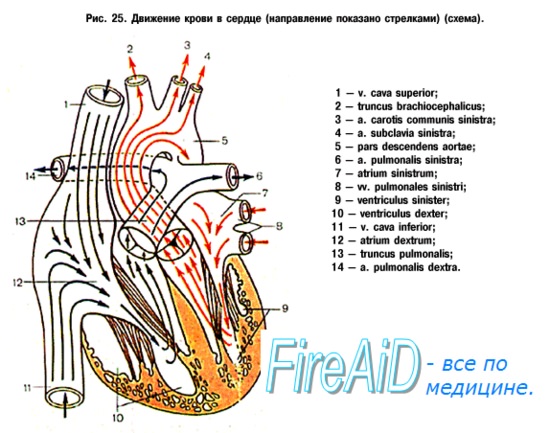
During physiological pregnancy, and especially in childbirth, such conditions of blood circulation arise, in which the load on the cardiovascular system significantly increases.
Pregnancy and childbirth make significant demands on the function of the heart due to an increase in blood mass and total weight of a pregnant woman, the emergence of a new link in the systemic circulation (uteroplacental circulation), changes in all types of metabolism, functions of the endocrine apparatus, and the central nervous system.
In the second half and especially towards the end of pregnancy, mechanical factors also become of considerable importance, to a certain extent hindering the normal functioning of the cardiovascular system, mainly the high standing of the diaphragm, which reaches the greatest extent by the 36th week of pregnancy. The high standing of the diaphragm, according to V.V.Saikova, lowers its work as an additional motor of blood circulation, reduces the vital capacity of the lungs, impedes pulmonary circulation and entails displacement of the heart; at the same time, the heart does not so much rise as it approaches the chest and at the same time rotates somewhat around its axis. The change in the position of the heart is accompanied by a relative "twisting" of the blood vessels that bring and carry blood, which also causes difficulty in pulmonary circulation.
The main changes in hemodynamics during pregnancy are reduced to an increase in the mass of circulating blood (plasma and erythrocyte volume), minute and stroke volumes, the number of heart contractions, and blood flow velocity.
The increase in the mass of circulating blood occurs gradually. At the same time, the volume of circulating blood at 28-32 weeks of pregnancy increases by about 30-40%, amounting to 5-5.3 liters in the first trimester of pregnancy, and 6.0-6.5 liters in the third. The amount of circulating blood increases mainly due to liquid (plasma), which leads to a decrease in the specific gravity of blood and the emergence of "pletora of pregnant women". While the amount of circulating blood during pregnancy increases by 30%, the hemoglobin content increases by only 15%; the hematocrit index decreases.
As the gestational age increases, the minute volume of blood also increases - from 5.5 liters at the beginning of pregnancy to 6.4-7 liters at 28-32 weeks of gestation.
The increase in the minute blood volume is mainly due to an increase in the stroke volume and, to a lesser extent, to an increase in heart rate. In this case, the systolic volume increases by 25-50%, reaching 70-80 ml versus 60-65 ml in non-pregnant women. The blood flow velocity in pregnant women, equal to 10 s at the beginning of pregnancy in the “hand - ear” section, slightly increases towards the end of it (11-13 s). The pulse rate in healthy pregnant women, even at rest, increases. In this case, tachycardia is observed in more than 50% of pregnant women.
When talking about the level of blood pressure during pregnancy and childbirth in women with a healthy cardiovascular system, two things must be kept in mind:
- you need to know the dynamics of blood pressure before pregnancy and from the very beginning. The degree of excitability of the vasomotor apparatus in different women is different, and in changes in blood pressure and in the state of vascular tone, the functional state of the body, its nervous system, due to both exogenous and endogenous factors, plays an important role;
- in the absence of pathological changes in the state of the cardiovascular system, blood pressure during pregnancy and even during childbirth changes relatively insignificantly.
In the first half of pregnancy, systolic, diastolic and pulse pressure decreases slightly, and from 6-7 months there is a tendency to increase it (especially diastolic). Many authors talk about a wave-like rise in maximum blood pressure, starting from about the 6th month of pregnancy, but it remains within the physiological norm.
Still, it must be emphasized that if women with a normal initial blood pressure of 110-120 / 70-80 mm Hg. Art. there is a rise in it in the second half of pregnancy over 130-135 / 80-90 mm Hg. Art., this should be regarded as a signal of the possible onset of a pathological state of the vascular system on the ground.
It should be remembered that during childbirth, there are often sharp fluctuations in hemodynamics, which is reflected in changes in the level of blood pressure.
After opening the fetal bladder, blood pressure usually drops, sometimes quite sharply. Therefore, V.V. Stroganov recommends early opening of the fetal bladder as a preventive method for treating eclampsia.
In the second and third stages of labor, there are rapid and abrupt changes in the rise and fall of blood pressure. The venous pressure in the upper limbs (in the elbow vein) does not change significantly with increasing gestational age, while in the femoral veins it increases markedly.
When assessing the state of the cardiovascular system in pregnant women, gas exchange rates should also be taken into account. With the development of pregnancy, the vital capacity of the lungs (VC) decreases, the maximum ventilation of the lungs and oxygen saturation of the arterial blood decrease, the amount of under-oxidized metabolic products increases (the content of lactic acid increases). At the same time, the minute volume of respiration (MRV) increases, the efficiency of using the oxygen of the inhaled air increases. In the body of pregnant women, the oxygen reserve is significantly reduced and the regulatory capabilities are extremely tense. Especially significant circulatory and respiratory changes occur during childbirth. An increase in heart rate, an increase in stroke and minute volumes, blood pressure, oxygen consumption by tissues, an increase in the concentration of lactic and pyruvic acids, etc.
Studies by Adams and Alexander have shown an increase in the work of the heart during contractions by 20%, and after discharge of the placenta - by 18%. During the birth act, the work of the heart increases by 5%! and more in comparison with the state of rest (V. Kh. Vasilenko). All of the above factors are the cause of the emergence and development of that symptom complex of complaints and clinical manifestations, which undoubtedly indicates some changes and known tension in the functions of the cardiovascular system in pregnant women. However, these changes in the body of a healthy pregnant woman are physiological. Their severity depends on the general condition of the pregnant woman's body, its ability to quickly and fully adapt to new, unusual conditions of the external and internal environment, from diseases suffered in the past. The central nervous system plays an important role in determining these abilities of the pregnant woman's body. The symptom complex of functional changes that occur in most pregnant women can be different, from barely noticeable, almost not causing any complaints, to those on the verge of significant dysfunctions of the cardiovascular system.
The most frequent complaints, especially in the second half of pregnancy, often presented by healthy pregnant women, are: shortness of breath, palpitations, general weakness, and sometimes dizziness. The pulse rate reaches 90-100 beats / min, increasing even more during childbirth, especially during the period of fetal expulsion. Immediately after the end of labor, most often in the first hours of the postpartum period, if there was no significant blood loss during labor, bradycardia is observed with a slowdown in the pulse rate to 60-70 beats / min.
Tachycardia in pregnant women - one of the usual reactions of the heart. In the vast majority of cases, tachycardia in pregnant women with a healthy cardiovascular system is temporary. It weakens and disappears as the woman's body adapts to new external and internal stimuli.
Tachycardia during labor can reach a significant degree, especially during the period of fetal expulsion. The reasons are as follows:
- great physical stress;
- pronounced negative emotions (pain, fear);
- increasing relative oxygen starvation towards the end of labor.
Relative hypoxemia , along with mechanical factors that impede the normal operation of the cardiovascular apparatus and reduce VC, causes shortness of breath, which, to a greater or lesser extent, many women complain about in the second half of pregnancy. Shortness of breath in healthy pregnant women may be due to metabolic disorders with a pronounced shift towards acidosis and relative hypoxemia. Since, in addition, a mechanical factor acts in the second half of pregnancy, shortness of breath in pregnant women should be classified as a mixed form. During contractions and especially attempts, blood oxygen saturation is significantly reduced, because in the process of childbirth, breath holding, strenuous muscular work and a significant depletion of the oxygen reserve are combined. All this is one of the prerequisites for the appearance of shortness of breath in pregnant women and women in labor.
However, the adaptive mechanisms of the body allow the overwhelming majority of women to adapt well to the inevitable functional changes that occur during pregnancy, and serious disorders in the activity of the cardiovascular system usually do not occur.
In pregnant women, there is a slight increase in the heart due to some hypertrophy and expansion of the left ventricle. It depends on a number of interrelated reasons: a) an increase in the total mass of blood, b) some difficulty in advancing a gradually increasing mass of blood. However, minor hypertrophy and enlargement of the heart develop slowly and gradually, and the heart has time to adapt to the increased demands on the cardiovascular system.
During pregnancy, the working capacity of the heart increases, which, according to the literature, increases by an average of 50% in comparison with the period before pregnancy.
A significant increase during pregnancy in the absence of valvular disease or inflammation in the myocardium indicates a decrease in the contractility of the heart.
Auscultation, as many authors point out, in some pregnant women (about 30%), especially in the second half of pregnancy, a soft blowing systolic murmur at the apex of the heart and on the pulmonary artery is determined. These noises can be heard with a perfectly healthy cardiovascular system and are purely functional in nature. So, the systolic murmur of the pulmonary artery depends on its temporary relative narrowing due to some inflection due to the high standing of the diaphragm, which changes the normal location of the heart and large vessels. A systolic murmur at the apex of the heart indicates a slight functional insufficiency of the mitral valve. These murmurs disappear soon after childbirth, which confirms their functional origin.
Features of blood circulation during pregnancy, mainly in the second half of it, cause the appearance of a number of clinical symptoms that cause diagnostic difficulties (displacement of the borders of the heart, the appearance of noise, an emphasis of the second tone on the pulmonary artery, extrasystole). It is often difficult to decide if they are a manifestation of organic heart disease or physiological changes caused by pregnancy.
To assess the functional state of the cardiovascular system in pregnant women, electrocardiography (ECG), vector cardiography (VCG), ballisto- and phonocardiography (BCG and PCG) are of particular importance. ECG changes in pregnant women are reduced to the appearance of a left type, a negative T wave in lead III, an increase in the systolic index, an increase in the QRST segment and a T wave in leads I and III. With an increase in the gestational age, certain changes in the PCG are noted, due to the difficulty of pulmonary circulation and an increase in pressure in the pulmonary circulation. They are reduced to an increase in the distance Q (R) of the ECG to the I tone of the PCG (from 0.035 to 0.05 s), a change in the II tone due to the increase in the amplitude of its second component, an increase in the distance T ECG - II tone of the PCG (from 0.03 to 0.05 s), the appearance of additional sound phenomena - systolic noise, an increase in the amplitude of the II tone on the pulmonary artery, its splitting and bifurcation.
During pregnancy, the vector cardiogram also changes - the area of \u200b\u200bthe QRS loop by the end of pregnancy increases by more than 40%.
The balli-stokardiogram also changes very significantly during pregnancy. In the second half of pregnancy, the K wave increases and deepens, which is associated with an increase in blood flow in the descending aorta, a large blood supply to the vessels of the small pelvis and abdominal cavity, an increase in pressure in them, and, consequently, a corresponding increase in peripheral resistance.
With an increase in gestational age, the amplitude of respiratory oscillations IJ increases, the ballistocardiographic index (BI) decreases, the respiratory index (RI) increases, there are changes in the 1st degree according to Brown and disturbances in the ratios of ballistocardiogram waves - JK / IJ, KL / IJ, KL / JK.
Changes in BCG in healthy women are the result of overflow of blood vessels in the pelvis, an increase in venous flow to the right heart, changes in the anatomical axis of the heart due to its horizontal position.
In the physiological course of pregnancy, there are noticeable changes in vascular permeability associated with a violation of the functional state of vascular membranes and changes in capillary circulation.
With capillaroscopic studies, an increase in the number of capillary loops, their expansion, mainly of the venous part, the presence of a more turbid background, pericapillary edema, and a slowdown in blood flow are found.
In recent years, it has been proven that an increase in the minute volume (and a change in other hemodynamic parameters) occurs from the beginning of pregnancy, increasing only until the 28-32th week, after which it gradually decreases.
As you know, the main load on the cardiovascular system is observed immediately after the expulsion of the fetus against the background of relative rest. Due to a sudden decrease in intra-abdominal pressure, an immediate restructuring of the entire blood circulation should occur. At this point, the vessels of the abdominal cavity quickly overflow with blood. There is a kind of bleeding into the vessels of the abdominal cavity. The blood flow to the heart decreases, and the heart works faster, but with a significant decrease in systolic volume - "half empty" (GM Salgannik et al.). Meanwhile, the intensified work of the heart at this moment is also required because during the period of exile, especially towards the end of it, the woman in labor necessarily develops a state of relative hypoxia; to eliminate her, the heart must work hard, with tension.
A healthy body, a healthy cardiovascular system have the ability to easily and quickly adapt to often significant and sudden changes in hemodynamics , in this connection, in a healthy woman in labor, as a rule, the necessary coordination in the circulatory system occurs quickly. However, with certain defects in the work of the heart, most often it is in the third stage of labor that its functional insufficiency can be revealed. It is possible and necessary to foresee and prevent the occurrence of circulatory insufficiency, for which it is necessary to study in advance the state of the cardiovascular system of each pregnant woman and to know at what pathological changes in this system dangerous disorders in childbirth occur.
In cases of an unclear diagnosis, a pregnant woman must be sent to a hospital (at the beginning of pregnancy - to a therapeutic one, in the third trimester - to) for in-depth clinical examination, observation and treatment.
Table of contents of the topic "Fetus in certain periods of development. Fetus as an object of childbirth. Changes in a woman's body during pregnancy.":1. Fetus in certain periods of development. Two (II) month old fetus. Development level of two (II) month old fetuses.
2. The level of development of a three to six month old fetus. Signs of a three to six month old fetus.
3. The level of development of a seven to eight month old fetus. The maturity of the newborn. Signs of maturity in a newborn.
4. The fetus as an object of childbirth. Fetal skull. Fetal skull sutures. Fetal fontanelles.
5. The size of the fetal head. Small oblique size. Medium oblique size. Straight size. Large oblique size. Vertical dimension.
6. Changes in a woman's body during pregnancy. The mother-fetus system.
7. The endocrine system of a woman during pregnancy.
8. The nervous system of a woman during pregnancy. Gestational dominant.
10. Respiratory system of a woman during pregnancy. Respiratory volume of pregnant women.
11. The digestive system of a woman during pregnancy. Liver in pregnant women.
During pregnancy there are significant changes in activities cardiovascular system of the mother... These changes allow the delivery of oxygen and a variety of nutrients and the removal of metabolic products necessary for the fetus.
The cardiovascular system functions during pregnancy with increased stress. This increase in load is due to increased metabolism, an increase in the mass of circulating blood, the development uteroplacental circle of blood circulation, a progressive increase in the body weight of a pregnant woman and a number of other factors. As the size of the uterus increases, the mobility of the diaphragm is limited, intra-abdominal pressure rises, the position of the heart in the chest changes (it is located more horizontally), and some women experience a mild functional systolic murmur at the apex of the heart.
Among the many changes of cardio-vascular system, inherent in physiologically ongoing pregnancy, first of all, an increase in the volume of circulating blood (BCC) should be noted. An increase in this indicator is noted already in the first trimester of pregnancy, and in the future it constantly increases, reaching a maximum by the 36th week. The increase in BCC is 30-50% of the initial level (before pregnancy).
Hypervolemia occurs mainly due to an increase in the volume of blood plasma (by 35-47%), although the volume of circulating erythrocytes also increases (by 11-30%). Since the percentage increase in plasma volume exceeds the increase in red blood cell volume, the so-called physiological anemia of pregnancy... It is characterized by a decrease in the hematocrit number (up to 30%) and hemoglobin concentration from 135-140 to 100-120 g / l. Since a decrease in the hematocrit number is observed during pregnancy, a decrease in blood viscosity occurs. All these changes, which have a pronounced adaptive character, ensure the maintenance during pregnancy and childbirth of optimal conditions for microcirculation (oxygen transport) in the placenta and in such vital organs of the mother as the central nervous system, heart and kidneys.
With a normal pregnancy, systolic and diastolic blood pressure decreases in the II trimester by 5-15 mm Hg. Peripheral vascular resistance is also usually reduced. This is mainly due to the formation of the uterine circle of blood circulation, which has low vascular resistance, as well as to the effect on the vascular wall of estrogen and progesterone of the placenta. A decrease in peripheral vascular resistance, together with a decrease in blood viscosity, greatly facilitates hemocirculation.
Venous pressuremeasured on hands healthy pregnant womendoes not change significantly.
During pregnancy, there is physiological tachycardia... The heart rate reaches its maximum in the III trimester of pregnancy, when this indicator is 15-20 per minute higher than the initial data (before pregnancy). Thus, the normal heart rate in women in late pregnancy is 80-95 per minute.
The most significant hemodynamic shift in pregnancy is the increase in cardiac output. The maximum increase in this indicator at rest is 30-40% of its value before pregnancy. Cardiac output begins to increase from the earliest stages of pregnancy, while its maximum change is noted at 20-24 weeks. In the first half of pregnancy, an increase in cardiac output is mainly due to an increase in the stroke volume of the heart, later - to a slight increase in heart rate. The minute volume of the heart increases partly due to the effect on the myocardium of placental hormones (estrogen and progesterone), partly as a result of the formation of the uteroplacental circulation.
Electrocardiographycarried out in the dynamics of pregnancy, allows you to detect a persistent deviation of the electrical axis of the heart to the left, which reflects the displacement of the heart in this direction. According to echocardiography, there is an increase in the mass of the myocardium and the size of individual parts of the heart. X-ray examination reveals changes in the contours of the heart, resembling the mitral configuration.
The processes of hemodynamics during pregnancy are greatly influenced, as already noted, has a new uteroplacental circle of blood circulation... Although the blood of the mother and the fetus does not mix with each other, changes in hemodynamics in the uterus are immediately reflected in blood circulation in the placenta and in the fetus and vice versa. Unlike the kidneys, central nervous system, myocardium and skeletal muscles, the uterus and placenta are unable to maintain their blood flow at a constant level with changes in systemic blood pressure. The vessels of the uterus and placenta have low resistance and blood flow in them is passively regulated, mainly due to fluctuations in systemic arterial pressure. In the later stages of pregnancy, the vessels of the uterus are maximally expanded. The mechanism of neurogenic regulation of uterine blood flow is mainly associated with adrenergic influences. Stimulation of alpha-adrenergic receptors causes vasoconstriction and decreased uterine blood flow. A decrease in the volume of the uterine cavity (prenatal rupture of amniotic fluid, the appearance of contractions) is accompanied by a decrease in uterine blood flow.
Despite the existence separate circles of blood circulation in the uterus and placenta (the placental membrane is on the way of the two blood flows), the hemodynamics of the uterus is closely connected with the circulatory system of the fetus and the placenta. The participation of the capillary bed of the placenta in the blood circulation of the fetus consists in the rhythmic active pulsation of the chorionic capillaries, which are in constant peristaltic movement. These vessels with varying blood volume cause alternate lengthening and contraction of the villi and their branches. This movement of villi has a significant effect not only on the fetal blood circulation, but also on the circulation of maternal blood through the intervillous space. Therefore, the capillary bed of the placenta can rightly be considered as the "peripheral heart" of the fetus. All these features of the hemodynamics of the uterus and placenta are usually combined under the name "uteroplacental circulation".
Is pregnancy possible with diseases of the cardiovascular system? It is possible, just before that you need to consult your doctor, especially if you suffer from rheumatism and rheumatic heart disease, he must give you permission to plan pregnancy. If you are feeling well, and you are tired, while shortness of breath and increased heartbeat rarely occur only with physical exertion, you will not have any problems with bearing and giving birth to a healthy child.
If you constantly, even when you are calm, shortness of breath appears and it begins to increase when you quickly begin to move, do light work. It is better not to take risks with pregnancy, it is very dangerous for both you and the baby. Even termination of pregnancy in this case is a dangerous procedure.
With the development of pregnancy, a lot of stress goes on a woman's cardiovascular system, because all systems work doubly, because a woman must provide the fetus with full-fledged life. A pregnant woman increases her body weight, blood also increases in volume, and the uterus, which is growing, begins to push the diaphragm upward, because of this, changes in the position of the heart occur. Changes in the hormonal background begin to occur in the body. Such changes in a woman's body load the cardiovascular system very strongly, when the term begins to increase, the load becomes even greater.
During labor, the cardiovascular system is very overstrained, especially when the second period of attempts begins. Also after childbirth, the cardiovascular system will have to endure stress. Because with the rapid emptying of the uterus, blood begins to redistribute, because of this, changes in hormones again occur.
What is the danger of cardiovascular disease for pregnant women?
Women begin to experience complications of a different nature during pregnancy, labor and the postpartum period; both the life of the woman and the child are threatened here. It is very dangerous that the fetus lacks blood circulation for the first month, especially this problem arises in the second half and during childbirth.
Is pregnancy possible in women with rheumatism?
Rheumatism is a toxic-immune disease that affects the joints and heart valves. Rheumatism appears due to B-hemolytic streptococcus, most often women suffer at a young age.
During pregnancy, the rheumatic process begins to worsen. Especially for the first time of a month then during childbirth. What complications occur in pregnant women with rheumatism?
1. Often, pregnancy is terminated prematurely.
2. Toxicosis continues on later lines.
3. The fetus lacks oxygen (hypoxia).
4. Disturbed uteroplacental blood flow.
Pregnancy with heart disease
Women who have a heart defect require urgent hospitalization, according to indications, it is obligatory three times per pregnancy:
1. At 12 weeks, a pregnant woman must undergo a complete cardiological examination in a hospital and here the question will be raised whether to leave the child or it would be better to terminate the pregnancy.
2. At 32 weeks, a woman should undergo a heart check, if necessary, then cardiac therapy, because it is during this period that the greatest stress on the heart falls.
3. The last heart check should be two weeks before
childbirth to prepare well for them.
A pregnant woman with cardiovascular problems should remember that the entire outcome depends on her behavior, especially on her lifestyle. If a woman receives the necessary drugs that support and facilitate the work of the heart, follows the regimen, listens to the doctor's recommendations, the pregnancy will end safely and the woman will be able to give birth without any problems.
What to do if pregnancy is contraindicated for a woman?
First, you need to cure the defect, perhaps with the help of a surgical method, often it helps the woman to return to a full life. But all the same, such a woman is at risk, therefore, it will be necessary to be observed by a cardiac surgeon throughout pregnancy.
Is pregnancy possible with hypertension
Up to 15% of pregnant women suffer from hypertension, high blood pressure. Often women do not even know that they have high blood pressure. For the first time months, most often it is reduced or normalized, this will complicate the task.
Hypertension is dangerous because up to 70% is complicated by toxicosis in the later lines. During childbirth, hypertensive encephalopathy may appear, with this disease a headache appears and vision is very impaired. Retinal detachment and cerebral hemorrhage are considered very dangerous complications.
How to prevent hypertension in pregnant women? Constantly and carefully monitored by a doctor, weekly. If the pressure is high, urgently go to the hospital in the maternity ward.
Also, hypertension can have its own stages of development, it depends on this whether it is possible to maintain a pregnancy:
Stage 1 - pregnancy is possible, gestation and childbirth are successful.
Stage 2 - pregnancy is allowed only if the woman has not previously experienced hypertension crises and her liver and kidneys are fully functional.
2 B and 3 stages of pregnancy are completely prohibited.
Pregnant women who suffer from hypertension are sent to the hospital in three weeks, where they should be provided with both physical and emotional rest.
So, pregnancy with cardiovascular disease is possible, but you need to be very careful here. Before planning, I was necessarily examined by a cardiac surgeon if it was necessary to undergo the necessary course of treatment. If you suddenly have a serious illness and in no case should you bear and give birth to a child, because this threatens both your health and the child, it is best to think about other methods. It's not worth the risk. It is very important for pregnant women who suffer from cardiovascular diseases to constantly keep their health under control, to undergo the necessary course of treatment and not to forget about preventive methods.
LECTURE No. 13.The incidence of diseases of the cardiovascular system in pregnant women is 5-10%.
The main complications in cardiovascular diseases: perinatal mortality and maternal mortality.
A complication of cardiovascular disease is:
Gestosis during pregnancy
Anemia
Premature birth
· abortion
Chronic uteroplacental insufficiency
Chronic fetal hypoxia
Pregnancy and childbirth contribute to the exacerbation of the rheumatic process, pulmonary edema and the progression of chronic cardiovascular failure, the appearance of visceral forms of the rheumatic process: nephritis, hepatitis, pleurisy, etc.
The structure of maternal mortality:
1.Extragenital pathology (the leading one is cardiovascular pathology)
2.gestosis
3.bleeding
4.puulent-septic diseases
Increased stress on the cardiovascular system during pregnancy:
1.increase in the weight of a pregnant woman (by 10-11%, that is, by about 10-11 kg)
2.growth of fetal weight (about 3000 g, but maybe more)
3.High standing of the diaphragm, which leads to a displacement of the axis of the heart in a horizontal state
4. clamping of large vessels
5.endocrine load
6. the emergence of a new placental circulation.
Hemodynamic changes:
1. change in minute volume and stroke volume of the heart. The minute volume increases by 20-30%, with normal pregnancy (by 26-30 weeks). starting from the second trimester, an increase in the minute volume of the heart begins, its peak is observed by 32-36 weeks.
2. an increase in the frequency of heart contractions.
3.increase in blood pressure and venous pressure
4. increase in BCC by 20 - 25%. The plasma volume is increased by 900 ml.
5.increased blood flow rate
6. an increase in the total peripheral vascular resistance.
7. Hematocrit and hemoglobin decrease during normal pregnancy.
In childbirth, there is an increase in the minute volume of the heart. Changes in the cardiovascular system require adaptation (increase in heart rate, minute volume). In childbirth, the work of the right and left ventricles is maximized, gas exchange and oxygen consumption increase (during attempts). With each contraction of the uterus, approximately 300 ml of additional blood flows to the heart.
In the postpartum period, blood redistribution occurs (due to a decrease in intrauterine and intra-abdominal pressure.
During lactation, there is also a load on the cardiovascular system (therefore, it is always necessary to decide on the admissibility of feeding).
80% of all diseases of the cardiovascular system in pregnant women are rheumatic defects. The management of such pregnant women is authorized by the therapist. Obstetrician-gynecologist, cardiologist and therapist jointly decide on the possibility of maintaining pregnancy.
The solution to this question depends on:
1.forms of the defect and its severity
2.stages of circulatory failure and functional state of the cardiovascular system
3.the presence of arrhythmia
4.the state of the most important organs and systems: liver, kidneys, lungs,
5. obstetric pathology
Indications for termination of pregnancy.
1. Active rheumatic heart disease, recurrent rheumatic heart disease.
2. Insufficiency of blood circulation 2A and 3 stages.
3. Atrial fibrillation, stenosis of the left atrioventricular opening and stenosis of the aortic opening.
Terms of hospitalization to resolve the issue of the admissibility of maintaining pregnancy:
1.Up to 12 weeks. Reveal the form of the defect, the degree of the rheumatic process, establish the pros and cons of the defect.
2. 26-32 weeks. (peak of the increase in BCC).
3. 35-37 weeks (choose and decide the method of delivery).
Congenital heart defects are quite common. some of them are formed during intrauterine development (influenza virus, rubella, herpes, respiratory virus). Formation of a defect at 4 to 8 weeks of ontogenesis in a future girl. The most common:
1.open arterial duct
2.triad, tetrad, pentad of Fallot
3.aortic stenosis, pulmonary artery stenosis
DIAGNOSTICS.
Recognition of the defect is difficult, since pregnant women may have shortness of breath, systolic murmur at the apex in 50% is normal.
The diagnostic criterion is the anamnesis: whether she had rheumatism, whether there was swelling of the joints, frequent exacerbations of chronic tonsillitis and flu-like conditions.
It is important to determine the degree of risk in pregnant women (criteria):
1.the woman's age
2.during previous pregnancy and childbirth
3.recency of the disease, myocardial condition
4.somatic chronic pathology
DEGREES OF RISK.
Grade 1 - pregnancy with heart disease, but without severe heart failure and without complications of the rheumatic process (subclinical manifestations).
2nd degree. Pregnancy with heart disease and with an initial picture of heart failure: shortness of breath, tachycardia, signs of rheumatism.
Grade 3 - pregnancy with decompensation of heart disease with a predominance of right ventricular failure in the active phase of rheumatism.
4 degree - decompensation, predominance of left ventricular failure or total heart failure in the active phase of rheumatism.
Continuation of pregnancy can be considered acceptable with 1-2 degrees of risk in the conditions of outpatient observation and inpatient treatment.
At the second degree of risk, the second stage of labor should be shortened by the imposition of obstetric forceps.
At 3-4 degrees, pregnancy is contraindicated, as well as contraindicated within 2 years after the attack of the transferred rheumatism.
Primary diagnosis: primiparous 25 years old. Pregnancy 1, term 28-29 weeks. Insufficiency of the mitral valve without pronounced signs of heart failure. Rheumatism active 1 degree. Last attack 1 year ago. Chronic tonsillitis. Risk degree 1-2.
When collecting anamnesis: whether the therapist was observed, how the previous pregnancies proceeded, whether she was hospitalized.
Laboratory data: C-reactive protein, dysproteinemia, ECG, phono-KG, echo-KG. Determination of the activity of the rheumatic process by traditional methods.
Fetoplacental insufficiency can be detected by ultrasound.
Indications for delivery regardless of the gestational age:
1.blue vices (Fallot's tetrad). Cyanosis is primary, secondary.
2. Congenital heart defects with high secondary hypertension.
3. The presence of a negative reaction at minimum load, manifestations of decompensation.
4. Ineffectiveness of complex treatment.
If up to 12 weeks - curettage of the uterine cavity, in the later stages, intravaginal delivery is performed (plus stimulation with oxytocin).
Abdominal delivery (caesarean section) is performed when:
1. coarctation of the aorta 2-3 degrees with the threat of cerebral hemorrhage.
2. Vascular aneurysm
3. a combination of somatic pathology with obstetric (congenital malformation + gestosis).
Treatment of cardiovascular insufficiency: stationary, bed rest, correction of fluid intake, balanced nutrition, physiotherapy exercises.
Cardiac drugs, antirheumatic, desensitizing, diuretic drugs.
The most widely used preparations of cardiac glycosides are strophanthin (slowly on glucose 0.3-0.5 ml after 5-10 minutes, the effect is maximum for 1-1.5 hours); diuretics: furosemide, veroshpiron, hyperbaric oxygenation, oxygen cocktails. Antibiotic therapy, taking into account the sensitivity of microbes (in 30% of pregnant women, group B streptococcus is found). Penicillin and its analogues are used.
Delivery in severe forms - they try to avoid abdominal delivery (they try to lead labor through the natural birth canal, even without switching off attempts). With an increase in hemodynamic disturbances, the 2nd period is shortened by the imposition of obstetric forceps.
Pain relief during labor: since labor pain depletes the regulatory capacity of the body, it leads to a disturbance in the heart rhythm, etc., to the appearance of heart failure, pulmonary edema.
Apply Promedol 1-2%, nitrous oxide with oxygen, GHB 10-20 ml intravenously; trilene, trichlorethylene, electroanalgesia, autoanalgesia.
Obstetric forceps are used to numb the pudendal block.
After childbirth: prevention of blood loss, including blood transfusion if there is post-hemorrhagic anemia), and. etc. Blood loss can be due to uterine hypotension, but it can also be coagulopathic, that is, due to changes in the coagulation system. To stop bleeding, use: uterotonics, drugs that affect blood clotting.
HYPERTONIC DISEASE.
Often there is hypertension before pregnancy. Pregnancy is a state of stress, increased stress. Hypertensive disease complicates pregnancy in 30%, against its background preeclampsia often develops, often hypertension is the cause of maternal mortality. At the beginning of pregnancy, blood pressure is normal, between 14-28 weeks there is even a decrease in blood pressure, and in the last 3 months there is an increase in blood pressure to high levels. In terms of identifying essential hypertension, early detection and registration of pregnant women with essential hypertension is necessary.
Stage 1 - phase A - latent, prehypertensive, transient reaction.
Stage 1 Phase B - an unstable, short-term increase in blood pressure, but reversible at rest.
2A - unstable increase in blood pressure.
2B - the increase in blood pressure is stable, but the function of all organs is preserved, there are no organ changes (organic).
3A - persistent increase in blood pressure, compensated, but there are dystrophic, fibrosclerotic changes
organs.
3B - decompensation, persistent increase in blood pressure, rarely occurs during pregnancy.
At stage 1, pregnancy is permissible, at the second stage, the issue is decided individually, after examination in a hospital. Stage 3 and malignant hypertension - pregnancy is contraindicated.
Clinic: pain in the region of the heart (cardio-neurotic), no cardiac pathology, dizziness, headaches in the back of the head.
Complex of neurotic complaints: hyperexcitability, head fights, palpitations, facial flushing, sweating.
Patients with essential hypertension are at risk (for both the pregnant woman and the fetus).
Risk levels:
1st degree: minimal. Complications occur in 20%, rarely pregnancy worsens the course of the disease.
Grade 2: severe extragenital pathology, accompanied by gestosis, spontaneous miscarriage. Fetal weight is reduced, the fetus is hypotrophic, an increase in perinatal mortality (20%).
Grade 3 - the maximum threat to the life of a pregnant woman.
1 degree of risk corresponds to hypertension 1 tbsp. 2nd degree corresponds to hypertension 2A, the third degree of risk - hypertension 2B, grade 3, malignant hypertension.
At grade 1, every 5 women have gestosis (nephropathy). Pregnancy is acceptable.
At grade 2, late toxicosis often occurs, at grade 3 it is difficult to act with antihypertensive drugs (contraindicated).
Indications (vital) for a caesarean section in hypertension:
1. premature placental abruption.
2. Retinal detachment
3.disorder of cerebral circulation
4. intrauterine fetal hypoxia.
Treatment: therapeutic and protective regimen, diet, antispasmodic intake (dibazol, papaverine, no-shpa, aminophylline; saluretics are usually rare, antihypertensive drugs - gangliblockers, clonidine.
Characteristic signs of pulmonary edema:
1. shortness of breath (respiratory rate up to 30 per minute).
2. Severe weakness.
3. Cold sweat.
4. Acrocyanosis.
5. Cough with mucous sputum.
6. Rapid pulse with little filling.
7.various rales over the lungs
8.foam, blood in sputum
The effect of antipsychotics, pipolfen, seduxen, promedol, GHB. IVL, oxygen through defoamers. Cardiac glycosides: intravenous digogxin 0.025% 0.5 ml, korglikon, strophanthin. Euphyllin. In 70%, pulmonary edema is fatal.