Whether healthy children were born with tvp 10. Collar space of a fruit. Examination of the collar space of the fetus. Other causes of deviation from the norm
During pregnancy, a woman undergoes an examination and passes many different tests, tests that allow you to monitor both the condition of the mother and the development of the child.
If any deviations occur, timely diagnosis allows you to prescribe treatment or terminate the pregnancy at a time when the threat to the life of the mother will be minimal. The screening and measurement procedure is carried out by every pregnant woman who is registered in the antenatal clinic.
While the fetus is still small, it has developmental features, such as the accumulation of fluid in the neck. As it develops, starting from the 2nd trimester, this feature disappears. This space between the shells (directly the skin and soft tissues of the cervical spine) is called the thickness of the collar space. This indicator is taken into account when screening the first trimester, which is carried out at 11-12 weeks of pregnancy.
The thickness of the collar space is an important indicator of many fetal pathologies associated with intrauterine development and chromosomal abnormalities. It is advisable to conduct such a study only from 10 to 14 weeks. Until the 10th week, the fetus is still too small and it is difficult to see all the subtleties using an ultrasound examination.
Starting from the 14th week, excess fluid in the tissues of the fetus begins to dissolve, so it becomes more difficult to determine the anomaly.
In addition to TVP, first trimester screening includes:
- Biochemical screening. Blood is taken from a woman to determine the level of protein and hormone. If this figure is too low, there is a possibility of spontaneous abortion. If too high, there is a risk of having a baby with.
- Assessment of the location and size of the fetus. Dimensions are evaluated by many indicators, one of which is TVP. If the size of the fetus is smaller than expected, they speak of a developmental delay or missed pregnancy.
- Definition of non-developing pregnancy. It is very important to notice a frozen pregnancy in time in order to remove the fetus from the uterus in a timely manner. If he stays in the body of a woman for a long time, complications and inflammatory processes may begin. As a rule, this is determined by searching for a heartbeat and assessing the size of the entire body of the fetus according to the term.
- Assessment of the fetal cardiovascular system. At this time, the fetal heartbeat is determined, and you can also see the work of his heart and blood vessels, which makes it possible to detect congenital heart defects in time.
It is worth remembering that most tests indicate the risk of a particular anomaly, but not always their result should be taken as a sentence.
Diagnosis and norm

Normal TVP is less than 3 mm
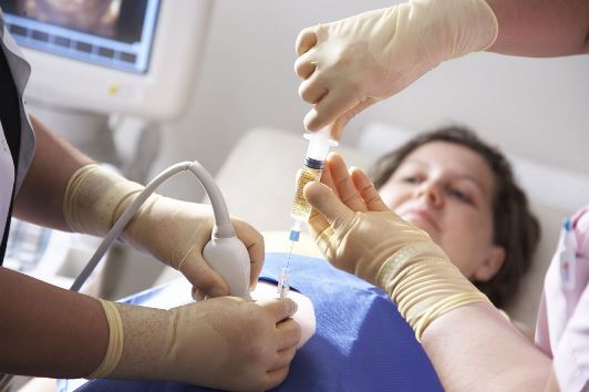
Diagnostics is carried out using . This procedure is absolutely safe for mother and child. Ultrasound is used several times during pregnancy and is safe at any stage.
The normal thickness of the collar space is normally from 0.7 to 2.7 mm, depending on which week of pregnancy the screening is performed. The collar space reaches its maximum size at the 14th week, and then begins to decrease.
The reliability of the procedure depends on many factors: the professionalism of the doctor, the equipment, the correctly determined gestational age, the location of the fetus, etc.
The ultrasound procedure itself for determining TVP takes place in several stages:
- performed either transvaginally or abdominally. If this is a transvaginal ultrasound, it is recommended to go to the toilet and empty the bladder before the procedure.
- Abdominal ultrasound requires reverse preparation. In order to better examine the fetus, a full bladder is needed, therefore, half an hour before the procedure, a woman is advised to drink a couple of glasses of pure non-carbonated water and not urinate this time. With an abdominal ultrasound, the examination is performed through the abdominal wall. The gel is applied to the skin of the abdomen. The doctor applies the sensor and drives it along the stomach, slightly pressing.
- With a transvaginal ultrasound, the woman undresses to the waist and lies on the couch on her back. The knees must be bent and spread. A special elongated ultrasound probe with a protective disposable nozzle is lubricated with gel and inserted shallowly into the vagina. Unpleasant or painful sensations should not arise. If there are pains in the lower abdomen, you must inform the doctor about it. A small amount of blood on the sensor is considered normal. The blood will stop smearing during the day.
The result is given to the patient in the form of a completed ultrasound form. It must be taken to the doctor. Biochemical screening is usually ready within a few days.
Chromosomal causes of increased TVP

With increased TVP, a woman is included in the risk group. The more the norm is exceeded, the more likely the development of a chromosomal abnormality in a child. It is worth remembering that we are talking about probability, that is, a guarantee that a child has a pathology is not always given.
Screening is carried out only after informing the woman. She can voluntarily refuse the examination.
If we talk about the chromosomal causes of the increased thickness of the collar space, then both trisomy (extra chromosome) and monosomy (lack of one chromosome) are likely.
Chromosomal abnormalities that can be detected by screening include:
- Down Syndrome. This is one of the most common chromosomal abnormalities, accompanied by the appearance of an extra chromosome. It is difficult to determine the causes of this pathology, the risk group includes women after 35 years of age, age primiparous. Children with Down syndrome have characteristic features of appearance: a round flat face, slanted eyes, small ears, short fingers. Also, these children are characterized by mental retardation and muscle weakness.
- Patau syndrome. In this disease, the 13th chromosome is copied. The survival rate of children with Patau syndrome is quite low. There is a high chance of intrauterine death. After birth, more than 90% of children with this pathology die before the year. Typical symptoms are multiple malformations, heart defects, hydrocephalus or microcephaly, cleft lip, palate.
- Turner syndrome. In this condition, one X chromosome is missing. The risk of this pathology is in no way related to the age of the mother. Children with Turner syndrome have pronounced sexual infantilism, a short neck, deformity of the joints, and often heart defects. Intellect is usually preserved, but oligophrenia also occurs.
With Down syndrome in a child, a woman is given a choice: to terminate the pregnancy or to keep it. If the chromosomal abnormality is incompatible with life, an abortion is recommended.
Other causes of deviation from the norm

The thickness of the collar space is not always associated with a chromosomal abnormality. It is possible to talk about pathology only on the basis of the results of ultrasound and biochemical screening in general. In some cases, the reasons for the deviation of TVP from the norm may be other pathologies of fetal development, as well as the state of the body of the mother herself.
The most common reasons for an increase in TVP are:
- Heart defects. Serious heart defects can be identified during the first screening. If they are incompatible with life, doctors recommend terminating the pregnancy, but treatment is possible in some cases. If the mother refuses to have an abortion, but the defect is serious, most likely, the pathology will lead to intrauterine death.
- Anomalies of connective and bone tissue. Various deviations in the formation of connective and bone tissue, leading to deformation of the skeleton, can be determined at the initial stages of pregnancy.
- . If CMV enters the body of a pregnant woman, the likelihood of infection of the fetus is very high. If the infection occurred during pregnancy, the virus begins to affect the development of the fetus, causing various abnormalities, provoking stillbirth, intrauterine death, miscarriages. After birth, the child may develop pneumonia, deafness, and heart defects are often observed.
- . This disease is transmitted through contact with pets and is dangerous only when infected during pregnancy. If a woman becomes infected in the first trimester, the probability of developing malformations that are incompatible with life is about 20%.
More information about Down syndrome can be found in the video:
After identifying the pathology, a further examination is prescribed: sampling of amniotic fluid for research, taking placental cells, embryonic villi. These procedures provide more reliable information regarding the existing deviations.
The choice regarding termination of pregnancy always remains with the parents. Doctors may recommend, but cannot force, a pregnant woman to have an abortion.
From 12 to 40 weeks, the fetal period of development of the unborn baby begins. At this time, all organ systems are not yet functionally developed. Week 13 - the period of manifestation of local motor reactions of the fetus. The nervous, respiratory, endocrine, and skeletal systems of the fetus continue to actively form. The features of your future baby become more expressive. The 13th week of pregnancy is the initial period of manifestation of the first emotional reactions of the unborn baby.
Fetal development at 12-13 weeks
To assess the development and diagnosis of fetal pathology, fetal fetometry is performed at 12 or 13 weeks.
Fetometry parameters and their norm for the fetus at the 13th week of pregnancy:
- biparietal - 24 mm;
- thigh length - 12 mm;
- chest circumference - 24 mm.
At 13 weeks, the embryo has a weight of 31 g, height - 10 cm.
TVP at 13 weeks
The thickness of the collar space or TVP is a parameter that doctors pay attention to during ultrasound screening at 13 weeks of gestation. The thickness of the collar space is the accumulation of fluid on the back of the fetal neck. The determination of this parameter is important for the diagnosis of genetic abnormalities in the development of the fetus, in particular in the definition of Down syndrome, Edwards, Patau.
TVP at 13 weeks - the norm
The normal physiological value of the thickness of the collar space is 2.8 mm at 13 weeks. A small amount of liquid is typical for all babies. An increase in the thickness of the collar space of more than 3 mm indicates the possible presence of Down syndrome in the unborn baby. To confirm the diagnosis, it is necessary to conduct additional invasive examinations, which can be dangerous for the baby. The risk of developing this pathology is especially increased during the first pregnancy after 35 years.
Remember that the diagnosis of an increased thickness of the collar space does not indicate one hundred percent presence, but only allows you to determine the risk group among pregnant women.
03-03-2009, 10:49
Help, calm down. :091:
HELP! :091:
03-03-2009, 10:53
I have not had this, but I want to support you!
@@@@@@@@@@@@@@@@@@@@@@@@@@@@@@ Everything will be ok! :flower:
The music of wind
03-03-2009, 11:48
I want to support you and say that we recently visited Lebedev, and he said that there are situations when a 5mm TVP is born and a healthy child is born. He said this to the fact that 1 marker is not an indicator of something terrible. Moreover, the biopsy showed that everything is in order.
I really understand your feelings, you can't even imagine, as far as I understand... But you are definitely all right, don't fill your head with negativity: flower: Good luck!
03-03-2009, 12:06
Thank you for your support, girls, but it's too scary for me. And the closer to childbirth, the worse.
Natahaha
03-03-2009, 12:14
The main thing is peace!!
03-03-2009, 12:30
Help, calm down. :091:
They measured TVP at 12 weeks as much as 5 mm! She went through all the circles of hell: chorion biopsy, ultrasound for defects (including hearts), blood. They say everything is fine. But such a kra-a-ayne is rare, so no one can say how everything will be in the end. :(Whoever had this, please respond ... How do your children feel, who had an increase in TVP with normal tests. Were they born healthy? Did this somehow affect the child in the future?
HELP! :091:
I just want to support you, our TVP was normal (or maybe because it was not Lebedev who was watching at that time), but everything else was just a song: an increase in the GM ventricles, the only umbilical artery, PAPP-A is several times higher than normal , which together speaks of the well-known syndrome oh how many times.
I sincerely wish you good luck.
03-03-2009, 12:50
My close friend has a girl with Down syndrome. In addition to this diagnosis, their health is all right, i.e. there is no heart disease, which is very common in such children, nor any other abnormalities. When she was pregnant, only at the last ultrasound they suspected something was wrong with TVP, and she was not told anything about the possibility of having a child with this syndrome. By the way, the triple test was normal for her. She gave birth and they told her ... At first it was hard to realize this and did not know how to continue to live ... My smart husband supported me. Now my daughter is 1.2, there is a developmental delay, but they do a lot and love her madly.
They didn't do a biopsy and they don't have a wonderful sonographer Lebedev in the city. If you have not been to him, I advise you to go for a consultation.
In general, I wrote this so that you would look at the situation differently. You already love the baby, even though he has not yet been born. After all, children are not always born the way we want them to be. But this does not prevent us from loving them and rejoicing in their every achievement ...
03-03-2009, 13:03
Yes, I love my child and really look forward to his birth. Thank God, I did an analysis to determine the chromosomes of the fetus and the analysis showed a good result. So Down, thank God, is excluded.
I don't know what courage it takes to raise a child with Down's syndrome.... I admire these people.
03-03-2009, 13:06
I just want to support you, our TVP was normal (or maybe because it was not Lebedev who was watching at that time), but everything else was just a song: an increase in the GM ventricles, the only umbilical artery, PAPP-A is several times higher than normal , which together speaks of the well-known syndrome oh how many times.
Chorionic biopsy and cordocentesis were not done, because There was a constant threat of termination throughout the pregnancy.
And Lebedev said that he feels that everything is in order with the child.
I was on my nerves the whole pregnancy, I accidentally saw a report on TV about people with diabetes in England, I roared all week.
As a result, the child is healthy, Apgar 9/10.
I sincerely wish you good luck.
And if it's not a secret, how much GM ventricular expansion did you have?
03-03-2009, 13:21
My child had an increase in TVP-6.8 mm, but they found it only on the second ultrasound at the MHC and the second time. I did the first ultrasound in a consultation, I didn’t have any data on TVP for a period of 12 weeks (either they measured it and didn’t write it down, or they didn’t measure it at all).
After a consultation with geneticists at the MHC, which ended in swearing, I went to Lebedev, he confirmed the increase in TVP, measured even more than it was, something about 8 mm. There were no other markers, he said that the presence of a neck fold is very serious. I decided to do cordocentesis. For a number of reasons, cordocentesis had to wait about a month. What I have experienced during this time, not to convey, so many tears and nerves. Everything went well, the results showed that the child is healthy. True, I gave birth at 37 weeks, but this is our family. The girl was born full-term and ttt, healthy. I was afraid that I was giving birth to a psycho, everything worked out, my experiences were not reflected in her character, thank God. Good luck to you. If the amnio showed that the child is healthy, then everything should be ok. I had a consultation with an immunologist at OTTO, he saw my condition, that I was like a zombie, said that you shouldn’t worry so much, said that your child has this structural feature, all people are different ...
Lebedev suggested the presence of a certain virus, which can also cause an increase in TVP, but I took an analysis for its presence, it was not confirmed.
By the way, when I went to the clinic with my daughter for a massage, the masseuse told me that the child had a long neck.
03-03-2009, 13:33
I understand that you did a biopsy, it showed that everything is fine. Right? What are you afraid of now?
On this topic. At 12 weeks, everything was fine and wonderful. And at 22 weeks, it’s like a butt on the head: the nasal bone is not visualized at all, the kidneys are enlarged and the cervical fold is higher than normal :(. The doctor who observed the pregnancy personally agreed with Lebedev and the next day he accepted us out of turn. Unfortunately, he confirmed everything, except for enlarged kidneys. But it didn’t give me hope anyway. My husband and I were in such shock that we decided to undergo cordocentesis. According to the results, everything turned out well: 091:. It seems that I should have calmed down. But I was worried until the end of pregnancy. to nothing, but could not do anything: 005: So try to abstract and not think about the bad.
The child was born healthy, but restless. But can't you tell me exactly why? From the nerves during pregnancy, or is it like that in itself?
03-03-2009, 13:43
03-03-2009, 13:51
03-03-2009, 13:54
I'm so glad that those who wrote to me with the cubs are all right. :)
03-03-2009, 13:55
One geneticist told me, thanks to him, consoled me, :wife: that this could indicate dozens of genetic syndromes that can be accompanied by a normal karyotype (that is, everything is in order with the chromosomes) and of course this is not visible on ultrasound either. However, these syndromes are rare. May this serve as consolation and hope.:005:
So I'm interested in this, for those who once encountered, how did the pregnancy end? Have any nasty diagnoses popped up after the birth of the baby.
If you think about all this, you can go crazy. Nobody is immune from this. Why get on your nerves again. It's definitely not good for you or your baby. So enjoy your position. Everything will be fine!
03-03-2009, 14:07
You know, it always seemed to me that TVP is informative only at 11-14 weeks, after this indicator does not mean anything. Doctors never focused attention on this to me during subsequent ultrasounds ....
I didn’t visit Lebedev, although I really wanted to visit him. When I found out about him, Puzik was already 22 weeks old, and Lebedev after 21 weeks. no longer looks (in any case, I was told so when I wanted to sign up with him). I was at Voronin's, he looked at me, supported me of course (because I was shaking all over like an aspen leaf), but I really didn’t find out anything about the future of the kids who had a big TVP.
There were no thoughts about viruses. And you will not prompt how this analysis to hand over and where?
I'm so-Ak glad that everything turned out well for you !!! You are such a lucky mom!
I am now expecting my second child. When she did an ultrasound in the LCD, she told the doctor about the increased TVP in the first child. She was very surprised that it was considered informative at 22 weeks. It's not clear to me either. Everywhere they write that TVP is informative for a period of 11-14 weeks. For me, it still remains a mystery, shrouded in darkness. Perhaps the doctors are reinsured and do not want to take such responsibility on themselves, if God forbid something is really wrong, but they missed it.
You can get to Lebedev, I came to him after 22 weeks, you just need to be more persistent with the administrator girls.
The virus was called parvovirus B19. Handed over in Laboratory of an immunology "Immunobioservice", at OTTO. I was also there for a consultation with Professor Selkov, a very good uncle.
You will definitely be fine, no need to dwell on it. They did a biopsy, everything is fine - and thank God, forget about it. Your child has such a feature of the structure. Moreover, Voronin did the ultrasound for you, he probably looked at the heart and everything else. My friend had 3 markers, she also had a biopsy, the girl is healthy.
03-03-2009, 14:09
Thank you, enjoy, unfortunately it does not work, but I try at least not to be very nervous. So already buckets of tears have been shed since 12 weeks....:flower:
03-03-2009, 14:18
03-03-2009, 14:39
Now rummaged in the Internet. It just became interesting: The neck fold should be no more than 6 mm in the 2nd trimester, and you had more. Apparently this was a wake-up call for doctors.
Lord, let at least your 2nd pregnancy be smooth and calm. So much patience is needed to pass all these tests.
Thanks, I hope so too. The first ultrasound had already passed, I was shaking, it was scary. I passed the double test, now I'm waiting for the results.
Everything will be fine with you too, don’t bother yourself with bad things, you can go crazy, I know from myself.
In order not to produce the same topic, I'll ask here! Please answer if anyone knows anything about this.
Good afternoon.
I am 29 years old, my husband is 31. The first pregnancy ended in childbirth in 2004, without complications, my daughter is 5 years old, there are no health problems, but there is a Minor anomaly in the development of the heart (if it matters) - an additional chord that "gives" noises, but on health and activity of the child does not influence in any way (were examined in the cardiocenter at 67 b-tse in Moscow). Among the relatives of Down's disease, there are no congenital deformities and mental retardation, there are no hereditary diseases either.
There were no miscarriages or abortions. Now the second pregnancy. The period according to the date of menstruation is 12 weeks 3 days. At a period of 7 weeks, ultrasound showed signs of partial detachment of the fetal egg, retrochorial hematoma 1.9 cm, there was no blood discharge. To this day, conservation therapy is being carried out, including taking duphaston and utrozhestan.
Now to the point of the issue.
Yesterday, December 8, 2009, I had an ultrasound scan by A.M. Stygar. at the Scientific Center for Obstetrics and Gynecology. Ultrasound results:
CTE 6.4 cm. The dimensions of the embryo correspond to 13 weeks and 3 days. The thickness of the collar zone is 3 mm, the visualization of the nasal bone corresponds to the gestational age. Anomalies detected at this gestational age were not detected. Chorion along the back wall of the uterus, thickness 1.4 cm. The umbilical cord is visualized. The length of the cervix is more than 4 cm, the internal os is closed, the cervical canal is not dilated.
Conclusion:
Embryo sizes correspond to 13 weeks and 3 days. pregnancy
Swelling of the collar space
A consultation with a geneticist is recommended.
I donated blood for screening only yesterday, the results are not yet available, they will be tomorrow.
Then, in the same place at the Center for Obstetrics, I went for a consultation with a geneticist. The geneticist began to say that it was necessary to do a biopsy of the chorion faster, only before the end of the week - later it was no longer possible. She said that the collar space is a very probabilistic indicator, it makes no sense to wait for the results of a blood test, because. in her opinion, it will still be bad, i.e. will show a high risk. To be honest, I was a little alarmed by this behavior of a geneticist, given that they do a biopsy and this procedure is expensive. But this is my opinion. She spoke about the possible risks associated with the procedure only when I asked her about it. Moreover, given the detachment and hematoma that were in this pregnancy, the risks for my child are increasing.
Now I'm interested in the following questions:
1. Does it make sense to wait for the results of a double test blood test or is it really not as important as the collar zone?
2. As far as I understand, amniocentesis is much safer? then maybe it makes sense to wait until 16 weeks and do it? Or all the same a biopsy is more preferable?
3. What is my risk of losing a child after a chorionic biopsy?
4. Another factor - my blood is Rh-negative, my husband's blood is Rh-positive. My daughter is Rh-positive, no antibodies were found during the first pregnancy, after giving birth on the first day I was injected with anti-Rh immunoglobulin. There are currently no antibodies in this pregnancy. Question: Is there a risk of Rh incompatibility in this pregnancy after the chorionic villus biopsy procedure or after amniocentesis?
I really appreciate your help and answers. It is very important for me.
Sorry for so many letters...
Planned ultrasound during pregnancy for a woman is not only a joyful "meeting" with the baby, but also excitement, anxiety, and fear. Future mothers hang on every word of the doctor, often getting scared when they hear new terms. Today we will talk about an important indicator - the thickness of the collar space of the fetus. Its measurement during an ultrasound examination is a mandatory procedure, so you should not be afraid of this. Measurements obtained by an ultrasound doctor can become a reason for the expectant mother's worries. What indicators indicate a deviation from the norm and what to do about it, you will learn in the article.
What is the collar space?
So, what is the nuchal space in the fetus? This is an ultrasound marker that can be used to suspect a chromosomal pathology in a baby. Its thickness (abbreviated as TVP) is the width of the space in the region of the cervical spine of a child filled with subcutaneous fluid. The term "collar space" was proposed in 1996, and the marker itself is used in screenings in almost all developed countries of the world.
A study of the collar zone of the fetus is carried out between the 11th and 13th weeks of pregnancy, that is, at the first. Only during this period it will be informative. Before 11 weeks, the study is not carried out, since at this time the fetus is still very small. Starting from the 14th week, the fluid will be absorbed by the baby's lymphatic system, and this indicator is no longer of value to the diagnostician.

If you heard this term from the lips of a specialist, do not immediately panic. The collar space is normally present in each fetus. What you need to pay attention to is its thickness. With a significant deviation from the norm, the doctor may suspect the baby, most often it is Down syndrome. However, only an increase in the collar zone is not enough for an accurate diagnosis.
TVP norms
 The indicator of the thickness of the cervical-collar zone in the fetus can be determined both with the help of transabdominal ultrasound - with it the sensor is located on the anterior abdominal wall of the woman, and with transvaginal examination - a special vaginal sensor is used here. Most often, transabdominal ultrasound is performed, and a vaginal probe is used if the first method does not allow assessing the structures of the fetus. Some professionals may use a combination of methods.
According to International standards, the norm of the collar space for a period from 11 to 13 weeks is up to 3 millimeters inclusive. If there are no pathologies, then from the 14th week this indicator will decrease, and will come to naught in the 2nd trimester of pregnancy. The greater the deviation from the norm, the higher the likelihood of developing a pathology in a child.
Important: the deviation of TVP indicators is not a 100% guarantee that the baby has a pathology.
What does the excess of the norm of the collar space indicate?
As already mentioned, an increase in the collar space in the fetus indicates the likelihood of developing chromosomal pathologies:
- in 50% of cases, this is Down syndrome - a disease caused by the presence of an additional chromosome 47, the causes of the pathology are not fully understood, but the likelihood increases if the mother's age is less than 18 and more than 35 years, and also if the parents are blood relatives;
- 25% - Edwards syndrome - the presence of a triple 18th pair of chromosomes, babies born with this pathology, in 90% of cases do not live up to one year of age;
- a 10% chance of developing Turner syndrome - a disease that occurs due to the absence or defects of one of the X chromosomes, often this pathology causes miscarriages in the early stages;
- 5% - Patau syndrome - a disease in which the 13th chromosome pair has an extra chromosome, infants with this pathology die in the first months of life in 95% of cases.
But there is also the possibility of having a perfectly healthy baby. You can find out the risks of developing pathologies in a child in the table below.

If the indicator is 4 millimeters, the probability of an unfavorable outcome is 27%, with an increase in the indicator to 6 millimeters, the risk increases to 49%, but even with 9 millimeters of TVP in 22% of cases, children are born healthy.

What to do with elevated TVP?
A woman should know that such a study as measuring the thickness of the collar space of the fetus is not specific. This means that if deviations from the norm are detected, it is necessary to conduct a series of tests, according to the results of which it is already possible to judge the presence of pathologies in the child.
Chorionic biopsy
If the result obtained during the ultrasound exceeds 3 millimeters, the pregnant woman is sent for a chorion biopsy. What is the procedure? The placenta at the initial stages of pregnancy is called the chorion and it has the same chromosome set as the fetus. A biopsy is a study of a small piece of chorion, during which you can find out about the chromosomal composition of the baby's cells, without affecting him. This is a very informative procedure that allows you to diagnose many chromosomal pathologies and hereditary diseases. However, it cannot be called safe, so the reasons for its implementation must be serious. The consequence of such a diagnosis in 1 case out of 100 is a miscarriage.

The most reliable result of a chorion biopsy can be obtained by conducting a study in the period from 11 to 13 weeks. The timing of the manipulation can be shifted within 10-19 weeks. The material is taken with a long thin needle, which is punctured through the abdominal wall, or with a probe that is inserted through the cervix. The choice of method depends on the location of the placenta. After that, a piece of chorion tissue is sent to the laboratory, the woman will know the results in about a week.
Amniocentesis
An alternative method of research is amniocentesis. This method can be used if the biopsy data was inaccurate or the chorion tissue was destroyed in the laboratory during the analysis. The essence of the method is the analysis of amniotic fluid - amniotic fluid. It surrounds the baby and his particles of skin, hair, waste products get into it, examining which determine the presence of pathologies.
In the early stages of pregnancy, between 10 and 14 weeks, amniocentesis is performed very rarely, since there is a high probability of harm to the baby due to the small amount of amniotic fluid. In order to diagnose chromosomal pathologies, the analysis is carried out in the period from 17 to 22 weeks. In this case, with unfavorable results, the woman may agree to terminate the pregnancy.
PPAP-A analysis
Another marker of chromosomal pathologies is a blood test of PAPP-A, a protein whose synthesis is activated with the onset of pregnancy. The PPAP-A protein is present in the blood of all people, but in small quantities. In pregnant women, this protein is produced by the outer layer of embryonic cells, with the help of which it is attached to the wall of the uterus. A blood test in a woman for the level of PPAP-A will be most informative from 11 to 14 weeks, although it can also be prescribed from 8 weeks. An analysis for PPAR-R is prescribed in combination with a test for.
If, according to the results of screening, an excess of the permissible norm of TVP was found, most importantly, do not panic! Worth a re-examination. Perhaps the doctor took inaccurate measurements or the baby took an uncomfortable position. For example, the data may be unreliable if the baby's chin was tightly pressed to the chest, or vice versa, the head was thrown back.