What is obstetric ultrasound with doppler. How is the procedure? What does fetal dopplerometry reveal?
The key to the normal development of the fetus in the womb is many factors, among which two main ones are worth highlighting: the health of the expectant mother and adequate placental blood flow. Lack of blood flow can cause pregnancy complications and lead to a delay, and in some cases, even a cessation of intrauterine development. Such a method of prenatal diagnosis as a Doppler study makes it possible to assess the intensity of blood flow in the umbilical cord and uterine vessels and prevent an unfavorable course of pregnancy in a timely manner.
The most optimal time for the study is the II and III trimesters, although visualization of blood flow using Doppler is possible already at the 6th week of development.
Ultrasound is performed using an abdominal sensor and does not require special preparation.
The study includes the diagnosis of blood flow at the following levels:
uterine arteries,
uteroplacental circulation,
umbilical cord vessels,
vessels of the fetal brain (middle cerebral artery).
As a rule, blood flow Doppler is prescribed to pregnant patients according to the indications of an obstetrician-gynecologist. In the network of antenatal clinics "Medok", the Doppler ultrasound service is included in the management of pregnancy, as it allows our doctors to more closely monitor the development of the fetus and exclude any pathologies. In order to most accurately assess the condition of the fetus, a Doppler study is sometimes combined with cardiotocography and echography.
Responsibility for a favorable course of pregnancy
Highly qualified obstetrician-gynecologists and medical personnel
Modern ultrasound equipment
Special atmosphere in clinics
Attention and special approach to each patient
The procedure is prescribed for the 2nd and 3rd trimester of pregnancy. No special preparation is required.
all pregnant women in the 2nd-3rd trimester of pregnancy in order to diagnose the normal development of the fetus and exclude pregnancy pathologies
premature maturation of the placenta
risk of intrauterine growth retardation
suspicion of fetal hypoxia and fetoplacental insufficiency
What is dopplerometry?
Doppler, dopplerography (dopplerography), ultrasound duplex examination of blood vessels is an ultrasound method that measures the speed of blood flow in the vessels.
Using the Doppler method of assessing blood flow, blood clots or atherosclerotic plaques that contribute to impaired blood flow can be detected.
When performing a duplex examination, these two methods (conventional ultrasound and Doppler) are used simultaneously. Conventional ultrasound provides an image of the structures of the blood vessels, while Doppler allows us to image and evaluate the characteristics of the flow of blood moving through the vessels. During a duplex examination, the doctor receives a color image of the vessel, with a certain encoding in color of the speed and direction of blood flow.
What types of Doppler are there?
There are the following types of Doppler (Dopplerography):
Doppler during pregnancy.
Dopplerography of cerebral vessels.
Dopplerography of the vessels of the neck.
Dopplerography of the vessels of the lower extremities.
Dopplerography of the arteries of the lower extremities.
Doppler (doppler) during pregnancy.
Doppler, Doppler (Dopplerography) during pregnancy is an additional ultrasound method that allows you to evaluate the placental circulation and the "mother - placenta - fetus" system.
Dopplerometry has an important diagnostic value in the examination of pregnant women, especially in patients with bleeding disorders. To adequately assess the condition of the fetus, dopplerography is often performed in conjunction with cardiotocography and echography. The data obtained during the study can significantly affect the management of pregnancy and childbirth.
High information content, safety and the possibility of fetal Doppler ultrasound even in early pregnancy make this method indispensable in a comprehensive system of prenatal diagnosis.
At what time is dopplerometry performed during pregnancy?
Despite the fact that visualization of blood flow using Doppler is possible already at the 6th week of fetal development, the most informative study will be in the II and III trimesters.
The first Doppler ultrasound is usually performed at 20-24 weeks. Special indications for research at this time are violations of hemostasis in the patient, as well as the risk of preeclampsia, intrauterine growth retardation, hypoxia and fetoplacental insufficiency.
Planned dopplerography is usually performed at 30-34 weeks and is a mandatory component in a comprehensive assessment of the functional state of the fetus.
What is the peculiarity of Doppler during pregnancy?
Dopplerometry is a modern leading research method in obstetrics, which allows you to assess the state of the fetoplacental system of the unborn child.
Distinctive features of Doppler (Doppler) during pregnancy:
the principle of operation of Doppler ultrasound is based on changes in the frequency of the ultrasound wave depending on the speed of blood in various vessels of the umbilical cord, the aorta and cerebral arteries of the fetus, and the arteries of the uterus;
Dopplerography (Doppler) during pregnancy is similar to the usual ultrasound procedure. The study is carried out using a special Doppler sensor, which is equipped with all modern ultrasound diagnostic devices;
if conventional ultrasound gives an idea only of the structure of blood vessels, then dopplerography also shows the movement of blood in the form of a two-dimensional color image.
Unlike conventional ultrasound, Doppler is capable of:
determine the health of the baby's heart;
listen to the heartbeat, determine the patency and lumen of the vessels of the umbilical cord of the fetus;
determine how well the vessels of the fetus are supplied with blood;
detect insufficient functioning of the placenta and fetal hypoxia in the early stages.
What is color doppler during pregnancy?
During Doppler (Dopplerography) during pregnancy, a high-resolution color apparatus is used. This allows the blood flow of the mother and child to be colored with different colors, which is especially important for the early diagnosis of childhood heart defects and developmental anomalies. Different colors are also used to depict different directions of blood flow.
Special indications for fetal dopplerography
When the size of the fetus does not correspond to the gestational age.
Abnormal amount of amniotic fluid.
When pathological conditions of the placenta are observed: premature maturation, etc.
If fetal abnormalities or chromosomal abnormalities are suspected.
When a woman has diseases such as diabetes, anemia, kidney disease, etc.
The presence of pathologies during previous pregnancies.
When the fetal heart rate is abnormal. If you suspect heart disease or other heart disease.
With premature contractions - they are visible on CTG (cardiotocography).
With gestosis pregnant.
With Rhesus conflict.
With high blood pressure in the mother.
With multiple pregnancy.
With the threat of miscarriage or premature birth.
What is Doppler Ultrasound? Today, ultrasonic diagnostic methods are becoming more and more widespread in practical medicine. Such popularity is explained by the high degree of informativeness of this type of research. Doctors are also attracted by the absolute safety of the procedure for the body of both a healthy person and a pregnant woman. There are quite a few ultrasound options designed for each individual system and structure of our body that are available to the machine's sensor.
Where is it applied?
Most of all, however, the use of ultrasound is typical for the pediatric and obstetric areas. This is due to the fact that only proven and safe research methods should be used in these areas. Reflected from internal structures, the radiation from ultrasound does not harm the patient. Doppler or UZDG is also based on these principles of work. It allows you to graphically evaluate the direction and speed of blood flow in the vessels. This method is used during pregnancy to check the circulation of the mother, fetus and placenta. What is it - Doppler ultrasound is interesting to many.
Some women, having received a referral for this type of study, are frightened, as doctors do not give explanations about how Doppler ultrasound is performed and what its essence is. The reality is that Doppler ultrasound is no different from conventional ultrasound in terms of the procedure itself. The main difference is in the parameters that the doctor pays attention to during the study.
Work principles
Before considering the varieties of Doppler ultrasound, it makes sense to understand the principles of its operation. Doppler ultrasound is based on radiation from ultrasound, but it differs from conventional ultrasound in how the return signal is perceived. This phenomenon in physics is called the Doppler effect, which gave the name to the technique. The mechanisms of action of vascular Doppler ultrasound and its difference from conventional ultrasound are as follows:
1. Standard ultrasound produces wave radiation at the same time intervals and, according to the same principle, perceives its reflection. With its help, it is convenient to study relatively immobile organs and structures that practically do not change over time. Doppler ultrasound of the neck is now very widespread.
2. It will not be possible to study the blood flow in this way, since the blood tends to be constantly in motion in the cavity of the vessel. As a result, a conventional ultrasonic emitter is not able to fix the blood flow velocity.

3. The principle of operation of the dopplerometer is similar to the radar. It is able to fix and register individual fluctuations, making measurements of indicators through reflected radiation.
4. The Doppler transducer, therefore, sends ultrasound waves at a certain speed, and receives back at a different one. Evaluation of such data became the basis of the methodology. That is, the quality of blood circulation in the vessel depends on how quickly the ultrasound wave returns.
The first study based on Doppler ultrasound (what it is, we explained) was spectral streaming Doppler. It evaluates performance in M-echo mode. The results obtained were verified by means of a diagram that changed in the presence of pathology. Subsequently, the study was supplemented by visualization and became known as "Doppler echocardiography" and "color mapping". These techniques are used to detect developmental pathologies in the fetus.
Flow Doppler
This method is unique and irreplaceable. This is the only study that makes it possible to assess the state of the blood flow of the uterus, placenta and fetus during pregnancy. It should be borne in mind that Doppler is performed only if there are certain indications. In other cases, conventional screenings in the amount of three pieces are sufficient.
If there are complications during pregnancy, the appointment of Doppler ultrasound can be done starting from the 20th week. However, the vast majority of the study is carried out after the 30th week. This is the period of a planned ultrasound examination, and it is convenient to combine it with vascular Doppler ultrasound.
Not only the speed of blood movement through the vessels of the uterus and placenta is checked, but also individual arteries of the fetus, including the umbilical cord. Such a thorough study is explained by the fact that the blood flow velocity is rarely disturbed simultaneously in several vessels.
All indicators are recorded in the M-echo mode. A diagram is displayed on the screen, showing the fluctuations in the return radiation. This type of study does not give a three-dimensional image, only waves that correspond to the movement of blood in the arteries. In addition to this, a sound image of the vibrations is also obtained. It enables the audio specialist to identify a change that is not visible on the diagram. Doppler ultrasound of the vessels of the neck is very informative.

Doppler echocardiography
The heart is the basis of the circulatory system, so checking its work is also an integral part of the diagnosis. The widespread use of echocardiographic techniques has made it possible to use them during pregnancy to assess the condition of the child's heart. However, at the initial stage, there were no sensors capable of detecting changes in such a tiny organ.
What opportunities does he have?
Modern devices, in particular, Doppler, are equipped with a three-dimensional image display mode. Combining echocardiography with Doppler has improved both methods, making the following possible:
1. Such visualization makes it possible not only to assess the direction and speed of blood movement in the vessels, but also to check their structure and position. Thus, the modern method replaced the previous ones and became the leading and most common. Doppler ultrasound of the lower extremities is required for thrombosis.
2. The study of blood vessels has ceased to be superficial. Now not only the blood flow in the aorta, umbilical cord and middle cerebral artery is being studied. A high degree of visualization allows you to see the vessels in individual organs of the fetus.
3. The final stage of the study is to check the heart. The dimensions of the cavities of the organ, the state of its individual structures are measured. It became possible to diagnose heart defects at an early stage.
Although this technique is highly informative, not every specialist diagnostician can carry it out. In addition, it is quite expensive. Nevertheless, everyone can do an ultrasound with Doppler.

color mapping
This is a new version of the study, which allows you to correctly and thoroughly check venous and arterial blood flow in different modes. This study makes it possible to describe several indicators of the circulatory system simultaneously. This can be both the external structure of the vessel, and indicators of individual blood flows. Thus, it turns out not only to give an assessment of the speed of blood flow, but also to exclude its incorrect movement.
Peculiarities
The study has several features:
1. Mapping in color makes it possible to fix the direction of blood flow in the vessels, digitize the obtained indicators and create a three-dimensional picture at the same time. Conduct ultrasound of the kidneys with Doppler.
2. The speed and direction of the vessels are indicated on the screen in different colors. This provides the possibility of simultaneous study of venous and arterial vessels.
3. Research is applied in structures where several vessels are closely spaced. This is what makes it convenient for checking blood flow in the veins and arteries of the fetus and placenta.
Indications for holding
In the normal course of pregnancy, the Doppler of the vessels of the placenta, fetus and uterus is not prescribed. In this case, regular scheduled ultrasound examinations are sufficient. Doppler may be needed in a situation where there is a potential or clear risk of complications during pregnancy. Indications for Doppler ultrasound can be various diseases not only of a pregnant woman, but also of a child:

Study preparation
When a woman is prescribed an ultrasound with Doppler, she has a question about preparing for this type of study. It should be understood that Doppler is just a type of ultrasound, so no specific preparation is required for its implementation.
1. Most hospitals use disposable sheets and napkins, but just in case, you can take a clean cloth with you.
2. After the procedure, you will also need a few paper towels to wipe off the gel.
3. You can not eat tightly and drink a lot before the procedure. This is due to the fact that pressing the sensor can cause discomfort in the abdomen.
4. Clothing should be loose so that it can be easily removed before the procedure.
The duration of the procedure depends on the diagnostic option and is up to 30 minutes.
Conducting research
In terms of external characteristics, a Doppler examination does not actually differ from a standard ultrasound examination. Most often they are performed on the same device with switching modes and using different sensors. The main difference lies in the very essence of the procedure. The study is carried out as follows:
1. The initial position of the woman is horizontally on her back. Closer to childbirth, a woman is advised to turn slightly to the left to relieve tension from the uterus.
2. A specialist using a special sensor identifies the arteries of the uterus and their branches, as well as the umbilical vessels. The following is a description of the quality of blood flow in the uterus and placenta.
3. Calculations are made based on the comparison and ratio of blood flow in each heart rhythm.
4. For each group of vessels, several indicators are calculated. They are pulsation index, blood flow velocity, systole-diastolic ratio and resistance index.
5. The obtained indicators are compared with the norm, then a conclusion is drawn up. What is the norm of Doppler ultrasound?

Evaluation of indicators
To determine the further strategy for monitoring and managing patients with deviations in Doppler parameters, a division into three degrees of severity of blood flow disorders is used. It should be borne in mind that the inclusion in one of the categories occurs regardless of the numbers received, since the localization of the detected deviations is important.
1. Degree 1A. The rate of blood flow is reduced only in the placenta and uterus. At the same time, the vessels connecting the fetus and the placenta are unchanged and there are no signs of abnormalities in the intrauterine development of the child. Doppler ultrasound should be interpreted only by a highly qualified specialist.
2. Degree 1B. With it, the blood flow between the uterus and the placenta is not disturbed, while in the placental-fetal there is a decrease in indicators. In this case, there are the first signs of intrauterine growth retardation.
3. The second degree is characterized by impaired blood flow in both directions, that is, both between the uterine and placental, and between the vessels of the fetus. However, it is still possible to save the life of the fetus.
4. The third degree is a sign of critical and irreversible changes in the blood flow. This condition can lead to fetal death.
The first degrees involve increased control over the condition of a pregnant woman. If the third degree of severity of blood flow disorders was initially detected, this is a signal for emergency childbirth.

Where to do ultrasound with doppler?
This procedure can be carried out in a large medical diagnostic center. For example, in Moscow you can sign up for the "Alexandra Med" center, visit the international center "On Clinic". The cost of the study is from 1800 rubles.
We examined what it is Doppler ultrasound.
During the 9 months of gestation, a woman must undergo several scheduled ultrasound diagnostics. This is a mandatory and safe procedure, with the help of which doctors will find out how the pregnancy is proceeding, in what position the fetus is and whether its development corresponds to the term. Doppler ultrasound (dopplerography, doplerometry) is a safe and informative research method that assesses the quality of blood flow in the "womb-placenta-fetus" system.
What is a pregnancy doppler and how is it done? What are the indications for dopplerography? What do the results of Doppler ultrasound during pregnancy show and how to decipher them?
What is a doppler ultrasound during pregnancy and why is it needed?
Dopplerography is one of the methods of prenatal examination, which works like a conventional ultrasound machine. This diagnostic method allows you to identify violations in the development of the fetus. After doplerometry, the doctor will identify the cause of the failure, determine the tactics of treatment, or decide on early delivery. This important study allows you to save the health of the expectant mother and the life of the fetus.
During the procedure, the frequency of the sound is assessed, which changes when reflected from a moving object (blood stream). As mentioned earlier, with its help, the state of blood flow in the "womb-placenta-fetus" system is assessed.
Doppler ultrasound differs from the usual in the following points:
Doppler during pregnancy responds to the speed of blood movement in different vessels (vessels of the umbilical cord, fetal aorta, arteries of the brain, vessels of the uterus).
During the scan, the doctor observes the movement of erythrocytes (red blood cells) in a two-dimensional image on the screen.
There are 2 types of doplerometry: duplex and triplex. Duplex mode allows you to assess the patency of blood vessels, to identify the causes of circulatory disorders. Triplex mode presents a color picture with the movement of red blood cells. Sonographers prefer triplex scanning, which shows the most accurate results.
Indications for dopplerography

After pregnancy is confirmed, a woman must register with a gynecologist. The doctor will monitor the course of pregnancy and develop an action plan for each patient individually. This takes into account the condition of the pregnant woman, the development of the fetus, chronic diseases and bad habits of the mother.
During the gestation of the fetus, each woman must carry out other mandatory procedures. Dopplerography is an important study that is prescribed twice for the entire period, if there are no additional indications.
As a rule, scanning is carried out in the following periods:
- From 22 to 24 weeks;
- From 30 to 34 weeks.
If during the next scheduled examination, the doctor suspects various deviations, then Doppler ultrasound is prescribed several more times.
Doppler is used in the presence of diseases in the expectant mother:
- Arterial hypertension;
- Anemia of a high degree (a significant decrease in the number of red blood cells or hemoglobin in the bloodstream);
- Large fibroids in the uterus;
- Sexual infections;
- Insufficiency of the respiratory organs (lack of full gas exchange);
- Functional failure of the heart.
With the pathological development of pregnancy or anomalies of the placenta, Doppler ultrasound is performed in the following cases:

Dopplerography is not contraindicated for expectant mothers.
How is a Doppler ultrasound performed?
Doppler does not require preparation from a woman in position. This procedure is almost the same as a conventional ultrasound. The only difference from a standard ultrasound examination is that a woman has the opportunity to listen to the heartbeat of the unborn child and the sound of blood flowing through the vessels.
You will be interested in:

As mentioned above, Doppler ultrasound is prescribed between weeks 20 to 22 or from weeks 30 to 32. This diagnostic method is extremely important if the doctor suspects the pathology of the development of pregnancy.
During dopplerography, the same device is used as with a standard ultrasound. In most cases, these studies are carried out at the same time.
Doppler progress:
- A pregnant woman comes at the appointed time (no need to prepare for the upcoming study);
- The patient is offered to lie on the couch on her back and expose her stomach. If it is difficult for a woman to be in a horizontal position for a long time or the fetus is in an atypical position, then she can lie on her side;
- The skin of the abdomen is treated with a special gel and an ultrasonic probe is driven over it to examine the blood vessels.
 During the procedure, the doctor assesses the condition of the arteries of the uterus, navel, as well as the middle cerebral artery of the fetus. If necessary, the specialist studies the blood flow in the venous duct, the aorta in the chest, the renal arteries, the veins of the umbilical cord, the inferior vena cava and the intracardiac blood flow of the unborn child.
With the help of a special ultrasound mouse, the studied vessel is detected and displayed on the monitor.. At first, it is colored gray, after activating the Doppler mode, the doctor studies the necessary characteristics of the blood flow and enters them into the protocol of the clinical study.
A Doppler ultrasound takes longer than a standard non-Doppler ultrasound scan. In addition, the duration of the procedure depends on the position and activity of the fetus. If the fetus behaves calmly, then the sonographer registers its blood flow faster.
Deciphering the results of ultrasound with Doppler during pregnancy
With the help of doplerometry, the doctor determines the condition of the fetus and the expectant mother.
To assess the movement of blood through the vessels, specially designed indices are used - the ratio of blood flow velocity during contraction and relaxation of the heart muscles.
Using the graph, the specialist identifies the maximum blood velocity in systole(heart contraction), end diastolic velocity (relaxation of the heart muscles), and average velocity per systole and diastole. Then the device calculates one or several markers by which blood flow parameters are evaluated: PI - pulsation index, IR - resistance index, SDO - systolic-diastolic ratio.
After receiving the results, the doctor compares them with the norm and determines the condition of the unborn child. The norms of the results obtained during Doppler ultrasound during pregnancy for different indicators are presented in the tables below.
The norm of LMS and IR of the uterine arteries at different stages of pregnancy is presented in the table:
| week of pregnancy | FROM TO | IR |
| 12 — 13 | 2 – 3.5 | 0.52 – 0.7 |
| 14 — 16 | 1.9 – 2.5 | 0.48 – 0.68 |
| 17 — 19 | 1.7 – 2.5 | 0.44 – 0.62 |
| 20 — 24 | 1.6 – 2.5 | 0.4 – 0.6 |
| 25 — 31 | 1.7 – 2.4 | 0.4 – 0.58 |
| 32 — 37 | 1.6 – 2.3 | 0.35 – 0.58 |
| 38 — 40 | 1.4 – 2 | 0.32 – 0.55 |
Norm of LMS and IR of umbilical cord vessels:
| week of pregnancy | FROM TO | IR |
| 14 — 15 | 5 – 8.4 | 0.8 – 0.88 |
| 16 — 17 | 4 – 6.8 | 0.74– 0.85 |
| 18 — 19 | 3 – 0.53 | 0.67 – 0.8 |
| 20 — 22 | 2.9 – 4.4 | 0.66 – 0.78 |
| 21 — 24 | 2.5 – 3.8 | 0.61 – 0.76 |
| 25 — 27 | 2.5 – 3.8 | 0.6 – 0.75 |
| 28 — 31 | 2.3 — 3 | 0.54 – 0.7 |
| 32 — 36 | 2 – 2.8 | 0.5 – 0.65 |
| 37 — 40 | 1.8 – 2.8 | 0.45 – 0.64 |
Normal indicators of LMS and IR of the middle artery of the brain:
| week of pregnancy | FROM TO | IR |
| 20 — 25 | 4.3 – 6.8 | 0.77 – 0.85 |
| 26 — 27 | 4.2 – 7.8 | 0.76 – 0.87 |
| 28 — 29 | 4 – 8.7 | 0.75 – 0.88 |
| 30 — 33 | 3.7 – 8.6 | 0.74 – 0.88 |
| 34 — 37 | 3.3 – 7.9 | 0.69 – 0.87 |
| 38 — 40 | 2.8 – 7.5 | 0.64 – 0.86 |
Most often, blood flow disorders in the placenta occur due to late toxicosis or hypertension in the expectant mother.
It is possible to identify circulatory insufficiency in the "uterus-placenta" system by the following parameters:
- Reducing the rate of diastolic relaxation below normal;
- Increased resistance index in the arteries of the uterus;
- Early diastolic notch on the graph of blood flow velocity in the uterine arteries.
Circulatory disorders in the "placenta-fetus" system can be judged by a decrease in velocity in the umbilical arteries and an increase in resistance indices for a certain period of pregnancy.
A classification that describes the degree of placental circulatory disorder:
- IA Art. - pathological blood flow in the uterine arteries;
- IB Art. - violation of blood circulation in the blood vessels of the umbilical cord, which does not reach critical numbers;
- II Art. - pathological blood flow in the arteries of the uterus and umbilical cord, not reaching critical values;
- III Art. - lack of blood flow or negative indicators at the moment of relaxation in the vessels of the umbilical cord.

Pathologies of blood flow in the middle cerebral artery of a child are detected by the following ultrasound signs:
- Deviation from the norm of IR and a high difference between systolic and diastolic pressure (below normal);
- Absence or negative indicator of blood flow at the time of relaxation of the heart muscles;
- Centralization of the fetal circulation.
Signs of disorder of intracardiac blood flow in functional heart failure in a child:
- Decreased blood flow through all valves;
- Tricuspid valve insufficiency;
- Diastolic blood flow through the adult-type tricuspid valve in the fetus.
Pathological pulsations and synchronous heart rhythm in the mother and fetus indicate a violation of blood circulation in the venous vessel of the umbilical cord.
To determine the degree of blood flow disorder in a child, it is necessary to take into account the violation of blood flow in different vessels:

In addition, at 11-14 weeks of pregnancy, venous blood flow is disturbed due to hereditary diseases or congenital pathologies of the fetal heart, which increases the likelihood of an adverse pregnancy outcome.
In the presence of two or more fetuses, Doppler ultrasound is also prescribed. At the same time, the arteries of the umbilical cord and the brain of both children are examined. This is necessary to prevent feto-fetal transfusion syndrome (a complication of multiple pregnancy, in which the blood flow of different fetuses is different).
If the indices in the arterial vessels of the umbilical cord in one fetus are greater than in the other, then this indicates that the first child has a lack of blood.
Based on the foregoing, Doppler ultrasound is an absolutely safe diagnostic method for a pregnant woman and fetus from 13 weeks. With the help of research, the gynecologist controls the course of pregnancy and quickly responds to any pathologies.
The Doppler effect is based on the change in the frequency of the signal when reflected from moving objects, compared to the original. In this case, a signal is recorded in the form of a Doppler spectrum, that is, oscillations with different frequencies are “counted” for a certain time period and displayed in the form of luminous points of different intensity, which depends on the number of particles moving at the same speed. Since the Doppler effect allows you to estimate the speed of movement with great accuracy, in ultrasound (US) diagnostics, it is used to assess blood flow in the vessels. Such a study is called dopplerometry, or Doppler ultrasound, and can be carried out in two modes:
- Permanent wave(there is a constant emission of ultrasonic signals)
- Pulse(radiation goes in cycles of pulses).
In addition, it is possible to use color doppler mapping (CDI), consisting in the registration of blood flow velocities, coded in different colors, and superimposed on a conventional two-dimensional ultrasound image. The resulting images are called cartograms.
information Dopplerometry is becoming more and more widespread in obstetrics, as it allows using non-invasive(atraumatic, bloodless) ultrasound procedure to determine the condition of a pregnant woman and a child.
Norms of Doppler ultrasound during pregnancy
Disturbances in the utero-placental-fetal system occur due to improper implantation of the fetal egg and further development of the placenta, when changes in the spiral arteries do not occur in full. Doppler ultrasound abnormalities in the uterine arteries appear as a decrease in the diastolic component (exceeding the 95th percentile of normal). An important advantage of dopplerography is the ability, based on IR, to predict violations of the fetal-placental blood flow in (that is, one can assume the development, etc., and carry out adequate prevention).
After studying the uterine arteries, the umbilical cord arteries and fetal vessels (aorta and middle cerebral artery) are examined. This is necessary for a cumulative assessment of the severity of blood flow disorders in the mother-placenta-fetus system, as well as understanding compensatory possibilities(adaptive reaction of the body in response to the action of a damaging factor). The middle cerebral artery is examined using color doppler. The indications for the study of fetal-placental blood flow are generally similar to the indications for the study of blood flow in the uterine arteries (plus, non-immune fetal dropsy, congenital malformations, abnormalities of the umbilical cord vessels, pathological types of cardiotocograms, and others). To assess the fetal-placental blood flow, a number of indices are used:
Normally, the blood flow is the same in both arteries of the umbilical cord (each artery carries blood to about half of the placenta, so the difference in indicators should alert the doctor in terms of unilateral disorders in the vascular network). Normal indicators of IR of the umbilical arteries are presented in the table.
Pregnancy period, weeks | 5 percentile | 50 percentile | 95 percentile |
Violations determined by dopplerometry
Violation of blood flow in the fetal-placental system with Doppler ultrasound is manifested by an increase in the vessels of the umbilical cord and aorta above normal values, while a study of blood flow in the middle cerebral artery of the fetus notes a decrease in indices below the standard values. This is explained centralization of blood flow(that is, the blood supply to the vital organs of the fetus in the first place - the brain, heart, adrenal glands). Thus, dopplerometry of the vessels of the fetal-placental part of the blood flow allows at an earlier stage to determine changes in blood flow and to conduct timely therapy or careful delivery in the absence of the effect of treatment.
Classification of disorders of the utero-placental-fetal blood flow (according to Medvedev):
Idegree:
BUT- violation of the uteroplacental blood flow while maintaining the fetal-placental;
B- violation of the fetal-placental blood flow with preserved uteroplacental blood flow;
IIdegree: simultaneous violation of the uteroplacental and fetal-placental blood flow, not reaching critical values;
IIIdegree: critical disorders of fetal-placental blood flow with preserved or impaired utero-placental blood flow.
There is a direct relationship between the degree of blood flow disturbance and the frequency and severity of complications (, intrauterine hypoxia), as well as the condition of the newborn. Each degree has its own characteristics of pregnancy management:
At I degree - dynamic monitoring and therapy that improves blood flow with mandatory control (cardiotocography - recording of the fetal heartbeat), ultrasound and dopplerometry 1 time in 5-7 days. In the absence of deterioration, the pregnancy is prolonged until the term of delivery. If the indicators worsen, daily monitoring of CTG and Doppler ultrasound is mandatory and, if necessary, early delivery. In the normal state of the fetus, childbirth is possible perviasnaturalis(through the natural birth canal).
At the II degree - CTG and dopplerometry are performed 1 time in 2 days, also with adequate therapy. With a deterioration in performance, the question of early delivery is raised.
III degree of violations is most often a direct indication for early delivery.
In addition to studying blood flow in the vessels, Doppler ultrasound is used to doppler echocardiography(study of blood flow in the fetal heart in utero). This method currently comes out on top in the study of hemodynamics in the fetal heart, while using color doppler and pulse Doppler with an assessment of three main parameters: speed, direction and nature (homogeneity, turbulence) of blood flow. This method allows you to identify the most complex congenital heart defects.
Doppler echocardiography is performed according to the following indications:
- fetus and other pathological conditions of the fetus, where the assessment of intracardiac hemodynamics is an important prognostic sign;
- Abnormal image of the heart on conventional ultrasound;
- Clarification ;
- Determining the nature and severity of hemodynamic disorders;
- Presence of cardiac arrhythmias;
- Expansion of the chambers of the heart during routine ultrasound.
Dopplerography is also used for suspected extracardiac (non-cardiac) anomalies:
- Aneurysm of the vein of Galen (large cerebral vessel);
- Congenital malformations of the lungs, abdominal organs and kidneys;
- placenta accreta(a pathology in which the placenta grows into the wall of the uterus and does not separate spontaneously in the third stage of labor);
- Vascular anomalies(single umbilical artery and vasa previa).
Color Doppler and pulse Doppler are also used to diagnose such a serious pathology as hydatidiform mole, which is a special case trophoblastic disease (TB). TB is one of the most dangerous pathologies that usually manifests itself in the first trimester of pregnancy and can lead to the appearance of a malignant neoplasm ( chorioncarcinoma), which previously led to very high mortality. With this pathology, the normal development of the embryo does not occur, and the placenta grows in the form of bubbles that are filled with fluid. The most serious in terms of predicting the development of a malignant tumor is invasive(invasion - penetration into surrounding tissues) hydatidiform mole when abnormal tissue grows into the wall of the uterus. Since these structures are well supplied with blood, CDI has become widely used for diagnosis, which makes it possible to establish a diagnosis at an earlier time and carry out the necessary treatment.
Is Doppler Ultrasound Harmful?
Currently, there is a trend towards the use of technologies in ultrasound diagnostics that require high radiation power (this also applies to Doppler studies). Therefore, the issue of ultrasound safety is very acute, especially in pregnant women. Each ultrasonic sensor in the accompanying documentation contains the characteristics of the device for each mode of operation. In addition, there are regulatory documents that reflect the maximum allowable impact on tissues of ultrasonic waves. Ultrasound specialists should be guided in their work by the principle ALARA(As Low As Reasonably Achievable - as low as reasonably achievable), that is, each specialist must understand the capabilities of the device, but use them when the benefits outweigh the possible harm. To do this, on a number of devices indicators are installed:
- Thermal index(warns of possible overheating of tissues during the study). This index is especially important in the study of bone tissue (the second and third trimester of pregnancy - the study of the bones of the skull, spine, limbs of the fetus), since it is most susceptible to heating.
- Mechanical index(This index evaluates non-thermal processes in tissues during ultrasound - cavitation, which can cause potential tissue damage).
There is no exact data on the dangers and safety of ultrasound and, in particular, dopplerometry, since studies are not conducted on humans, but in the aquatic environment and on experimental animals. Therefore, the potential risk of conducting a study should be less than the useful information obtained.
Additionally The Doppler effect and techniques based on it are widely used in obstetric practice, as they allow not only to detect pathology in the mother-placenta-fetus system, but also to predict possible pregnancy complications.
Most women do not know about such a study as Doppler until the onset of the third trimester, and from that moment on, Doppler for pregnant women becomes quite a familiar procedure.
Doppler- this is one of the ultrasound diagnostic methods that allows you to assess the intensity of blood flow in various vessels, for example, in the vessels of the uterus and umbilical cord. It is most informative after 30 weeks, but in case of deviations during pregnancy (for example, if the fetus is lagging behind in development), Doppler ultrasound can be prescribed earlier - starting from 20 weeks.
Doppler indications
Adequate placental blood flow ensures the normal course of pregnancy. Violation of blood flow can lead to intrauterine growth retardation (IUGR), so the main reason for prescribing dopplerometry during pregnancy is precisely the discrepancy between the size of the body and / or organs of the baby.
Not necessarily with impaired blood flow, the child will lag behind in development, just the risk of an unfavorable course of pregnancy increases significantly. And vice versa, if there is a suspicion of a fetal lag in development, but the blood flow is not disturbed, then in most cases this indicates that the woman is carrying a small but healthy child.
Dopplerometry is also prescribed for:
- premature maturation of the placenta;
- severe oligohydramnios or polyhydramnios;
- umbilical cord anomalies;
- Rhesus conflict;
- gestosis (late toxicosis, complicated by vomiting, severe swelling and increased pressure in a pregnant woman);
- the future mother has kidney disease, hypertension, diabetes mellitus;
- suspected chromosomal pathology;
- non-immune dropsy of the fetus;
- uneven development of babies during multiple pregnancy (when there is a difference in their body weights of more than 10%).
If the fetus has heart problems, Doppler is performed together with CTG, the so-called Doppler echocardiography.
With fetoplacental insufficiency, dopplerometry is carried out systematically every 2-3 weeks.
Also, with the development of complications during the previous gestation of the fetus, a Doppler ultrasound may be prescribed during a subsequent pregnancy.
Preparation for the study and how it is carried out
Dopplerometry in pregnant women is carried out according to indications, and is not a mandatory examination during the normal course of pregnancy. But more and more often in antenatal clinics, all women, without exception, undergo Doppler ultrasound at 30-34 weeks as an assessment of the condition of the fetus.
This procedure is painless and harmless to both mother and fetus. The principle of Doppler ultrasound is the same as conventional ultrasound during pregnancy: a special Doppler sensor is driven across the abdomen, which is equipped with every modern ultrasound diagnostic device. Therefore, this type of research does not require special preparation.
Doppler- this is a visual assessment of blood flow (when a color and graphic image of curves of blood flow velocities is observed from the monitor screen).
dopplerography- this is the same dopplerometry, only the indications are additionally recorded on the tape in order to track the change (improvement / deterioration) in blood flow after the treatment.
Interpretation of dopplerometry indicators
Uterine arteries (a. uterina dextra - right and a. uterina sinistra - left uterine arteries, respectively). The uzist must determine the nature of the blood flow both in the left and in the right uterine artery, because with gestosis it can be disturbed in only one artery. Thus, assessing the blood flow in only one artery, you can give a false conclusion, which will negatively affect the health of the baby and the expectant mother.
There is such a scientific theory that if blood flow is disturbed in only one (mainly in the right) uterine artery, a woman has a high risk of late toxicosis (preeclampsia) with all the negative consequences.
With gestosis, the blood flow in the uterine artery is first disturbed, and when the situation worsens, the blood flow in the umbilical cord arteries worsens. Therefore, in case of violation of blood flow in the uterine arteries, it is necessary to periodically re-doppler to control the situation.
To assess blood flow in the uterine arteries, the resistance index (IR or RI) is calculated.

Often, pregnancy-induced hypertension develops due to impaired uterine blood flow. The body of the expectant mother independently increases blood pressure to increase blood flow to the intervillous space. So mom, without realizing it, helps the baby. Thus, it is necessary to improve blood flow and hypertension will disappear on its own.
Violation of blood flow in the uterine arteries is when the value of IR, PI or LMS is greater than normal.
The pulsation index (PI) of the uterine arteries should be within the following limits.

Indicators in the right and left uterine artery may differ slightly from each other. If both indicators are within the normal range, then such a picture is not considered a negative phenomenon.
Deviation of blood flow parameters from the norm in two uterine arteries at once indicates a violation of the uteroplacental circulation. This situation requires specific treatment - move more (regularly go for swimming or gymnastics for pregnant women).
Violation of blood flow in only one uterine artery indicates asymmetry of the uteroplacental blood flow. If the pregnancy proceeds normally, and the baby develops in accordance with the term, then the placenta is performing its functions.
You should be aware that at 18-21 weeks there may be a temporary violation of blood flow in the uterine arteries. This phenomenon is explained by the fact that the adaptive physiological process of cytotrophoblast invasion has not yet been finally completed. Therefore, if abnormalities in the uterine arteries are detected, a second Doppler ultrasound should be performed after 2-3 weeks, i.e. observe blood flow in dynamics.
The systolic-diastolic ratio (SDO) in the uterine arteries should be:

Umbilical cord arteries (a. umbilicalis). To obtain true results, the study should be carried out only at a time when the baby is at rest, and only when his heart rate is in the range of 120-160 beats per minute. Indeed, physiologically it is so laid down that with an increase in heart rate, there is a decrease in IR in the umbilical artery, and vice versa, with a decrease in heart rate, an increase in IR occurs.
Measurement of blood flow in the umbilical arteries should be carried out when the pregnant woman is lying on her back! An assessment of the severity of umbilical cord blood flow disorders cannot be objective when the future mother is located “on the left side”.
The umbilical cord must have two arteries and one vein. If there is an anomaly (the only artery of the umbilical cord), then the fetus may suffer from a lack of oxygen and nutrients, due to which there is a lag in its mass and growth. But it happens that the fetus adapts to such an existence and does not experience a deficiency of the necessary substances. Such children are born with low weight, but absolutely viable. Therefore, if there is one umbilical artery and the blood flow in it is not disturbed, then there is no cause for concern. But if the blood flow in a single artery is disturbed, inpatient treatment should be carried out to improve blood flow and, if necessary, early delivery (if the fetus is far behind in development).
The most widely used in assessing the nature of blood flow in the arteries of the umbilical cord was the resistance index. The readings in both umbilical cord arteries should be almost the same.

A blood flow disorder in the umbilical cord is when the value of IR, PI or LMS in the umbilical cord arteries is higher than normal.
The pulsation index (PI or PI) of the umbilical arteries must comply with the following standards:

Pathological is the registration of zero and reverse values of diastolic blood flow. This means that the fetus is in critical condition.
Only 2-3 days remain from the moment the permanent reverse values appear to the death of the fetus, therefore, it is necessary to carry out a caesarean section as soon as possible in order to save the life of the baby. This is possible only from the 28th week, when the baby is viable.
Systolic-diastolic ratio (SDO) in the umbilical arteries:

If the blood flow in the umbilical cord is disturbed, then, as a rule, there is a delay in the development of the fetus. If now there is no developmental delay, and the blood flow in the umbilical cord is disturbed, then later, without treatment, a fetal developmental lag may be observed.
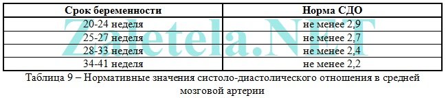
Middle cerebral artery of the fetus (a. cerebri media). When the fetus suffers, there is increase in the values of PI, SDO and speed in SMA.

Maximum speed (aka V max) in the fetal middle cerebral artery:

Systolic-diastolic ratio (SDO) for the middle cerebral artery:
fetal aorta. It comes out of the left ventricle of the heart, goes along the spine and ends in the lower abdomen, where the aorta divides into two iliac arteries, which provide blood supply to the legs of a person.
Deviations in the blood flow of the aorta can be detected only after 22-24 weeks of pregnancy.
The disruption of blood flow is increasing the values of IR, PI and SDO. Critical (talking about the death of the fetus) is considered registration of extremely low values up to their complete disappearance.

Changes in the aorta characterize the severity of intrauterine fetal hypoxia.


Systolic-diastolic ratio (SDR) for the fetal aorta:

Venous duct (VP). It is studied in the extended Doppler assessment of blood flow.
During the study, it is necessary not to take into account episodes of hiccup-like respiratory movements of the child and active movement.
Indexes are not used to assess the venous duct.
The diagnostic criterion for the pathological condition of the fetus is the presence of negative or zero blood flow values in the phase of atrial contraction. Zero or reverse values are recorded with fetal malnutrition, congenital malformations of the right heart, non-immune dropsy of the fetus.
Even with critical blood flow in the arteries of the umbilical cord, but with preserved blood flow in the venous duct in the phase of atrial contraction, it is possible to extend the gestation to the optimal time for childbirth.
Description of blood flow disorders and their treatment
1 degree
1 A degree- violation of blood flow in the uterine arteries, while in the umbilical cord the blood flow remains normal.
This degree of blood flow disturbance is not dangerous for the fetus.
Medical treatment of this condition is ineffective. Doctors still prescribe therapy with Actovegin and Curantil. Do not see on occasion!
In fact, if there is a violation of blood flow in the uterine arteries, it is more expedient to simply walk in the fresh air more often (breathing deeply) + eat right + move more (hiking, special exercises for pregnant women, morning exercises, yoga, swimming). And do not sit for hours at the computer! That's all treatment.
1 B degree- violation of blood flow in the arteries of the umbilical cord, and in the uterine arteries hemodynamics is normal.
This degree of blood flow disturbance requires the use of blood-thinning drugs to avoid developmental delay and fetal hypoxia.
In this case, a treatment is prescribed aimed at improving blood circulation (Placenta compositum, Curantil or Trental). Actovegin is prescribed as an antihypoxant, which improves the supply of oxygen to the fetus.
A blood test for coagulation ability (coagulogram) is also prescribed. With increased blood clotting, it is necessary to take stronger blood-thinning drugs than Curantil (for example, heparin or an agent that includes acetylsalicylic acid).
I degree of violation does not lead to the death of the fetus. There is a systematic monitoring of the nature of the blood flow (every 2 weeks) "plus" the control of fetal CTG (after 28 weeks of pregnancy). In addition, be sure to monitor blood pressure in a pregnant woman.
2 degree- simultaneous violation of blood flow in the uterine arteries and in the umbilical cord, which does not reach critical values (when the blood flow is preserved in the venous duct).
In this condition, medication is mandatory prescribed in a hospital, where round-the-clock monitoring of the fetal condition is provided. It is also necessary to monitor the state of blood flow by conducting Doppler + CTG every 2 days.
In degree II hemodynamic disturbances are rare, but there may be cases of intrauterine death.
3 degree- critical disorders of blood flow in the umbilical cord with intact or impaired blood flow in the uterine arteries. A critical violation is understood as the registration of reverse diastolic blood flow or its absence at all.
III degree of violation poses a danger to the health of the child, because in half of the cases, intrauterine death of the baby occurs. Therefore, when a 3rd degree of blood flow disturbance is detected, it is necessary to urgently perform a caesarean section in order to save the life of the baby, because at this stage of the disorder, treatment is not effective.
Conservative (natural) childbirth at grade 3 can lead to perinatal death of the child.
The cost of a doppler ultrasound in a private clinic is about 1,200 rubles.