Violations of blood flow in the uterine arteries, umbilical cord, placenta during pregnancy (nmpk). Violation of uteroplacental blood flow during pregnancy
During the period of bearing a child, the body of the expectant mother undergoes strong hormonal changes. In this regard, it is very important to constantly monitor the woman's health and the condition of the fetus. In medical practice, very often there is a violation of blood circulation in women who are waiting for an addition to the family. The appearance of an additional circle of blood circulation in the mother's body requires frequent examination by a specialist. After all, if the blood flow during pregnancy is disturbed, then there is a risk of fetal death, and at different periods of its bearing.
Blood flow during pregnancy: normal
Many women, especially those who are carrying their first child, are unaware of the existence of such a study as dopplerometry. It consists in ultrasound diagnostics, which is able to assess the intensity of blood flow in different vessels. This research is mainly done in the third trimester of pregnancy. But in some cases, they resort to it after the twentieth week of bearing a child. Doppler is considered a serious study that allows you to diagnose vascular pathology in the uterus and in the placenta, in the cerebral and carotid arteries and the aorta of the fetus. Comparing the obtained figures and blood flow rates during pregnancy, the specialist determines whether the child in the womb suffers from a lack of oxygen or not.
There are approved standards for Doppler measurements starting from the second trimester of pregnancy. These are the norms of the resistance index of the vessels of the uterus, umbilical cord, aorta and cerebral artery of the fetus. Doctors recommend not to try to decipher the results obtained on your own. There is a certain formula for the exact calculation of the vascular resistance index - this procedure should only be done by a doctor.
Impaired blood flow during pregnancy: degrees
For many expectant mothers, this diagnosis leads to panic and confusion. Should I be nervous? Could this pathology have any consequences for the child? What are the degrees of this disease? Let's try to find answers to these questions.
There are three degrees of disturbances in blood circulation through the blood vessels during gestation. The first is characterized by impaired blood flow that does not reach critical values (in the umbilical cord and artery). In this case, a positive state of fetal hemodynamics is observed. In both ventricles of his heart, there is a decrease in the diastolic function index, as well as an increase in the maximum velocity of blood flow through all heart valves. The first degree of the disease is divided into 1-a, in which only the uteroplacental blood flow is disturbed, and 1-b degree, in which there is an inferior fetal-placental blood flow.
In the second degree, there is a violation of the hemodynamics of the fetus. In 50% of cases, the maximum blood flow rate through all heart valves decreases. It should be noted that this phenomenon is less pronounced in the left sections. Disorders of blood movement are observed both in the fetus and in the uterine arteries. The second degree often turns into the third, and in a very short period.
The third degree signals a critical state of the fetal blood supply. At this stage, a deeper restructuring of intracardiac hemodynamics occurs. It is directly related to the centralization of blood circulation. Fetal hypoxia is not excluded. It is also possible a decrease in diastolic blood flow in the aorta, up to its disappearance. There is a simultaneous inadequate movement of blood in the aorta and carotid artery.
What threatens a violation of blood flow during pregnancy: the consequences
This pathology leads to placental insufficiency, which is observed in 25% of pregnant women.
It is known that the placenta is the main organ during the gestation of a future baby, with the help of which it breathes and nourishes, as well as excretion of its waste products. It is in the placenta that two systems of blood vessels converge, between which a membrane is located, which provides a kind of barrier between the body of the child and the mother. Thanks to the membrane, the blood of the mother and the unborn child does not mix. The placenta is also a protective shield against viruses and bacteria. It has an immune function to protect the fetus.
With placental insufficiency, the uteroplacental and fetal-placental blood flow is disturbed, and also the placenta itself is inadequate maturation. In connection with these changes, the unborn child does not receive sufficient amounts of nutrients and oxygen. For this reason, its development and growth slows down, the existing complications of pregnancy are aggravated.
Naturally, due to the fact that the blood flow during pregnancy is low, such serious changes can even lead to the death of the fetus. But this happens on rare occasions. Often, this pathology is detected at an early stage and can be successfully treated.
Violation of uteroplacental blood flow
In medical terminology, a violation of the uteroplacental blood circulation is denoted by degree 1a. The emergence of this pathology indicates a dangerous complication of pregnancy. It usually occurs at a later date.
Defective blood movement occurs between the uterus and the placenta. This condition contributes to a significant deterioration in the metabolism between the woman's body and the fetus. Naturally, this condition leads to certain consequences.
There are reasons provoking the development of such a condition. These include increased maternal blood pressure, diabetes mellitus, pneumonia and kidney disease in a pregnant woman, and infection in the fetus itself. It should be noted that the timely identification of possible risk groups is a serious preventive measure. Therefore, it is important for a pregnant woman to monitor her health and seek medical help even if she is slightly unwell.
What is the danger of a violation of blood flow in a child
In a single functional system mother-placenta-fetus, defective fetal-placental blood flow leads to placental insufficiency. After all, the placenta supplies the unborn baby with food and oxygen. It is she who is the connecting link that unites two complex systems - maternal and fetal. When such a pathology occurs, a violation of the blood flow in the child is observed. It should be noted that inadequate blood flow in the vessels of any degree leads to malnutrition of the fetus. His condition also depends on the stage of blood flow disturbance. Naturally, the third degree signals the critical condition of the child.
In case of early detection of this pathology, the doctor must determine the need for treatment in a hospital or at home. It all depends on the specific case and the duration of pregnancy.
In medical terminology, a violation of the fetal-placental blood flow is designated as grade 1b.
How to treat impaired blood flow during pregnancy
For the treatment of defective blood flow during the period of bearing a child, various drugs are used that help increase the resistance of the fetal brain to hypoxia, improve blood microcirculation and reduce blood coagulability. If necessary, the doctor prescribes antibacterial and antiviral drugs, as well as immunomodulators.
A good remedy that improves cerebral circulation, heart function and metabolism in a child with hypoxia is the drug Instenon. It is used in combination with other drugs.
The expectant mother is also credited with the use of Actovegin, a drug that helps to increase the resistance of fetal tissues and brain to hypoxia. It also improves metabolism, stimulates cell renewal of the unborn baby, improves blood circulation in the complex system - mother-placenta-fetus. As a result of treatment with Actovegin, blood flow parameters are improved, and the active growth of the child in the mother's womb is observed.
It is known that with placental insufficiency, as a rule, a chronic blood clotting disorder is observed. In this regard, experts recommend the use of drugs that prevent the formation of blood clots in blood vessels (for example, Curantila).
In the case of the 3rd (most difficult) degree of blood flow disturbance, experts cause premature birth.
Especially for - Ksenia Manevich
Violation of blood flow during pregnancy is determined using dopplerometry. The examination shows the movement of blood cells through the vessels from the mother through the placenta to the fetus. The further gestational period depends on these parameters, the doctor receives information about the lack of oxygen or nutrients, which is so important for the normal formation of the baby.
Blood flow during pregnancy
The circulatory system between the mother and the fetus is quite complex, therefore in medical practice it is customary to divide it into levels in order to understand the degree of damage. The main compartment of the entire blood flow system is the placental membrane, which supplies the embryo with all the necessary substances. At the same time, there is a hematoplacental barrier, due to the presence of special cells that do not allow various harmful cellular structures to pass through.The next part of the uterine blood flow is the spiral arteries, in which there is no muscle layer and, in the event of significant blood loss, will not be able to contract properly. And the last structure of blood circulation between a woman and an embryo is called the vessels in the umbilical cord. There are only 2 arteries and 1 vein that connect the placenta with the fetus. Violation of blood flow during pregnancy in this particular fetal-placental circle causes the most serious consequences.
Causes of impaired blood flow during pregnancy
There are no reliable reasons that lead to impaired blood flow in the uterine artery. The only known factors that predispose to failures in blood circulation between intrauterine development and the mother:- severe diseases of the endocrine system, diabetes mellitus or abnormalities in the work of the adrenal glands and thyroid gland;
- insufficient functioning of the kidneys and lungs;
- cardiovascular pathology and hypertension;
- anemia caused by a lack of iron, vitamins and anemia;
- increased thrombus formation;
- infectious diseases can cause inflammation and swelling of blood vessels, which threatens miscarriage at the beginning of gestation.
The degree of impaired blood flow during pregnancy
An analysis of blood flow during pregnancy, as well as an ultrasound scan with a Doppler, help to determine several degrees of deviations of blood circulation in all vessels between a woman and a baby. At the same time, doctors pay attention to infectious lesions, to abnormalities in the composition of the amniotic fluid, to the state of the placenta and other factors affecting the diagnosis.Violation of blood flow occurs:
1 degree - the uteroplacental circle functions with minimal deviation, which gives time for doctors to use drug therapy and restore the working capacity of blood circulation. Moreover, the 1st degree has two subspecies:
- the functioning of the blood flow between the fetus and the placenta is stable, and the uteroplacental circle is malfunctioning, which characterizes the 1a degree of impairment;
- violations for grade 1b are characterized by the opposite situation, when a lack of functioning is observed in the fetal-placental circle.
Grade 3 is characterized by critical marks in the course of diagnostic measures. If poor blood flow is associated with the placenta, then this will be called placental insufficiency, and may be caused by premature detachment or early aging of the membrane.
Thus, by classifying disorders, doctors can see a complete diagnostic picture of the state of blood flow and prescribe appropriate treatment.
Blood flow rate during pregnancy - table
To assess the movement of blood in various vessels, several indicators are used:- IR or resistance index is the difference between blood flow rates over the entire study period;
- PI or pulsation index - a comparison of the speeds for one cycle of blood circulation;
- LMS or systolic-distal ratio implies an assessment of the speed between the contraction of the heart and during the "rest" period.
| Vessel | Index | 28 weeks | 32 weeks | 36 weeks | 40 weeks |
|---|---|---|---|---|---|
| Artery of the umbilical cord | FROM TO | 3,1-3,7 | 2,8-3,4 | 2,4-3,0 | 2,2-2,5 |
| IR | 0,65-0,73 | 0,60-0,67 | 0,55-0,63 | 0,51-0,59 | |
| Pi | 1,08-1,09 | 0,95-0,96 | 0,83-0,84 | 0,73-0,74 | |
| Fetal aorta | FROM TO | 6,0-7,6 | 5,7-7,3 | 5,4-7,1 | 5,2-6,8 |
| IR | 0,82-0,88 | 0,80-0,86 | 0,77-0,83 | 0,75-0,81 | |
| Pi | 1,79-2,24 | 1,76-2,20 | 1,74-2,17 | 1,72-2,13 | |
| Internal carotid artery | FROM TO | 5,6-6,5 | 4,7-5,6 | 4,0-4,8 | 3,3-4,1 |
| IR | 0,78-0,88 | 0,74-0,84 | 0,71-0,81 | 0,69-0,78 | |
| Pi | 1,98-2,39 | 1,7-2,06 | 1,44-1,77 | 1,22-1,51 | |
| Uterine artery | FROM TO | 1,7-1,9 | 1,7-1,9 | 1,7-1,9 | 1,7-1,9 |
| IR | 0,46-0,55 | 0,69-0,86 | 0,69-0,86 | 0,69-0,86 | |
| Pi | 0,69-0,86 | 0,69-0,86 | 0,69-0,86 | 0,69-0,86 |
If the norm is exceeded in the arteries of the uterus, then this indicates an insufficient supply of oxygen to the baby, in the umbilical cord - indicates the presence of preeclampsia and vascular insufficiency. When observing abnormalities in the aorta, a diagnosis of an abnormal state of intrauterine development is made, additional tests will be required to normalize the vital signs of the fetus.
To improve blood flow, a drug is used that relaxes the muscles of the uterus on an individual basis, most often Ginipral, Magne-B6 or Euphyllin are prescribed. Additionally, Verapamil or Isoptin is used to reduce tachycardia.
To normalize blood flow, it is also required to balance the woman's diet, food and, in general, the daily menu should contain the maximum amount of vitamins and nutrients. It is necessary to reduce the emotional and physical workload of the body so that the blood circulation parameters are stabilized.
Most women do not know about such a study as Doppler before the third trimester, and from that moment on, Doppler for pregnant women becomes a completely common procedure.
Dopplerometry- This is one of the methods of ultrasound diagnostics that allows you to assess the intensity of blood flow in various vessels, for example, in the vessels of the uterus and umbilical cord. It is most informative after 30 weeks, but in case of deviations during pregnancy (for example, when the fetus is lagging behind in development), an ultrasound Doppler can be prescribed earlier - starting from 20 weeks.
Indications for Doppler
Adequate placental blood flow ensures the normal course of pregnancy. Violation of blood flow can lead to a delay in intrauterine development of the fetus (IGRP), therefore, the main reason for the appointment of dopplerometry during pregnancy is precisely the discrepancy between the size of the body and / or the baby's organs to the norms.
It is not necessary that if the blood flow is disturbed, the child will lag behind in development, it is just that the risk of an unfavorable course of pregnancy increases significantly. Well, and vice versa, if there is a suspicion of a fetal developmental delay, but the blood flow is not disturbed, then in most cases this indicates a woman carrying a small, but healthy child.
Also, dopplerometry is prescribed for:
- premature maturation of the placenta;
- severe lack of water or polyhydramnios;
- anomalies of the umbilical cord;
- Rh-conflict;
- preeclampsia (late toxicosis, complicated by vomiting, severe swelling and increased pressure in a pregnant woman);
- if the expectant mother has kidney disease, hypertension, diabetes mellitus;
- suspected chromosomal abnormality;
- non-immune dropsy of the fetus;
- uneven development of babies with multiple pregnancies (when there is a difference in their body weights by more than 10%).
If the fetus has heart problems, a Doppler is performed together with CTG, the so-called Doppler echocardiography.
With placental insufficiency, Doppler is performed systematically every 2-3 weeks.
Also, with the development of complications during the previous gestation of the fetus, a Doppler ultrasound scan may be prescribed during subsequent pregnancy.
Preparation for the study and how it is carried out
Doppler testing in pregnant women is carried out according to indications, and it is not a mandatory examination in the normal course of pregnancy. But more and more often in antenatal clinics, all women, without exception, at 30-34 weeks undergo a Doppler ultrasound scan as an assessment of the condition of the fetus.
This procedure is painless and harmless to both the mother and the fetus. The principle of Doppler analysis is the same as for conventional ultrasound during pregnancy: a special Doppler sensor is used in the abdomen, which is equipped with every modern ultrasound diagnostic apparatus. Therefore, this type of research does not require special preparation.
Dopplerometry- this is a visual assessment of blood flow (when a color and graphical representation of the curves of blood flow velocities is observed from the monitor screen by an ultrasound device).
Doppler ultrasonography- this is the same dopplerometry, only in addition, the readings are recorded on the tape in order to track the change (improvement / deterioration) of blood flow after the treatment.
Decoding of indicators of dopplerometry
Uterine arteries (a. Uterina dextra - right and a. Uterina sinistra - left uterine arteries, respectively). The Uzist must determine the nature of the blood flow both in the left and in the right uterine artery, because with gestosis it can be disturbed only in one artery. Thus, assessing the blood flow in only one artery, it is possible to give a false conclusion, which will negatively affect the health of the baby and the expectant mother.
There is such a scientific theory that if blood flow is disturbed in only one (mainly in the right) uterine artery, a woman has a high risk of late toxicosis (gestosis) with all the negative consequences.
With preeclampsia, at first, the blood flow in the uterine artery is disturbed, and when the situation worsens, blood flow in the umbilical arteries also deteriorates. Therefore, if the blood flow in the uterine arteries is disturbed, it is necessary to periodically repeat the Doppler to control the situation.
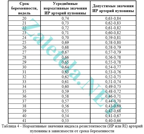
To assess the blood flow in the uterine arteries, the resistance index (IR or RI) is calculated.

Often, pregnancy-induced hypertension develops due to impaired uterine blood flow. The body of the expectant mother independently increases blood pressure to increase blood flow into the intervillous space. So mommy, without realizing it, helps the baby. Thus, it is necessary to improve blood flow and hypertension will disappear on its own.
Violation of blood flow in the uterine arteries is when the value of IR, PI or LMS is more than normal.
The pulsation index (PI) of the uterine arteries should be within the following limits.

Indicators in the right and left uterine artery may differ slightly from each other. If both indicators are within the normal range, then such a picture is not considered a negative phenomenon.
Deviation of blood flow indicators from the norms in two uterine arteries at once indicates a violation of the uteroplacental circulation. This situation requires specific treatment - to move more (regularly go swimming or gymnastics for pregnant women).
Violation of blood flow in only one uterine artery indicates the asymmetry of the uteroplacental blood flow. If the pregnancy is proceeding normally, and the baby develops in accordance with the term, then the placenta is performing its functions.
You should be aware that at 18-21 weeks there may be a temporary disturbance of blood flow in the uterine arteries. This phenomenon is explained by the fact that the adaptive physiological process of cytotrophoblast invasion has not yet been finally completed. Therefore, if abnormalities in the uterine arteries are detected, repeated Doppler ultrasound should be performed in 2-3 weeks, i.e. observe blood flow in dynamics.
The systole-diastolic ratio (SDR) in the uterine arteries should be:

Arteries of the umbilical cord (a. Umbilicalis). To obtain true results, the study should be carried out only at a time when the baby is at rest, and only when his heart rate is in the range of 120-160 beats per minute. Indeed, physiologically, it is so laid down that with an increase in heart rate, a decrease in IR in the umbilical artery occurs, and vice versa, with a decrease in heart rate, an increase in IR occurs.
Measurement of blood flow in the arteries of the umbilical cord should be done while the pregnant woman is lying on her back! Assessment of the severity of umbilical cord blood flow disturbance cannot be objective when the future mother is located “on her left side”.
The umbilical cord must have two arteries and one vein. If there is an abnormality (the only artery of the umbilical cord), then the fetus may suffer from a lack of oxygen and nutrients, which is why it lags behind in mass and growth. But it happens that the fetus adapts to such an existence and does not experience a deficiency of the necessary substances. Such babies are born with low weight, but absolutely viable. Therefore, if there is one artery of the umbilical cord and the blood flow in it is not disturbed, then there is no cause for concern. But, if the blood flow in a single artery is impaired, inpatient treatment should be carried out to improve blood flow and, if necessary, early delivery (if the fetus will greatly lag behind in development).
The most common in assessing the nature of blood flow in the arteries of the umbilical cord is the resistance index. The indicators in both arteries of the umbilical cord should be practically the same.
Violation of blood flow in the umbilical cord is when the value of IR, PI or LMS in the arteries of the umbilical cord is higher than normal.
The pulsation index (PI or PI) of the umbilical arteries must comply with the following standards:

Registration of zero and reverse values of diastolic blood flow is pathological. This means that the fetus is in critical condition.
From the moment of the appearance of constant reverse values to the death of the fetus, only 2-3 days remain, therefore, in the shortest possible time, it is necessary to carry out a cesarean section in order to save the life of the baby. This is possible only starting from the 28th week, when the baby is viable.
Systole-diastolic ratio (SDR) in the arteries of the umbilical cord:

If the blood flow in the umbilical cord is impaired, then, as a rule, fetal growth retardation is also observed. If now there is no delay in development, and the blood flow in the umbilical cord is disturbed, then without treatment, there may be a delay in the development of the fetus.
Middle cerebral artery of the fetus (a. Cerebri media). When the fetus is suffering, increase in the values of PI, LMS and speed in the CMA.

Maximum speed (also known as V max) in the fetal middle cerebral artery:

Systole-diastolic ratio (SDR) for the middle cerebral artery:

Fetal aorta. It leaves the left ventricle of the heart, goes along the spine and ends in the lower abdomen, where the aorta is divided into two iliac arteries, which provide blood supply to the human legs.
Abnormalities in the blood flow of the aorta can only be detected after 22-24 weeks of pregnancy.
Violation of blood flow is increase in the values of IR, PI and LMS... Critical (talking about fetal death) is considered registration of extremely low values up to their complete disappearance.

Changes in the aorta characterize the severity of intrauterine fetal hypoxia.


Systole-diastolic ratio (SDR) for the fetal aorta:

Venous duct (VP). It is studied in extended Doppler blood flow assessment.
During the study, it is necessary to disregard episodes of hiccups-like respiratory movements of the child and active movement.
Indices are not used to assess the ductus venosus.
The diagnostic criterion for the pathological state of the fetus is the presence negative or zero blood flow values in the phase of atrial contraction. Zero or reversible values are recorded in case of fetal malnutrition, congenital malformations of the right heart, non-immune dropsy of the fetus.
Even with critical blood flow in the arteries of the umbilical cord, but with preserved blood flow in the ductus venosus in the phase of atrial contraction, it is possible to prolong gestation until the optimal time for childbirth.
Description of blood flow disorders and their treatment
1st degree
1 A degree- violation of blood flow in the uterine arteries, while in the umbilical cord blood flow remains normal.
This degree of blood flow disturbance is not dangerous to the fetus.
Medical treatment of such a condition is ineffective. Doctors still prescribe therapy with Actovegin and Curantil. Do not see each other on the occasion!
In fact, if the blood flow in the uterine arteries is disturbed, it is more advisable to simply walk more often in the fresh air (breathing deeply) + eat right + move more (walking, special exercises for pregnant women, morning exercises, yoga, swimming). And do not sit for hours at the computer! That's all the treatment.
1 B degree- violation of blood flow in the arteries of the umbilical cord, and in the uterine arteries hemodynamics are normal.
This degree of impaired blood flow requires taking blood-thinning drugs in order to avoid developmental delay and fetal hypoxia.
In this case, treatment is prescribed to improve blood circulation (the drug Placenta compositum, Curantil or Trental). Actovegin is prescribed as an antihypoxant that improves oxygen supply to the fetus.
A blood test for coagulation (coagulogram) is also prescribed. With increased blood clotting, it is necessary to take stronger blood thinning drugs than Curantil (for example, heparin or a drug that contains acetylsalicylic acid).
I degree of violation does not lead to fetal death. Systematic monitoring of the nature of blood flow (every 2 weeks) "plus" control of CTG of the fetus (after 28 weeks of pregnancy) is carried out. In addition, be sure to monitor the blood pressure of the pregnant woman.
2nd degree- simultaneous disturbance of blood flow in the uterine arteries and in the umbilical cord, which does not reach critical values (when blood flow is preserved in the venous duct).
In this condition, medical treatment in a hospital is mandatory, where round-the-clock monitoring of the condition of the fetus is provided. It is also necessary to monitor the state of blood flow by performing a Doppler + CTG every 2 days.
With II degree, hemodynamic disturbances are rare, but cases of intrauterine mortality can be observed.
Grade 3- critical disturbances of blood flow in the umbilical cord with preserved or disturbed blood flow in the uterine arteries. Critical violation is understood as registration of reverse diastolic blood flow or its absence at all.
III degree of violation carries a danger to the health of the child, because in half of the cases, intrauterine death of the baby occurs. Therefore, if 3 degrees of blood flow disturbance are detected, it is necessary to urgently carry out a cesarean section in order to save the baby's life, because at this stage of the violation, the treatment is not effective.
Conservative (natural) childbirth at grade 3 can lead to perinatal death of the child.
The cost of a Doppler ultrasound scan in a private clinic is about 1,200 rubles.
Why is exercise during pregnancy so necessary?
1.Decreased activity (physical inactivity) during pregnancy increases the risk of impaired blood flow in the placenta, which reduces the supply of oxygen and nutrients to the baby.
2.Sport during pregnancy allows you to improve uteroplacental blood flow, activating metabolic processes in the mother's body, due to which the child develops well
3. Regular exercise during pregnancy helps to normalize blood pressure, especially if a woman suffers from a decrease (in the first half of pregnancy, pressure tends to decrease, and in the second half - to increase).
4. Headaches decrease, muscles of the legs, back, pelvic floor become stronger, which is an excellent prevention of varicose veins, hemorrhoids.
5. Sport during pregnancy helps to normalize intestinal motility, which is the prevention of constipation.
6. After small physical exertion, mood rises, a surge of energy appears, stress is relieved and sleep is normalized.
7. Sport during pregnancy reduces the risk of edema, since movement improves blood flow in the kidneys and increases their excretory function.
Stop factors
After physical exertion during pregnancy, shortness of breath, profuse sweating, tachycardia, dizziness and other uncomfortable phenomena should not occur.
If the upper blood pressure rises in the expectant mother above 130 and / or lower above 90 mm Hg. Art., then you can play sports during pregnancy only after consulting a doctor, so as not to provoke the development of complications.
It is important that after the workout, the expectant mother feels more energized than before and does not feel very tired.
What is the right way to play sports during pregnancy so that it will only benefit mom and baby?
Advice 1. Perform exercises for pregnant women only when you feel well.
An expectant mother should not be overloaded if she does not feel very well. In such cases, it is better to just take a walk in the fresh air.
.jpg)
Tip 2: Avoid Excessive Exercise During Pregnancy
We are talking about strength training and lifting weights, especially above shoulder level, exercises that require prolonged tension of the muscles of the legs and abdomen, as well as running and jumping. All this can cause an increase in the tone of the uterus and the risk of termination of pregnancy.
Do not overuse stretching exercises for pregnant women, as while carrying a baby, the ligaments soften and it is easy to get a dislocation or other injury.
Intensive aerobic exercises (dancing, shaping, step aerobics) can be continued up to 16-18 weeks, that is, until the period when the fetus becomes large and the belly begins to grow significantly. Elements that require balancing (for example, dancing pirouettes) are best skipped.
Advice 3. Physical activity during pregnancy should be limited if there is a violation of the course of pregnancy or exacerbation of a chronic disease
A violation of the course of pregnancy includes, for example, the threat of termination or its termination earlier in the same period, multiple pregnancy, polyhydramnios, oligohydramnios, severe toxicosis of the first or second half of pregnancy, anemia, increased uterine tone, bleeding, placenta previa, etc. It is clear that you will have to give up training during an exacerbation of any chronic diseases (for example, pyelonephritis, sinusitis, osteochondrosis, etc.) or the appearance of acute colds, especially with an increase in body temperature (ARVI, tonsillitis, etc.), as well as if there are diseases such as diabetes mellitus, thyroid dysfunction, which affect metabolic processes in the body, reducing the overall adaptive capacity. In such cases, exercise for pregnant women increases the load on the body and can provoke an aggravation of the course of the disease or the development of complications of pregnancy.

Tip 4. Choose physical activity for pregnant women with a low injury rate
It is not necessary to "drive with the ball" during pregnancy. Basketball, volleyball are not classes for expectant mothers. This also includes alpine skiing, horse riding, snowboarding, sledding, ice skating, rollerblading, cycling, scuba diving and underwater diving, diving.
What you can:
- Cross-country skiing. With this type of load, there are practically no shock effects on the joints and spine, and the muscles of the back, arms, legs are well trained. The main thing is to ski at a moderate pace. With good support for the legs and arms, the probability of falling is practically zero.
- Swimming is one of the safest sports during pregnancy, but cool water should be avoided, especially if the uterine tone is increased, which further increases it, as well as swimming in natural bodies of stagnant water (ponds, lakes), as there is a high risk the occurrence of an infection of the genitourinary tract due to the presence of local microorganisms. While swimming, you should not raise your head high above the water, as the lower back arch and the neck muscles tense (blood vessels supplying the brain may be pinched). It is best for expectant mothers to swim breaststroke, on their backs, resting their heads on a swimming board and working with their legs.
- In the first trimester, you can swim and do water aerobics for 40-50 minutes (together with a warm-up). The water will massage the calves, facilitating venous drainage. Overcoming water resistance, muscles work without tension, but with maximum effect.
- Optimal. It combines gymnastics, breathing exercises, the ability to effectively concentrate and relax. All movements in yoga are soft and fluid, which makes them safe and effective.
- If the expectant mother spends most of the time while waiting for the baby (especially if she is in a static sitting or standing position for a long time), then it is advisable to periodically during the day (1 time in 1.5-2 hours) do a warm-up - get up, walk and perform some simple exercises for pregnant women.
Tip 5. Exercise regularly during pregnancy
If the expectant mother did not exercise regularly before pregnancy, especially in the first trimester, it is best to go to workouts three times a week for 30 minutes. If you do this more often and intensively, you can provoke a violation of the course of pregnancy or an exacerbation of any chronic disease, since the load will be a stress factor for an unprepared organism. By the way, training can be replaced by regular walking (including on a treadmill). Before starting exercises during pregnancy, a warm-up is necessary - light stretching for the arms and legs, as well as rotational movements of the head and tilting of the body from side to side.
Tip 6. Control your pulse
The pulse should not exceed more than 130 beats per minute, since at a higher frequency, proteins in the body are burned, and this is a building material for a baby, as well as an increased load on the cardiovascular system as a whole.
During exercise, it is necessary to correctly calculate the heart rate. To do this, subtract age from 220 and find 70% from the resulting figure, For example, if the expectant mother is 20 years old, then 220 - 20 = 200 is the maximum heart rate that should be during exercise at this age; out of 200 we calculate 70%, we get 140 - this is the heart rate that it is desirable to adhere to so that the classes are as effective as possible and at the same time do not harm the mother and the baby. But if the expectant mother went in for sports before pregnancy, then more active and frequent exercises are possible under the supervision of a coach.
The role of the transport system, which supplies nutrients with oxygen from the mother to her baby, is played by the placenta. It acts as a special link connecting two separate vascular systems - uterine and umbilical cord blood flow - together. The well-being and safety of the baby depends on the quality of the work of this unique organ. That is why, during the ultrasound examination, not only the level of the physical development of the fetus is checked, but also possible violations of the uterine blood flow during pregnancy are determined. What indicators should the normal blood flow in the placenta meet, what symptoms can be used to suspect a pathology, and how can this disease be treated?
Violation of blood flow during pregnancy. The structure of the uteroplacental blood flow
Blood flow between a woman and a fetus is provided not only through the placenta. In addition to it, a complex network of blood vessels is involved in the uteroplacental circulation system, working in conjunction with the placenta, which help it to fully supply the fetus with everything necessary and at the same time prevent oxygen starvation of the fetal tissues.
The circulatory system between mother and baby consists of three levels, which change with the increase in gestational age and perform specific functions. Under the influence of many factors, "breakdowns" can occur on any of them. And depending on their location and severity, the entire subsequent treatment plan and tactics of childbirth depends.
- The central link of the system is the placenta. As it grows, its villi grow tightly into the uterine walls and "suck" from the mother's blood a set of all the necessary elements for the development of the fetus. In this case, incest between the mother and the baby does not occur. The multilayer hematoplacental barrier plays the role of a solid "sponge" that allows only useful substances to pass through, and filters out all dangerous compounds and viruses and sends them back into the mother's venous bloodstream.
- The second level of blood flow is considered to be a branch of the spiral arteries of the uterus. They are intended solely to support pregnancy and are dormant until conception. When the embryo is four weeks old, the arteries gradually begin to lose muscle tissue and the ability to contract. Closer to the fourth month of gestation, they fill with blood and connect to the placenta. Violation of blood flow in the uterine artery during pregnancy leads to a blockage of blood circulation at all other levels.
In addition to the benefits for the baby, these arteries carry a certain risk to the mother's life. Their rupture can cause severe bleeding during delivery as they lose their ability to contract.
- The third level of blood flow is formed by the umbilical cord vessels. The vein and two arteries connect the embryo and the placenta and therefore play the most important role in the development of the baby. Violation of the fetal-placental system is most often the cause of congenital abnormalities in the development of the child.
Violation of blood flow during pregnancy: causes
Factors of primary failure are:
- Genetic predisposition.
- Infection with viral or bacterial microflora.
- Endocrine disorders (corpus luteum inferiority, ovarian dysfunction, hypothyroidism).
- Lack of tissue that serves as the basis for the further development of the ovum and placenta.
All these factors lead to abnormal development and attachment of the placenta, and its subsequent inferiority.
The causes of secondary placental insufficiency include:
- Obstetric diseases (fibroids, endometriosis).
- Endocrine system diseases (diabetes, hyperthyroidism).
- Chronic diseases (hypertension, renal pathologies, thrombophlebitis).
- Complications of pregnancy (abnormal placenta previa, Rh-conflict, multiple pregnancy).
- Factors of the external unfavorable environment.
- Bad habits.
- Social and living conditions.

Violation of blood flow during pregnancy. Placental insufficiency classification
Violation of blood flow is a common pathology that is caused by abnormalities in the structure of the placenta and umbilical cord, and in 60% it causes a delay in the mental and physical development of the baby. The generally accepted and most common are the following classifications.
Depending on the time of appearance, there are:
- Primary placental insufficiency - manifests itself before the 16th gestational week and is associated with a violation of the mechanism of implantation of the ovum and the subsequent malformation of the placenta.
- Secondary placental insufficiency - diagnosed after 16 weeks of pregnancy, when the placenta is already fully formed. The cause of the violation is the influence of various negative factors of external origin.
In accordance with the clinical picture of the course of the disease, placental insufficiency is divided into the following forms:
- Compensation - metabolic disturbances in the work of the placenta are recorded, but the blood flow between the uterus and the placenta or between the placenta and the baby is not disturbed. In this condition, there may be a partial blockage of blood flow, for example, a violation of the umbilical cord blood flow through one of the vessels or a violation of the right blood flow of the uterus during pregnancy. The female body is able to fully compensate for the baby's oxygen deficiency by increasing blood flow in other ways. This ensures optimal development of the child without the risk of hypoxia. The baby develops normally and is born on time without congenital anomalies.
- Subcompensation - the mother's body cannot resume the flow of oxygen to the baby, since all the links of the blood flow system do not fully function. This causes some difficulties due to oxygen deficiency, which worsens the condition of the fetus and can provoke congenital malformations.
- Decompensation - during the Doppler diagnosis, a complete violation of blood flow is determined, which is difficult to eliminate with drug treatment. This condition often results in complex heart defects or postnatal mortality.
Violation of placental blood flow during pregnancy can be acute and occurs against the background of premature placental abruption or its incorrect location in the uterus. Chronic impairment of blood flow can fail at any stage of gestation and is most common.
According to ultrasound, three degrees of fetal hemodynamic disorders are distinguished.
Grade 1 - provides for minor changes in blood circulation between a woman and her baby, which is effectively treated with the help of special medications. At this stage, only one part of the circulatory system is disturbed, for example, only in the uterine artery. Timely diagnosis of pathology and its subsequent treatment completely eliminates the risk of disorders in the child.
Depending on which part of the blood flow is affected, two varieties of the first degree are distinguished:
- impaired blood flow of grade 1a during pregnancy suggests that the permeability between the uterus and the placenta is reduced, while the connection between the placenta and the fetus is not broken. Improper treatment in 90% of cases may be accompanied by a slight delay in fetal development, which is characterized by insufficient body weight and general growth indicators.
- impaired blood flow of 1b degree during pregnancy indicates that the uteroplacental blood flow is normal, and the fetal-placenta circulatory system is impaired. In 80% of future women in labor, pregnancy proceeds with signs of a delay in the normal development of the fetus.
Grade 2 - it is diagnosed if there is placental insufficiency at all levels. In this case, it is almost impossible to compensate for the oxygen deficiency, since the fetal aorta, uterine artery and umbilical cord artery are unable to fully pass blood flow. This degree is detrimental to the baby and often becomes the cause of his death.
Violation of blood flow of the 2nd degree during pregnancy is very unstable and in the shortest possible time passes into the last most critical degree.
Grade 3 is characterized by the centralization of the blood flow. The baby's condition becomes critical, since its intracardiac hemodynamics is completely disrupted. Reverse diastolic blood flow is often recorded with Doppler imaging. This degree is often diagnosed with clear signs of developmental delay and is practically not amenable to drug treatment.

Diagnosis of placental insufficiency
For the prophylactic detection of possible problems with blood flow, all women in the position undergo diagnostics three times, which includes Doppler measurements. Recommended times for ultrasound: from 11 to 14 gestational weeks, from 20 to 24, and from 32 to 34 weeks.
Obstetric examination
Women in a position that are at risk for the formation of placental insufficiency are subject to regular clinical observation. Particular attention is paid to such indicators:
- Woman's weight. Exceeding this value often indicates latent gestosis.
- The circumference of the abdomen and the height of the fundus of the uterus. A deviation from the norm of these indicators in 50% of cases indicates a delay in fetal development.
- Uterine tone and bleeding.
- Fetal movement and heart rate. A decrease in these indicators speaks of possible fetal hypoxia.
Laboratory research
Such a diagnosis is used to determine the condition of a pregnant woman in the third trimester with compensated impairment of blood flow. For this, the hormonal status of a woman is monitored, provided that the fetus has no signs of malnutrition (intrauterine lag).
Laboratory tests include the following:
- Calculation of the amount of alkaline phosphatase in venous blood.
- Determination of the level of oxytocin.
- Study of the concentration of estradiol in urine.
Doppler studies
Such a complex name belongs to a painless diagnostic procedure that allows:
- measure the speed of blood flow in the veins and arteries that connect the mother and child;
- determine the direction of blood flow;
- diagnose pathological changes before the appearance of external symptoms.
Additionally, you can evaluate the following indicators:
- The degree of aging of the placenta.
- Lots or low water.
- Possible malformations.
- Fetal hypoxia.
- Genetic abnormalities.
- Signs of intrauterine infection.
- Refinement of the placenta.
Such an examination is carried out on additionally equipped devices for ultrasound examination or special portable devices. Thanks to sensors that measure indicators, all data is reproduced on the monitor. In the course of diagnostics, a dopplerogram is compiled, which shows the systolic-diastolic ratio of blood flow. In other words, it shows the difference between the sent and back received blood flow, which helps to determine the degree of patency of the vessels that connect the uterus, placenta and baby.
The most favorable position for Doppler imaging is lying on your side. You can also conduct a study on the back, but in this position, many women may exhibit uterine hypertonicity, which significantly distorts the results obtained.

What symptoms indicate impaired blood flow during pregnancy?
It is very difficult to suspect problems with blood flow in the placenta without examination. But there are several symptoms, in the event of which it is better to consult a doctor:
- Pathological physical activity of the baby. If he is overly active or, conversely, practically does not move, this signals hypoxia.
- Severe toxicosis in the second half of pregnancy. Late gestosis often accompanies pathology of placental blood flow.
- Too slow increase in abdominal circumference. It is difficult to notice it on your own, therefore, as a rule, the gynecologist pays attention to this during a planned visit to the patient.
- Bloody vaginal discharge. This is the most dangerous symptom that indicates placental abruption.

Violation of blood flow during pregnancy: consequences
Even the smallest deviations in blood circulation between mother and fetus reduce the amount of incoming nutrients, vitamins and oxygen. If this "fasting" is prolonged, the following complications may occur:
- Pathological termination of pregnancy.
- Fetal hypoxia.
- Congenital heart defects.
- Increased risk of intrauterine and perinatal death.
- Detachment of the placenta or premature aging.
- Gestosis.
When diagnosing the first degree of compensated impairment of blood flow during pregnancy, the consequences for the child are uncritical and over time, a slight lag in growth or development is smoothed out, and the baby is catching up with his peers.
If a woman is diagnosed with a more severe diagnosis of decompensated placental insufficiency of the last degree, the prognosis is less optimistic and pregnancy often ends in fetal fading or the birth of a defective child.

Impaired blood flow during pregnancy: treatment
The most favorable period for conservative treatment is the first degree of pathological blood flow. The second degree is extremely rarely amenable to correction, but the third is a direct indicator for emergency delivery using a cesarean section, if the gestational period allows.
The tactics of treatment is based on the pathogenesis of the disease, and provides for a complex effect on all links of the blood flow chain:
- To improve microcirculation with minor deviations, women are prescribed the homeopathic drug Hofitol, and if it does not have the desired effect, medications with more active active ingredients are prescribed, for example, Pentoxipharm or Actovegin.
- If a woman has a history of thrombophlebitis, she is shown drugs to restore the properties of blood to pass through the vessels. Such means include Curantil.
- Drotaverin or No-Shpa is used as a vasodilator.
- To reduce uterine tone and improve blood flow, magnesium is prescribed in the form of droppers and magnesium B6 is administered orally.
- To provide an antioxidant effect, the intake of vitamin C and tocopherol is necessarily indicated.
If the woman's condition does not improve and, according to the results of control studies, blood flow is not restored, the woman is offered hospitalization. This provides a closer control over the condition of the fetus.
To avoid possible complications from the side of the uteroplacental blood flow, it is better to engage in the prevention of this pathology. Walk more in the fresh air, eat right, give up bad habits and, most importantly, go through all scheduled examinations in a timely manner. Then you will be able to warn, and if it appears, start treating blood flow disorders in time.