Screening of the first trimester of pregnancy - what you need to know about the norms and results. Why is needed and when the first, second and third screening is carried out during pregnancy screenshots as accurately determines pathologies
Some time ago, pregnant women and knew did not know about such a procedure as Prenatal or perinatal . Now such a survey is held all future mothers.
What is screering during pregnancy, why are it carried out and why is its results so important? Answers to these and other exciting many pregnant women questions about perinatal screening we tried to give in this material.
To exclude in the future any misunderstanding of the information provided, before proceeding directly to consideration above the designated topics, it is worth identifying to some medical terms.
Prenatal screening - This is a special kind of such a standard procedure, as screening. This comprehensive examination consists of Ultrasound diagnosis and laboratory study in this particular case biochemistry of mother serum. Identifying at the early stages of some genetic deviations - This is the main task of such an analysis during pregnancy, like screening.
Prenatal or perinatal means antenatova, and under the term screening Medicine implies a number of studies of a large formation of the population, which are carried out in order to form the so-called "risk group", subject to one or another diseases.
Happens universal or selective screening .
It means that screening research Make not only pregnant, but also other categories of people, such as children of the same age to establish diseases characteristic of this period.
With help genetic screening doctors can learn not only about the problems in the development of the baby, but also to react to complications in the course of which a woman may not even suspect.
Often the future mother, having heard that they had to go through this procedure several times, they begin to panic and worry in advance. However, there is nothing to fear here, you just need to ask the gynecologist in advance, why need screening For pregnant women, when and, most importantly, how this procedure is done.
So, let's start, perhaps with the fact that screening spend three times for the whole pregnancy, i.e. in each trimester . Recall that trimester - This is a period consisting of three months.
What it is screening 1 trimester ? To begin with, will answer the common question about how many weeks first trimester of pregnancy . In gynecology there are only two ways to significantly establish a deadline for pregnancy - calendar and obstetric.
The first is based on the bottom of the conception, and the second depends on menstrual cycle preceding fertilization . therefore I trimester - This is the term that on the calendar technique begins the first week from conception and ends with the fourteenth week.
In accordance with the second way I trimester
- This is 12 obstetric weeks. Moreover, in this case, the term is counted from the beginning of the last menstruation. Recently screening
Not appointed pregnant women. 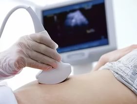
However, now many future mothers are interested in passing such a survey.
In addition, the Ministry of Health urgently recommends appointing research to all future mothers without exception.
True, this is done voluntarily, because No one may force a woman to go through any analysis.
It is worth noting that there are categories of women who are simply obliged to pass due to certain reasons screening, for example:
- pregnant from thirty-five years and further;
- future mothers, a history of which is present information about the presence of a threat spontaneous ;
- women who in the first trimester moved infectious diseases ;
- pregnant women who are forced by the state of health take on early darling drugs for their position;
- women who were fixed in previous pregnancy various genetic deviations or Anomalies in the development of the fetus ;
- already giving birth previous women of children with any deviations or Defaults in development ;
- women who diagnosed froker or Regressing pregnancy (termination of the development of the fetus);
- suffering from narcotic or women;
- pregnant women, in the family of which or in the family of the future of the child were recorded cases hereditary genetic deviations .
What time do prenatal screening 1 trimester ? For the first screening during pregnancy, the period is set in the interval, starting from 11 weeks to 13 obstetric weeks of pregnancy and 6 days. Previously, there is no sense above the marked sentence, since its results will be non-informative and absolutely useless.
The first ultrasound at the 12th week of pregnancy makes it no coincidence. Since it is on this time that embryonic and begins fetal or fetal the period of development of the future person.
This means that the embryo turns into a fruit, i.e. There are obvious changes that speak about the development of a full-fledged living human body. As we said earlier, Screening research - This is a set of events, which consists of ultrasound diagnostics and blood biochemistry.
It is important to understand that screening Ultrasound In 1 trimester during pregnancy plays the same important role as laboratory blood tests. Indeed, in order for genetics to make the right conclusions following the results of the examination, they need to be studied as the results of the ultrasound and the biochemistry of the patient.
How many weeks the first screening is held, we talked, we now move on to decipher the results of a comprehensive study. It is really important to consider in more detail the norms established by doctors of the results of the first screening during pregnancy. Of course, only a specialist in this area, which has the necessary knowledge, and most importantly, will be able to give a qualified assessment of the results of the analysis.
We believe that any pregnant woman it is advisable to know at least general information about the main indicators prenatal screening and their regulatory values. Indeed, for most future mothers, it is characteristic of being too diverse in relation to everything that concerns the health of their future Chad. Therefore, they will be much calmer if they will know in advance what to expect from the study.
Decoding screening 1 trimester on ultrasound, norms and possible deviations
All women know that during pregnancy they have to undergo more than once an ultrasound study (hereinafter ultrasound), which helps the doctor to track the intrauterine development of the future child. In order to screening ultrasound Gave reliable results, you need to prepare in advance to this procedure.
We are confident that the overwhelming majority of pregnant women know how to make this procedure. However, we will not even repeat that there are two types of research - transband and transabdominal . In the first case, the instrument sensor is inserted directly into the vagina, and in the second it is in contact with the surface of the front abdominal wall.

For a transvaginal type of ultrasound, no special preparation rules are provided.
If you have a transabdominal study, then before the procedure (approximately within 4 hours before the ultrasound) should not go to the toilet "on the little", and half an hour is recommended to drink up to 600 ml of ordinary water.
The thing is that the survey must be carried out necessarily on the liquid filled bladder .
In order for the doctor to receive a reliable result Ultrasound screening The following conditions must be respected:
- the term of the survey - from 11 to 13 an obstetric age;
- the position of the fetus should allow a specialist to carry out the necessary manipulations, otherwise the mammy will have to "influence" the baby so that he overturned;
- copchiko-parmer size (Further, the CTR) should not be less than 45 mm.
What is a CTR during pregnancy on ultrasound
When conducting an ultrasound, a specialist is necessarily examining various parameters or fetus sizes. This information allows you to determine how well the baby is formed, and whether it is developing correctly. The rules of these indicators depend on the period of pregnancy.
If the amount obtained as a result of the ultrasound, the value of a parameter is deviated from the norm in a large or smaller direction, this is considered to be a signal of any pathologies. Copchiko-parmer size - This is one of the most important initial indicators of the proper intrauterine development of the fetus.
The value of the CTR is compared with a mass of the fetus and a period of pregnancy. Determine this indicator measurement of the distance from the dice of the child's pattern before its tailbone. As a general rule, the greater the CTR indicator, the greater the period of pregnancy.

When this indicator slightly exceeds or vice versa is slightly smaller than the norm, then there are no reasons for panic. This speaks only about the peculiarities of the development of this particular child.
If the value of the CRT deviates from the standards to the most side, this signals the development of the fetus of large sizes, i.e. Presumably the weight of the child at birth will exceed the averaged norms of 3-3.5 kg. In cases where the CTR is significantly less than regulatory values, it may be a sign that:
- pregnancy Does not develop as it should, in such cases, the doctor must carefully check the palpitation of the fetus. If he died in the womb, the woman needs urgent medical care ( scraping of the uterine cavity ) to prevent a possible threat to health ( development of infertility ) and life ( infection, bleeding );
- the body of a pregnant woman produces an insufficient number, as a rule, that can lead to spontaneous miscarriage. In such cases, the doctor prescribes a patient an additional examination and discharges medicines containing hormones ( , Dufston );
- mother is ill infectious diseases , including venereal;
- fetal has genetic deviations. In such situations, doctors prescribe additional studies along with, which is part of the first screening analysis.
It is also necessary to emphasize that there are often cases when the low CTR speaks about the incorrectly established period of pregnancy. This applies to the option of the norm. All that a woman needs in such a situation is to pass a repeat ultrasound examination after some time (usually after 7-10 days).
BPR fetal (biparity size)
What is this BPD on ultrasound during pregnancy? When conducting an ultrasound of fetal research in the first trimester, doctors are interested in all possible characteristics of the future child. Since their study gives specialists a maximum of information on how the intrauterine development of a small little man occurs and everything is in order with his health.
What is it BPR Futin ? First, we will decipher the medical abbreviation. BPD - this is biparial fetal head . Distance between the walls dumpless bones of the skull , Simply the size of the head. This indicator is considered one of the main to determine the normal development of the child.
It is important to note that the BPD shows not only how good the baby develops correctly, but also helps doctors to prepare for the upcoming delivery. Because if the size of the head of the future child deviates from the norm in the right direction, he simply will not be able to go through the generic paths of the mother. In such cases, scheduled caesarean sections are prescribed.

When the BPD deviates from the established norms, it may indicate:
- about the presence of the fetus of such incompatible pathologies like brain hernia or tumor ;
- about the large size of the future child, if other major fetal parameters are ahead of the established development standards for several weeks;
- about hoping development, which will return to normal after some time, provided that other major fetal parameters fit into the norm;
- on the development of the fetus brain resulting from the presence of infectious diseases in the mother.
The deviation of this indicator in a smaller side indicates that the baby's brain develops incorrectly.
Thickness of the collar space (TVP)
PLP FRUT - what it is? Collar space Fetal or size cervical fold - This is a place (more precisely, the oblong formation), which is between the neck and the upper skin sheath of the infant body, in which the cluster of the fluid is observed. The study of this value is carried out when screening the first trimester of pregnancy, since it is on this time that it is possible for the first time to measure TVP, and then analyze it.
Starting from 14 weeks of pregnancy, this formation gradually decreases in the amount and by 16 week almost disappears from visibility. For TVP, certain norms are also installed, which are directly dependent on the period of pregnancy.
For example, norm thickness of collar space At 12 weeks should not go beyond the range from 0.8 to 2.2 mm. Thickness of collar space at 13 weeks should be between 0.7 to 2.5 mm.
It is important to note that for this indicator, experts establish averaged minimum values, the deviation from which indicates the thinning of the collar space, which is the same as the extension of the TVP is considered an anomaly.
![]()
In the event that this indicator does not correspond to the tables designated in the above table of 12 weeks and at other times of pregnancy, the following result, most likely, indicates the presence of the following chromosomal deviations:
- trisomy 13. , disease known as syndrome Patau, characterized by the presence of an additional 13 chromosome in human cells;
- Trisomy on 21 chromosome, known to all as down syndrome , genetic human disease in which karyotype (i.e., a complete set of chromosomes) is represented instead of 46 chromosomes 47th;
- monosomy by X-chromosome , genomic disease, named after the scientists who have discovered sherosezhevsky-Turner syndrome it is characterized by such anomalies of physical development, as lowland, as well as sexual infantilism (immaturity);
- trisomy in 18 chromosome - This is a chromosomal disease. For edwards syndrome (The second name of this disease) is characterized by the multiplicity of malformations that are incompatible with life.
Trisomy - This is an option aneuploidy . Change karyotype in which there is an additional third in a person's cell chromosome instead of normal diploid set.
Monosomy - This is an option aneuploidy (chromosomal deviation) at which there are no chromosomes in a chromosomal set.
What are the rules for trisomy 13, 18, 21 installed during pregnancy? It happens that in the process of cell division, the failure occurs. This phenomenon got a name in science. aneuploidy. Trisomy - This is one of the varieties of Aneuploidy, in which instead of a pair of chromosomes in the cage there is an extra third chromosome.
In other words, the child inheritss its parents an additional 13, 18 or 21 chromosomes, which in turn entails genetic deviations that impede normal physical and mental development. Down syndrome according to statistics, this is the most common disease due to the presence of 21 chromosomes.
Children born with Edwards syndromes, as in the case of syndrome Pataau. , usually do not live up to a year, unlike those who are not lucky to be born with dauna syndrome . Such people can live to deep old age. However, this life can be called by existence, especially in the countries of the post-Soviet space, where these people are considered outcasts and try to avoid and not notice them.
In order to eliminate such anomalies, pregnant women, especially from a risk group, should necessarily undergo a screening survey. Researchers argue that the development of genetic deviations is directly dependent on the age of the future mother. The younger woman is, the less likely that her child will have any anomalies.
For the establishment of trisomy in the first trimester of pregnancy is studying collar Petroda With ultrasound. In the future, pregnant women periodically pass the blood test in which the most important indicators are for genetics. alpha Fetoprotein (AFP), Inhibin-A, Chorionic Gonadotropin (HCG) and Estor .
As mentioned earlier, the risk of genetic deviations in a child depends primarily on the age of the mother. However, there are cases when trisomy is fixed in young women. Therefore, doctors during screening are studying all possible signs of anomalies. It is believed that an experienced ultrasound specialist can identify problems during the first screening survey.
Signs of Down syndromes, as well as Edwards and Pataau
For trisomy 13 is characterized by a sharp decline in level Papp-A (Papp Associated with pregnancy protein (protein) A-plasma ). Also, the marker of this genetic deviation is. The same parameters play an important role in the definition of the presence of the fetus edwards syndrome .
When there is no risk of trisomy 18, normal indicators PAPP-A and B-HCG (free beta subunit hCG)
fixed in biochemical blood test. If these values \u200b\u200bare deviated from the standards set for each specific period of pregnancy, then, most likely, the child will find genetic defects of development. 
It is important to note that in the case when the specialist records signs indicating the risk during the first screening trisomy A woman is sent to a further examination and advice on genetic groups. For the formulation of the final diagnosis of the future mother will have to go through such procedures as:
- biopsy Chorione . obtaining a sample of chorion tissues for the diagnosis of anomalies;
- amniocentesis - this is puncture of amniotic shell To obtain the sample spindle water In order to further study them in the laboratory;
- placentzetsis (biopsy placenta) , with a given invasive diagnostic method Specialists are selected sample placental fabric with the help of a special puncture needle that is pierced front abdominal wall ;
- cordocentsis , the method of diagnosing genetic deviations during pregnancy, at which the analysis of the fetal bodies is being analyzed.
Unfortunately, if a pregnant woman passed any of the above studies and supplied during bioskrining and ultrasound The diagnosis of presence in the fetus of genetic abnormalities was confirmed, the doctors will offer to interrupt pregnancy. In addition, in contrast to standard screening research data invasive examination methods they can provoke a number of heavy complications up to spontaneous miscarriage, so doctors resort to them quite rare amounts.
Nasal bone - It is a bit elongated, quadrangular, convex in front of a pair of person. With the first screening on the ultrasound, the specialist determines the length of the bone of the baby's nose. It is believed that in the presence of genetic deviations, this bone is developing incorrectly, i.e. Its osenation occurs later.

Therefore, if the nasal bone is absent or its size is too small when conducting the first screening, this indicates the possible availability of various anomalies. It is important to emphasize what the nasal bone length of 13 weeks or 12 weeks. When screening at 11 weeks, a specialist checks only its presence.
It should be emphasized that when the nasal bone is inconsistent with the norms established, but according to other key indicators, there are really no reasons for anxiety. This state of affairs may be due to the individual characteristics of the development of this particular child.
Heart Frequency (CSS)
Such a parameter as Heart rate It plays an important role not only in the early periods, but also throughout the entire pregnancy. Constantly measure and follow fruit heartbeat frequency It is already necessary only to notice the deviations and, if you need, save the baby's life.
I wonder what though myocardium (Cardiac Muscle) It begins to shrink on the third week after conception, hear the heartbeat can only be starting with the sixth midstime week. It is believed that at the initial stage of the development of the fetus, the rhythm of its heart abbreviations should correspond to the mother's pulse (on average it is 83 stroke per minute).

However, in the first month of the intrauterine life, the number of cordiality of the baby will gradually increase (about 3 strikes per minute every day) and by the ninth week of pregnancy will reach 175 beats per minute. Determine the CSS of the Fetal with the help of an ultrasound.
When holding the first ultrasound, experts pay attention not only to the heart rate, but also look, as the baby's heart develops. For this use the so-called four-chamber slice . Methods of instrumental diagnostics of heart rate defects.
It is important to emphasize that the deviation from the standards of such an indicator as CSS indicates the presence defaults in the development of the heart . Therefore, doctors are carefully studied on the cut structure atrial and Heart ventricles fruit . In case of detection of any deviations, experts send pregnant on additional research, for example, on echocardiography (ECG) with dopplerography.
Starting from the twentieth week, the gynecologist of the female consultation will listen to the heart of the child at the power of a special tube with each planning visit to the pregnant woman. Such a procedure as auscultation of the heart not applied in earlier deadlines due to its inefficiency, because The doctor just can't hear the heartbeat.
However, as the baby develops, his heart will listen every time everything is more distinct. Auscultation helps a gynecologist to determine the position of the fetus in the womb. For example, if the heart is better listening at the level of the mother's navel, then the child is in a cross position, if the navel left or lower, then the fruit in head preservation , and if above the navel, then in tazov .
With 32 weeks of pregnancy to control heartbeat use cardiotokography (Abbreviated Ktr ). When conducting the above types of surveys, the specialist can fix the fetus:
- bradycardia . Abnormally low heartbeat frequency which is usually temporary. This deviation can be a symptom of the presence of the mother autoimmune diseases, anemia, , as well as the expressing cord, when the future child does not receive enough oxygen. The cause of bradycardia can be congenital heart defects To eliminate or confirm this diagnosis, a woman is mandatory sent to additional surveys;
- . High heart rate. Such a deviation specialists fix highly rarely. However, if the heart rate is much higher than provided by the norms, it says about the mother or hypoxia Development intrauterine infections, anemia and genetic deviations Fetal. In addition, medical drugs that make a woman can influence the heart rate.
In addition to the above characteristics during the first screening ultrasound research, experts also analyze the data:
- about symmetry hemispheres of the brain fetal;
- about the size of the circumference of his head;
- on the distance from the occipital to the frontal bone;
- on the length of the bones of shoulders, hips and forearm;
- on the structure of the heart;
- on the location and thickness of Chorion (placenta or "orphanage");
- on the amount of water (spindle);
- on the state of zev cervical cervix mother;
- on the number of vessels in umbilical cord;
- about absence or availability hypertonus uterus .
As a result of the ultrasound, in addition to the above-mentioned genetic deviations ( monosomy or Sherezhevsky-Turner Syndrome, Trisomy on 13, 18 and 21 chromosm , namely down syndromes, Patau and Edwards ) The following pathologies can be revealed:
- nervous tube , eg, spine Development Pulk (Meningomyelice and Meningocele) or card-brain hernia (Encephancele) ;
- core De Lange Syndrome , anomaly at which multiple defective defects are fixed, entailing both physical deviations and mental backwardness;
- triploidy , genetic malfunction, in which the chromosomal set fails, as a rule, the fruit in the presence of such pathology does not survive;
- ommophalcela , embryonic or cord hernia, the pathology of the anterior abdominal wall, in which some organs (liver, intestines and others) develop in the hernial bag outside the abdominal cavity;
- syndrome Smith-Outia , genetic deviation that affects the processes that later leads to the development of a set of heavy pathologies, for example, or mental retardation.
Biochemical Screening 1 trimester
Let's talk in more detail about the second stage of the integrated screening survey of pregnant women. What it is biochemical screening of 1 trimester, And what rules are installed for its main indicators? Actually, biochemical Screening - it's nothing but biochemical analysis The blood of the future mother.
This study is carried out only after ultrasound. This is due to the fact that thanks to the ultrasound examination, the doctor establishes the exact period of pregnancy, from which the normative values \u200b\u200bof the basic indicators of blood biochemistry are directly dependent. So, remember that you need to go to biochemical screening only with ultrasound results.
How to prepare for first screening during pregnancy
About how they do, and most importantly, when the screening ultrasound is done, we talked above, now it is worth paying attention to the preparation for a biochemical analysis. As in the case of any other analysis of blood, this study needs to be prepared in advance.
If you want to obtain a reliable result of biochemical screening, you will have to follow the following recommendations as accuracy:
- blood for conducting biochemical screening is strictly on an empty stomach, doctors do not recommend even drinking water, not to mention any eating;
- a few days before screening, you should change your familiar diet and start following a gentle diet, at which it is impossible to eat too fat and spicy dishes (so as not to raise level), as well as seafood, nuts, chocolate, citrus fruits and other allergens products, even If you have not previously had an allergic reaction to anything.
Study compliance with these recommendations will make it possible to obtain a reliable result of biochemical screening. Believe me, it is better to suffer some time and abandon your favorite delicas, then not to worry about the results of the analysis. After all, any deviation from the established norms of doctors will be interpreted as pathology in the development of the baby.
Quite often on all kinds of pregnancy and childbirth forums, women are talking about how the results of the first screening are expected with such an excitement, and they were forced to do all the procedures again. Fortunately, as a result, pregnant women received good news about the health status of their babies, as the adjusted results talked about the absence of any deviations in development. 
It was the fact that future mother was not prepared, as follows to the passage of screening, which ultimately led to the receipt of unreliable data.
Imagine how many nerves were spent and shed bitter tears, while women waited for new survey results.
Such colossal stress does not pass without a trace for the health of any person, and even more so for a pregnant woman.
Biochemical Screening 1 trimester, decoding results
When conducting the first biochemical screening analysis, the main role in the diagnosis of any deviations in the development of the fetus are played by such indicators as free β-subunit of human chorionic gonadotropin (Further Hgch. ), as well as PAPP-A (plasma protein A associated with pregnancy) . Consider in detail each of them.
Papp-A - What is it?
As mentioned above, Papp-A. - This is an indicator of a biochemical analysis of the blood of a pregnant woman who helps specialists to establish the presence of genetic pathologies for the development of the fetus. The full name of this magnitude sounds like pRGNANCY Associated Plasma Protein A that in the literal translation into Russian means - associated with pregnancy Plasma protein A .
It was the protein (protein) A, which produced during pregnancy the placenta is responsible for the harmonious development of the future child. Therefore, such an indicator as the PAPP-A level calculated in 12 or 13 weeks during pregnancy is considered a characteristic marker to determine genetic anomalies.
Based on the analysis to verify the PAPP-A level:
- pregnant older than 35 years old;
- women who have previously gave birth to children with genetic deviations in development;
- future mothers, in the family of which there are relatives with genetic deviations in development;
- women who have suffered such diseases like , or shortly before pregnancy;
- pregnant women who had complications or spontaneous miscarriages earlier.
Regulatory values \u200b\u200bof such an indicator as Papp-A. Depend on the term of pregnancy. For example, the PAPP-A norm of 12 weeks ranges from 0.79 to 4.76 honey / ml, and at 13 weeks - from 1.03 to 6.01 honey / ml. In cases where, as a result of the test, this indicator deviates from the norm, the doctor prescribes additional research.
If the analysis revealed a low level of PAPP-A, then it can talk about chromosomal deviations in the development of a child, for example, down syndrome, also this signals the risk of spontaneous miscarriage and regressing pregnancy . When this indicator is raised, then this is most likely the result of the fact that the doctor could not calculate the term of pregnancy.
That is why the biochemistry is passed only after the ultrasound. However, high Papp-A. It may indicate the likelihood of developing genetic anomalies in the development of the fetus. Therefore, with any deviation from the norm, the doctor will send a woman for an additional examination.
Scientists gave this name to this hormone, because it is thanks to him that it is possible to reliably learn about pregnancy for 6-8 days after fertilization occurred egg cell It is noteworthy that Hgch. Begins to be produced chorion Already in the first hours of pregnancy.
Moreover, its level is growing rapidly and already by 11-12 weeks of pregnancy exceeds the initial values \u200b\u200bof thousands of times. Then Gradually gives his position, and its indicators remain unchanged (starting from the second trimester) until childbirth. All test strips to help determine pregnancy contain hCG.

If level chorionic gonadotropin of man Increased, it may indicate:
- about the presence of the fetus down syndrome ;
- about multiple pregnancy ;
- about the development of the mother;
When the level of hCG is lower than the standards provided, it says:
- about possible syndrome Edwards in the fetus;
- on risk miscarriage ;
- about placental insufficiency .
After a pregnant woman has passed an ultrasound and biochemistry of blood, a specialist should decipher the results of the survey, as well as calculate the possible risks of the development of genetic anomalies or other pathologies using a special PRISCA computer program (Prsk).
In the form with final screening data will contain the following information:
- about age risks anomalies in Development (depending on the age of pregnant, possible deviations change);
- on the values \u200b\u200bof the biochemical indicators of the blood analysis of a woman;
- on risk of possible diseases;
- IOM coefficient .
In order for as far as possible to calculate the possible risks of development in the fetus of certain deviations, experts calculate the so-called Multiple of Median (Multiple of Median) coefficient. To do this, all the obtained screening data is introduced into the program that builds a graph of the deviation of each indicator of the analysis of a particular woman from the averaged norm set for most pregnant women.
Normal is the IOM, which does not go beyond the range of values \u200b\u200bfrom 0.5 to 2.5. At the second stage, this coefficient is adjusted for ages, races, presence of diseases (for example, diabetes ), bad habits (for example, smoking), the number of previous pregnancies, ECO and other important factors.
At the final stage, a specialist makes a final conclusion. Remember, only the doctor can correctly interpret the screening results. In the video below, the doctor explains all key points associated with the first screening.
Creation price 1 trimester
The question of how much this study is and where it is better to go through, worries many women. The thing is that not in every state clinic you can make such a specific examination for free. Based on the reviews left on the forums, many future mothers do not trust free medicine at all.
Therefore, you can often find a question about where to do in Moscow or other cities screening. If we talk about private institutions, then in a rather well-known and well-proven laboratory invitro biochemical screening can be made for 1600 rubles.
True, this cost does not include an ultrasound, which will definitely ask for a specialist before conducting a biochemical analysis. Therefore, it is necessary to separately undergo an ultrasound examination elsewhere, and then go to the laboratory for blood delivery. And do it need it in the same day.
Second screening during pregnancy, when to do and what is included in the study
According to the recommendations of the World Health Organization (hereinafter WHO), each woman is obliged to go through three screenings throughout the entire period of pregnancy. Although in our time, gynecologists guide all pregnant women to this examination, there are those who are missing screenshots for any reason.
However, for some categories of women, such a study must be mandatory. This applies primarily to those who have previously gave birth to children with genetic deviations or malformations. In addition, it is mandatory to pass the screening:
- women aged from 35 years, since the risk of developing various pathologies in the fetus depends on the age of the mother;
- women who in the first trimester took medicines or other prohibited preparations for pregnant women;
- women who have previously moved two or more miscarriage;
- women who suffer from one of the following diseases transmitted by the child inheritance - diabetes mellitus, diseases of the musculoskeletal system and the cardiovascular system, as well as oncopathology;
- women who have the risk of spontaneous miscarriage.
In addition, the screening must necessarily have future mothers if they or their spouses were exposed to radiation before conception, and also moved immediately before pregnancy or during her bacterial and infectious diseases . As with the first screening, the second time, the future mother should also do an ultrasound and donate a biochemical blood test, which is often called a triple test.
Dates of the second screening during pregnancy
So, will answer the question of how many weeks do the second screening
during pregnancy. As we have already identified, the first study is carried out in the early periods of pregnancy, namely, from 11 to 13 weeks of the first trimester. The following screening research is carried out in the so-called "golden" period of pregnancy, i.e. In the second trimester, which begins from 14 weeks and ends 27 weeks. 
The golden second trimester is called, because it is during this period of time all the initial ailments associated with pregnancy ( nausea, weakness, and others) retreat, and the woman can fully rejoice at its new state, because it feels a powerful tide of strength.
A woman should visit his gynecologist every two weeks to keep track of pregnancy.
The doctor gives the future mother to recommendations regarding its interesting position, and also informs a woman about what surveys and at what time it should pass. Standardly pregnant with urine analysis and overall blood test before each visit to the gynecologist, and the second screening takes place from 16 to 20 week of pregnancy.
Ultrasound Screening 2 trimester - what is it?
When conducting a second screening first, undergo an ultrasound to determine the exact period of pregnancy so that later specialists could correctly interpret the results of the biochemical blood test. On the Ultrasound The doctor studies the development and size of the internal organs of the fetus: the length of the bones, the amount of chest, head and abdomen, the development of cerebellum, lungs, brain, spine, heart, bladder, intestines, stomach, eye, nose, and the symmetry structure of the face.
In general, the analysis is exposed to all that is visualized by the help of ultrasound surveys. In addition to studying the main characteristics of the development of the kid, experts check:
- how the placenta is located;
- the thickness of the placenta and the degree of its maturity;
- the number of vessels in umbilical cord;
- state of walls, appendages and cervix;
- the quantity and quality of the accumulating waters.
Norms on ultrasound screening 2 trimester of pregnancy:
Decryption of triple test (biochemical blood test)
In the second trimester, experts pay special attention to such three markers of genetic deviations as:
- chorionic gonadotropin - this is produced by the chorion of the fetus;
- alpha Fetoprotein ( further Afp ) - this is plasma protein (protein), Initially produced yellow body And then produced liver and fetal gastrointestinal ;
- free estriol ( next hormone E3. ) Is a hormone that is produced in placenta , as well as fetal liver.
In some cases, the level is also studying ingina (hormone, produced Foliculas) . For each week of pregnancy, certain standards have been established. Optimal is considered to hold a triple test at the 17th week of pregnancy.
When the HCG level in the second screening is overestimated, it may indicate:
- on multiple pregnancy ;
- about sugar diabetes Mother;
- on the risk of development down syndrome if two other indicators are below the norm.
If the hCG is reduced on the contrary, then it says:
- on risk Edwards syndrome ;
- about frozen pregnancy;
- about placental insufficiency .
When the level of AFP is high, i.e. risk:
- availability of anomalies in development kidney ;
- defects nervous tube ;
- deviations in Development abdominal wall ;
- damage brain ;
- malovodia ;
- fetal death;
- spontaneous miscarriage;
- occurrence resh conflict .
Redued AFP may be a signal:
- edwards syndrome ;
- sugar diabetes mother;
- low location placets. .
At a low level is a high risk:
- development anemia in the fetus;
- adrenal and placental insufficiency;
- spontaneous miscarriage ;
- presence down syndrome ;
- development intrauterine infection ;
- delays of physical development of the fetus.
It is worth noting that the level Hormone E3. Some drugs have an influence (for example,), as well as the incorrect and unbalanced nutrition of the mother. When E3 is raised, doctors diagnose diseases kidney or multiple pregnancy, and also predict premature genera, when the level of estriol is sharply up.
After the future mother will be held two stages of screening surveys, the doctors analyze the information received using a special computer program and calculate all the same the coefficient of traffic police , as well as in the first study. In conclusion, risks will be indicated on any kind of deviation.
The values \u200b\u200bare indicated in the form of a fraction, for example 1: 1500 (ie, one case is 1500 pregnancies). The norm is considered if the risk is less than 1: 380. Then in the conclusion it will be indicated that the risk is below the cut-off threshold. If the risk is above 1: 380, then the woman will guide for additional advice to genetics or will be offered to undergo invasive diagnostics.
It is worth noting that in cases where the first screening biochemical analysis corresponded to the standards (indicators were calculated HCG and PAPP-A ), then in the second and for the third time a woman is enough to make only ultrasound.
Last screening survey Future mom passes in third trimester . Many wonder what they look at the third screening and when this study should be passed.
As a rule, if pregnant women were not diagnosed with any deviations in the development of the fetus or during pregnancy in the first or second survey, it remains only a ultrasound study, which will allow a specialist to make final conclusions about the state and development of the fetus, as well as His positions in the womb. 
Determining the position of the fetus ( head or pelvic ) It is considered an important preparatory stage before childbirth.
So that the delivery is successful, and the woman could give birth independently without surgery, the child must be in the head prepay.
Otherwise, doctors plan a cesarean section.
The third screening includes such procedures as:
- Ultrasound which are all pregnant without exception;
- dopplerography - this is a technique that focuses mainly in the state of the vessels placets. ;
- cardiotokography - a study that allows you to determine the child's heartbeat frequency in the mother's womb;
- biochemistry of blood , when conducting attention to focus on such markers of genetic and other deviations, as the level Hgch, ɑ-fetoprotein and PAPP-A .
The timing of the third screening during pregnancy
It is worth noting that only the doctor makes a decision on how many weeks 3 screening should pass a woman based on the individual characteristics of this particular pregnancy. However, it is considered optimal when the future mother passes the planned ultrasound at 32 weeks, and then immediately gives a biochemical blood test (if there is indications), and also uses other necessary procedures.
However, according to medical testimony to conduct dopplerography or Ktg The fetus can be since 28 weeks of pregnancy. Third trimester Begins from 28 weeks and ends by the birth for 40-43 weeks. The last screening ultrasound is usually assigned to 32-34 weeks.
Decoding ultrasound
At what time is the third screening ultrasound, a pregnant woman passes, we figured out, now let's talk more in detail about the decoding of the study. When conducting an ultrasound in the third trimester, the doctor pays special attention to:
- on the development and structure of cardio-vascular system child to eliminate possible pathology of development, for example,;
- on proper development brain , abdominal organs, spine and urogenital system;
- on the cranial cavity vienna Galen. which plays an important role in the proper functioning of the brain to exclude aneurysm ;
- on the structure and development of the child's face.
In addition, ultrasound allows a specialist to evaluate accumulating waters, appendages and uterus mother and check and the thickness of the placenta . In order to exclude hypoxia and pathology in the development of the nervous and cardiovascular system , as well as reveal the features of blood flow in vessels of the uterus and baby, as well as in the umbilical cord, spend dopplerography .
As a rule, this procedure is carried out only by testimony simultaneously with the ultrasound. In order to exclude hypoxia fruit and determine Heart rate spend Ktg . This type of research is focused exclusively at the work of the heart of the baby, so cardiotokography prescribed in cases where the doctor has concerns about the state cardiovascular child systems.
The ultrasound in the third trimester of pregnancy allows you to determine not only the prelation of the child, but also the maturity of its lungs, from which the readiness depends on the birth. In some cases, hospitalization may be required to preserve the life of the child and the mother with the aim of early delivery.
| Index | The average rate for 32-34 weeks of pregnancy |
| Placenta thickness | from 25 to 43 mm |
| Amniotic index (spindle) | 80-280 mm |
| Degree of placental maturity | 1-2 Ripening degree |
| Tone uterus | is absent |
| Matchy Zev | closed, length is at least 3 cm |
| Frup growth | on average 45 cm |
| Power weight | average 2 kg |
| Plots of the abdomen of the fruit | 266- 285 mm |
| BPD | 85-89 mm |
| Full thigh length | 62-66 mm |
| Breast girth of fruit | 309-323 mm |
| Future forearm size | 46-55 mm |
| Fetal bone | 52-57 mm |
| Length of the shoulder of the fruit | 55-59 mm |
According to the results of the biochemical analysis of blood MOM coefficient should not spare from the range from 0.5 to 2.5. The risk value for all possible deviations must match 1: 380.
Almost every pregnant woman heard something about the screening of the first trimester of pregnancy (prenatal screening). But often even those who have already passed, do not know why it is appointed specifically.
And the future mothers who are still to have, this phrase at all sometimes seems frightening. And it scares it only from the fact that the woman does not know how this is done, how to interpret the results later, why do you need a doctor. These are on many other questions regarding this topic, you will find the answers in this article.
So, more than once it was necessary to deal with the fact that the woman hearing the incomprehensible and unfamiliar word screening, began to draw terrible paintings in her head, who scared her, causing her desire, to abandon this procedure. Therefore, the first thing we will tell you what the word "screening" means.
Screening (eng. Screening - Sorting) is various methods of research, which in view of its simplicity, safety and accessibility, can be used massively in large groups of persons to identify a number of signs. Prenatal, means prenatal. Thus, it is possible to give the following definition by the concept of "prenatal screening".
The screening of the first trimester of pregnancy is a complex of diagnostic studies used in pregnant women under a certain period of pregnancy, to identify gross fetal developmental defects, as well as the presence or absence of indirect signs of pathologies for the development of the fetus or genetic anomalies.
A permissible time for screening 1 trimester is 11 weeks - 13 weeks and 6 days (see). Earlier or later screening is not carried out, since in this case the results obtained will not be informative and reliable. The most optimal period is considered to be 11-13 obstetric weeks of pregnancy.
Who goes to screen the first trimester of pregnancy?
According to Order No. 457 of the Ministry of Health of the Russian Federation of 2000, prenatal screening is recommended to hold all women. A woman can refuse him, no one forced her will lead to these studies, but it is extremely ragly and speaks only about the illiteracy of a woman and a negligence attitude towards himself and above all to his child.
Risk groups to which prenatal screening should be carried out at mandatory:
- Women whose age is 35 years and more.
- The presence of a threat to the interruption of pregnancy in the early stages.
- Spontaneous (e) miscarriage (s) in history.
- Measuring (IE) or regressive pregnancy (s) in history.
- Availability of professional harm.
- E previously diagnosed e chromosomal anomalies and (or) malformations of the fetus on the results of screening in past pregnancy, or the presence of born children with such anomalies.
- Women who suffered an infectious disease in early pregnancy.
- Women who took medicines forbidden to receive pregnant women in early pregnancy.
- The presence of alcoholism, drug addiction.
- Hereditary diseases in the family in a woman or in the family of a child's father.
- I am also a good connection between the mother and father of the child.
Prenatal screening for a period of 11-13 weeks of pregnancy, consists of two research methods - this ultrasound screening 1 trimester and biochemical screening.
Ultrasound Research in Screening
Preparing for research: If the ultrasound is carried out transvaginally (the sensor is entered into the vagina), then no special preparation is required. If the ultrasound is carried out transabdominal about (the sensor is in contact with the front abdominal wall), then the study is carried out with a complete urinary bubble. To do this, it is recommended for 3-4 hours to it not to urinate, or for an hour and a half before the study, drink 500-600 ml of water without gas.
Prerequisites for obtaining reliable data ultrasound. According to the norms of screening of the first trimester in the form of ultrasound, it is carried out:
- Not earlier than in 11 obstetric weeks and no later than 13 weeks and 6 days.
- CTR (Copchiko-Durmer Size) of the fetus is not less than 45 mm.
- The position of the child should allow the doctor to adequately carry out all the dimensions, otherwise, it is necessary to pash, to move, to like some time so that the fetus change its position.
As a result of ultrasound The following indicators are investigated:
- CTR (Copchiko-Especially Size) - Measured from Dark Bones to Copper
- Head circumference
- BPR (Biparity Size) - Distance between Dumplings
- Distance from the frontal bone to the occipital bone
- Symmetry Hemispheres of the brain and its structure
- TVP (thickness of the collar space)
- Heart rate (cardiac frequency) of the fetus
- Length of the shoulder, femur bones, as well as bones of forearm and shin
- The location of the heart and stomach of the fetus
- Dimensions of the heart and large vessels
- The location of the placenta and its thickness
- Number of water
- Number of vessels in umbilical cord
- The state of the internal zea cervix
- Availability or absence of a hypertonus of uterus
Decoding the data obtained:
What pathologies can be identified as a result of ultrasound?
According to the results of ultrasound screening 1 trimester, we can talk about the absence or availability of the following anomalies:
- - Trisomy of 21 chromosome, the most common genetic disease. Prevision of detection of 1: 700 cases. Thanks to the prenatal screening, the birth rate of children with Down syndrome decreased to 1: 1100 cases.
- Pathology of the development of the nervous tube (Meningocele, Meningomelice, Encephansele and others).
- Ommopalcela - pathology, in which part of the internal organs is under the skin of the front abdominal wall in the jewelry bag.
- Syndrome Patau - Trisomy on 13 chromosome. Meeting frequency on average 1: 10000 cases. 95% of born children with this syndrome die for several months due to severe damage in the internal organs. On the ultrasound - the rapid heartbeat of the fetus, the violation of the development of the brain, the Ommophalcela, the slowdown in the development of tubular bones.
- - Trisomy in 18 chromosome. Meeting frequency 1: 7000 cases. More often occurs in children whose maternity over 35 years old. The ultrasound of the fetal heartbeat, ommopalcela, are not visible, one cord artery instead of two, is observed.
- Triploidy is a genetic anomaly at which a triple set of chromosomes is observed, instead of a double set. Accompanied by multiple defects in the fetus.
- Cornelia de Lange syndrome - Genetic anomaly, in which the fetal has various malformations, and in the future, mental retardation. Meeting frequency 1: 10000 cases.
- Syndrome Smith-Outia - Autosomal-retest An external genetic disease manifested by a metabolic violation. As a result, the child has multiple pathologies, mental retardation, autism and other symptoms. Meeting frequency on average 1: 30000 cases.
Read more about Dauna Syndrome Diagnostics
Mainly, an ultrasound study for a period of 11-13 weeks of pregnancy is carried out to identify Down syndrome. The main indicator for diagnostics becomes:
- The thickness of the collar space (TVP). TVP is the distance between the soft tissues of the neck and the skin. An increase in the thickness of the collar space can speak not only about increasing the risk of birth of a child with Down syndrome, but also that other genetic pathologies are possible in the fetus.
- In children with Down syndrome, the nasal bone is not visually visualing for a period of 11-14 weeks. Face contours are smoothed.
Up to 11 weeks of pregnancy, the thickness of the collar space is so small that it is impossible to appreciately and reliably evaluate it. After 14 weeks, the fetal is formed a lymphatic system and this space may normally be filled with lymph, so the measurement is also not reliable. The frequency of occurrence of chromosomal anomalies in the fetus, depending on the thickness of the collar space.
When deciphering the data of screening 1 trimester, it should be remembered that only the indicator of the thickness of the collar space is not a guide to action and does not indicate a 100% probability of the presence of a disease in a child.
Therefore, the next stage of screening 1 trimester is carried out - blood take to determine the level of β-hCG and RARR-A. Based on the obtained indicators, the risk of the presence of chromosomal pathology is calculated. If the risk based on the results of these studies is high, then they are offered to conduct amniocentesis. This is the taking of arrogant water for carrying out more accurate diagnosis.
In particularly difficult cases, the cordocentsis may be required - taking umbilical umbilical blood for analysis. Also can use biopsy Vorce Horion. All these methods are invasive and conjugate with risks for mother and fetus. Therefore, the decision on their conduct is solved by a woman and her doctor together, taking into account all the risks of holding and refusing the procedure.
Biochemical screening of the first trimester of pregnancy
This stage of the study is carried out after an ultrasound. This is an important condition, because all biochemical indicators depend on the period of pregnancy up to day. Every day, the norms of indicators change. A ultrasound allows you to determine the term of pregnancy with the accuracy that is necessary for the proper study. At the time of blood delivery, you must have ultrasound results with the specified pregnancy period based on the CTR. Also, a frozen pregnancy, regressive pregnancy, can be revealed to the ultrasound, in this case, it does not make sense.
Preparation for research
Blood taking on an empty stomach! It is undesirable to even drink water in the morning of this day. If the study is carried out too late, it is allowed to drink some water. It is better to take with me food and snack immediately after the blood fence, rather than violate this condition.
2 days before the designated day of the study, all products that are strong allergens should be excluded from the diet, even if you never had allergies on them - these are chocolate, nuts, seafood, as well as very fat dishes and smoked.
Otherwise, the risk of getting unreliable results increases significantly.
Consider what the deviations from the normal indicators of β-hCG and RARR-A may be indicated.
β-hgch - chorionic gonadotropin
This hormone is produced by chorion ("shell" of the fetus), due to this hormone it is possible to define pregnancy in early periods. The level of β-hCG gradually rises in the first months of pregnancy, its maximum level is observed in 11-12 weeks of pregnancy. Then the level of β-hCG gradually decreases, remaining unchanged during the second half of pregnancy.
| Normal indicators of the level of chorionic gonadotropin, depending on the period of pregnancy: | The increase in the level of β-hCG is observed in the following cases: | The decrease in the level of β-hCG is observed in the following cases: | |
| Weeks | β-hgch, ng / ml |
|
|
| 10 | 25,80-181,60 | ||
| 11 | 17,4-130,3 | ||
| 12 | 13,4-128,5 | ||
| 13 | 14,2-114,8 | ||
PAPP-A - Protein-A associated with pregnancy
It is a protein that is generated by the placenta in the body of a pregnant woman is responsible for the immune response during pregnancy, and is also responsible for the normal development and functioning of the placenta.
IOM coefficient
After receiving results, the doctor assesses them, calculating the MOM coefficient. This coefficient shows the deviation of the level of indicators in this woman from the average normal size. The rate of the MOM coeption is 0.5-2.5 (with multiple pregnancy to 3.5).
The data of the coefficient and indicators may differ in different laboratories, the hormone and protein level can be calculated in other units of measurement. You should not use the data in the article as the norms to your research. It is necessary to interpret the results with your doctor!
Next, using the PRISCA computer program, taking into account all the indicators received, the age of a woman, its bad habits (smoking), the presence of diabetes mellitus and other diseases, the weight of a woman, the amount of fruit or the presence of ECO - is calculated by the risk of a child with genetic anomalies. High risk is a risk of less than 1: 380.
Example: If the conclusion is indicated high risk 1: 280, this means that of 280 pregnant women with the same indicators, one will have a child with genetic pathology.
Significance when indicators may be different.
- Eco - the values \u200b\u200bof β-hCG will be higher, and RARR-A is below average.
- With obesity in a woman, the hormone level can increase.
- With multiple pregnancy β-hCG higher and norms for such cases have not yet been established accurately.
- Diabetes mellitus can cause increased hormone levels.
A mass examination of a practically healthy population aimed at identifying persons suffering from any diseases is desirable - in the early stages. Diagnostic methods that are used for screening must be fast, convenient, cheap, have sufficient sensitivity to identify early stages, when the person himself has not yet places complaints, but also not lead to great hyperdiagnosis.
In medical practice, under the word "screening", various surveys and tests are implied to pre-identify people, among which are above the chance of the presence of a certain illness or state than other people in this study group.
Screening results do not confirm and do not refute the diagnosis. Screening is only the first step of the survey of a group of people, who are needed with a positive response to finish to finally make a diagnosis or remove it.
Screening is very important during pregnancy and screening of a newborn, because they make it possible to identify pathological conditions and illnesses at the stage of intrauterine development or in the first month of life. It is necessary to properly prevent patients information about the importance of screening, screening standards and deviations from it. Screening for certain weeks pregnancy allows you to identify the characteristic problems in these periods.
The main indicators of the tests used for screening are sensitivity and specificity, as well as prognostic significance and efficiency. The sensitivity of screening is determined by the ability to unmistakably reveal people who have a detectable disease. Specificity of screening is characterized by the ability to identify those who do not have this disease.
The prognostic significance of screening is determined by the probability of the presence of the disease, in the condition that the result of screening is known. The effectiveness of screering tests is estimated on the basis of the attitude of the likelihood. It summarizes specificity, sensitivity, as well as the prognostic significance of the positive and negative screening response.
Screening during pregnancy
The risk that the future child may be born with any chromosomal pathology or a congenital disease is always. It is spilled for all women. Allocate basic risks and individual. Basic risk is also called source. Its value depends on how many years a pregnant woman and on what time of pregnancy it is. Individual risk is calculated after testing and testing tests, given the basic risk data.
Screening during pregnancy is differently called prenatal diagnostics. These tests are conducted in most developed countries.
These include:
- Biochemical blood test;
- Ultrasound diagnostics (ultrasound screening);
- Invasive diagnostics (survey of chorion vane, fence amniotic fluid, cord blood, placenta cells for research).
Why do you need screening for weeks during pregnancy?
Screening for weeks during pregnancy plays a significant role in the diagnosis of anomalies for the development of the future child and genetic abnormalities. Screening during pregnancy makes it possible to identify persons in risk groups to develop the above problems. In the future, there is an in-depth examination of pregnant women to confirm or refutate the alleged diagnosis.
Each screening is carried out on certain weeks of pregnancy, the direction to which the district obstetrician-gynecologist is discharged. After receiving a positive screening result, the family proposes invasive intervention to obtain the genetic material of the future child. They can be chorion abion and amniocentesis. Amniocentesis implies a fence of ammunition waters, which are in their composition solid cells of the epithelium of the fetal. Chorionbiopsy is called the fence of the cells of the village of Chorion.
Reaffirming the serious illness of the fetus, the family consults on the possible interruption of pregnancy. Consultation of a genetics doctor with full provision of information about the disease, its forecasts, existing treatment methods. If the family makes the decision to enter the child with certain defects or genetic disabilities, the woman is sent to childbirth into an appropriate hospital, which specializes in the conduct of such patients.
Screenings for weeks in pregnant women make it possible to identify deviations from normal values \u200b\u200bin the development of the fetus and complications of the flowing pregnancy. Allocate 3 screenings for weeks of pregnancy.
- 1 screening (10-14 weeks);
- 2 screening (15-20 weeks, 20-24 weeks);
- 3 screening (32-36 weeks).
Screering what trimester is the most important?
The first screening during pregnancy can be considered the most significant. Ultrasound of the developing fetus allows you to confirm the fact of pregnancy, evaluate how many children are expected in the family. Evaluation of the structure of the fetus and the identification of developmental anomalies is extremely important in these weeks. In addition to the ultrasound, the future mother will have to pass blood from the vein on the analysis ─ screening for possible chromosomal disorders.
1 screening gives preliminary results indicating the health of the child. If necessary, the woman is then sent to additional surveys.
First screening during pregnancy
Scronging 1 trimester is a very exciting event for mothers in anticipation of kids. It is the most important of all three screenings on weeks of pregnancy. It was at this stage that mother for the first time hears the conclusions of the doctors about how a child develops, and whether he has health problems. Sometimes the results of the conducted studies are disappointing, which leads to a deeper examination of a pregnant woman. These surveys make it possible to solve a difficult question about prolongation or interruption of this pregnancy. Optimally, the first screening in 12 weeks of pregnancy (± 2 weeks) passed. Screening standards will tell the attending physician.
What time spending screening 1 trimester?
Screening 1 trimester is carried out on 10 - 14 weeks of gestation, it is best to 12 weeks of the obstetric period of gestation. Therefore, it is necessary to accurately determine the term of pregnancy as possible, so as not to make the first screening early or on the contrary late. The future mother should understand the need for appointed procedures and do not hurry to do the ultrasound of the fetus in private clinics at its discretion.
This is due to the fact that the 7 trimester screening includes not only the child's ultrasound study, but also the study of biochemical blood parameters. They must be done in one day. Often the full screening of 1 trimester can be passed only in certain clinics of the city. It is done for free. Read more about Screening 1 trimester will tell the district gynecologist, he will give the necessary directions for research. In the future, according to the results of screening 1 trimester, additional analyzes of a pregnant woman and consulting it by various experts will be required.

The first screening is recommended from 10 to 14 weeks gestation, but many doctors try to appoint screening before the expiration of 12 weeks. In this period, the most optimal to evaluate the studied blood indicators and avoid extra false-positive results. It is equally important to examine the woman additionally as soon as possible when the positive results of screening obtained up to 12 weeks. Perhaps pregnancy will have to interrupt. The earlier it will be done, the less complications will be touched by a future mother.
What includes 1 screening?
1 Screening in pregnant women are called a combined test. It combines the study of biochemical parameters (markers) of blood and data ultrasound.
The test indicators of blood biochemistry includes: the value of B-hgch (free β-subunit of human chorionic gonadotropin) and placental protein (protein) associated (related) with pregnancy. For ultrasonic signs (markers) 1 screening include the measurement of the thickness (values) of the collar space (TVP) in a developing child.

Ultrasound Screening is used on all screenings for weeks in future mothers. 1 Screening during pregnancy certainly implies an ultrasound study of the fetus. The doctor assesses where the fruit egg is located (in the uterus or not), how many embryos are developing in the uterus, what is the activity of the work of the heart of the embryo and its motor activity, whether all organs, limbs are properly laid. In addition, the following structures are estimated: a gusty bag, chorion, uteros, amnion. You can see if there is no threat of interruption of developing pregnancy, there is no accompanying pathologies of the uterus and ovaries (features of development, tumor, etc.)
Ultrasonic marker, which is used when deciphering screening ─ thickness (quantity) of the collar space (TVP) in a child. Characterizes this indicator the accumulation of fluid under the skin in the child in the neck area from the back side.
It is best to measure the magnitude of the collar space for 11-14 weeks of gestation. In this case, the size of the embryo from the tailbone to the pattern (Kopchiko-Dummer Size ─TCTr) ─ 45-84 mm. As the CTR increases, with proper development of the fetus, TVP should increase.
Based on the magnitude of the collar space and the initial risk of mother, an individual risk is calculated by the presence of deviations from the fetus. A TVP When conducting ultrasound screening, it is necessary to measure very carefully to the tenths of a millimeter. Therefore, modern high-quality equipment for performing 1 screening should be used.
An increase in the sizes of a TVP with ultrasound screening is associated with the risk of trisomy 18 and 21 chromosomes, terner syndrome and other genetic diseases and congenital developmental abnormalities.
With a down syndrome in a child on a screen of 1 trimester, the value of B-hgch in the blood flow of women is increased, and the content of placental protein, on the contrary, is less than the norm. False-positive test results are in 5% of cases. With trisomy 13 and 18 chromosomes at the same time, the concentration of both proteins in the bloodstream of the future mother is reduced.
There is a two-stage screening method 1 trimester. The first stage includes ultrasound screening and study of the necessary biochemical blood parameters described above. After calculating individual risks, a pregnant woman decides on further pregnancy. That is, if the risk of genetic (chromosomal) disorders are high (more than 1%), the family is proposed to study the chromosomal set of a developing child (karyotype). With low risk (less than 0.1%), standard maintenance of pregnant women continues.
It happens that the risk of chromosomal rearrangements is estimated as an average (0.1-1%). Then it is best to go through another ultrasound of the fetus. On such an ultrasound, the following parameters are studied: the size of the nose bone, the speed of blood in the venous protocol, the speed of blood flow through a three-rich valve. If the ultrasound do notes that the nose bones in the child are not viewed, reverse blood flow (reverse) in venous duct and regurgitation on a three-rolled valve, then a fetal karyotyping is shown.
Such passage of screening during pregnancy helps to recognize most chromosomal pathologies in a child, while false positive results are only 2-3% of cases.
Blood chemistry
The test indicators in the blood flow of the mother at the first screening are B-hCG and placental protein associated with pregnancy (RARR-A). Expanding screening should only be engaged in specialists who are trained. Interpret the results of the study of biochemical blood parameters can not be independently. In different population groups their norms of norm.
- β-subunit hgch
Chorionic human gonadotropin (HCG) ─ is glycoprotein, which consists of two parts (a and b of the subunit). The first is the component of different hormones of the human body. These include luteinizing hormone, follicle-stimulating hormone and thyrotropic hormone. But the second (B-subunit) is part of only hCG. Therefore, it is precisely it is determined to diagnose pregnancy and its complications.
HCG is synthesized in Trofoblast fabric, which is involved in the formation of the placenta. After the day after the introduction of a fertilized egg, the synthesis of HCG begins in the endometrium. This glycoprotein is needed in order to help form progesterone with a yellow body at the most initial pores of embryonic development. Another hCG increases the formation of testosterone in the embryos of the male and has an impact on the bark of adrenalities of the embryo.
The HCH person can increase not only when entering the child, but also with certain tumors. Therefore, it is possible to increase the level of hCG even in men, which indicates such disadvantaged in the body.
HCG is the basis of pregnancy tests. During pregnancy, the level of hCG gradually grows to 60-80 days after the last menstruation. Then its level is reduced to 120 days, after which it remains stable up to childbirth.
In the bloodstream future mothers, whole hCG molecules and free A and B subunits are circulated. In 1 trimester, the free B-HCH content is 1-4%, and 2 and 3 trimesters ─ less than 1%.
If the fetus has chromosomal anomalies, then the content of free B-hCG increases faster than the total value of hCG. This makes the study of the content of B-HCG acceptable in 1 trimester of pregnancy (in 9-12 weeks).
In the Down syndrome, the amount of free hCG chain increases. This is celebrated in 1 trimester. The content of the Dimer Form of the HCG is marked only in 2 trimester. In some diseases, the content of HCG decreases. These include Edwards Syndrome and other genetic disorders.
It may increase the level of B-hCG not only in genetic deviations in a child, and with other problems and conditions of pregnancy: tooling twins or triple, pronounced toxicosis, reception of some medicines, diabetes mellitus, etc.
- Placental protein associated (related) with pregnancy
Placental protein associated with pregnancy ─ is a protein that is synthesized by trophoblast. Throughout the pregnancy, the content of this protein increases to the birth themselves. By 10 weeks gestation concentration increases 100 times. If the first screening is determined by the normal magnitude of the placental protein, then with a probability of 99%, it can be said that the outcome in pregnancy will be good. With a fetal floor and its weight, the content of this protein is not connected.
In the 1st trimester and early 2 trimester in genetic disorders, the child significantly reduces the content of placental protein associated with pregnancy. In 10-11 weeks of gestation, this is especially clearly traced. Thus, a sharply reduced concentration of this protein at the first screening is observed in trisomes 18, 21, and 13 chromosomes. A little less than this is pronounced with the aneuploids on the gender chromosomes and trisomy 22 chromosomes.
Low concentration of placental protein associated with pregnancy, and in other situations. These include: miscarriage, delay in the development of the fetus, childbirth ahead of time, stillbirth.

The second screening during pregnancy is very important for the prenatal diagnosis of congenital anomalies for the development of the fetus and detecting chromosomal diseases. The determination of the risk of chromosomal anomalies of the future child must be done, given the data of the first screening in 12 weeks of pregnancy (± 2 weeks).
What time spend screening 2 trimester?
Screening 2 trimester is carried out during pregnancy, starting from 15 weeks. From 15 to 20 week of pregnancy, a woman gives blood from Vienna. From 20 to 24 week, gestation is held the second ultrasound of the fetus. The direction for the second screening gives an obstetrician-gynecologist who will be trained in pregnancy. As a rule, 2 screening is carried out in the same medical institution where a woman is observed. If necessary, a woman gives a referral to the appropriate medical institution. The second screening is carried out for free.
What includes 2 screening?
Screening 2 trimester includes a biochemical blood test and ultrasound of the fetus. In the blood, the content of alpha-fetoprotein (AFP), the chorionic gonadotropin of a person (HCG) and non-conjugated estriol is investigated.
Alpha Fetoprotein
Alpha fetoprotein ─ protein, which is produced in the yolk bag of the embryo, the liver of the fetus and its organs of the gastrointestinal tract. The kidney of the fetus remove AFP into amniotic fluid, from there it enters the blood flow of the mother. This process begins with 6 weeks of pregnancy. Since the end of the first trimester, the concentration of AFP in the blood of the mother is growing, reaching the greatest values \u200b\u200bto 32-33 weeks of pregnancy.
If AFP is reduced when conducting a second screening, and the level of hCG is high, then the risk of trisomy in the fetus (including Down syndrome). The high level of AFP at 2 screening can also indicate disadvantages of the fetus, in particular about the high risk of developing the vices of the nervous tube, kidneys, the malformations of the esophagus, intestine and the anterior abdominal wall.
Unconjugated estrilla
Unconjugated estrilla ─ is one of the estrogen who play a big role in the female body. This hormone is formed in the liver of the fetus, adrenal glands and placenta. Only a small part of non-conjugated estriot is formed in the parent organism.
Normally, the level of non-conjugated estriot grows together with the term of gestation. His reduced level during screening 2 trimester can be under Down syndrome, the absence of a fetal brain. Sometimes it decreases before the threat of abortion or before childbirth ahead of time.
After examining only AFP and HCG during the second screening, in 59% of cases, it is possible to identify Down syndrome by the fetus. If you include an unconjugated estrilla in this analysis, screening will be effective in 69% of cases. If 2 screening included only AFP, then it would be three times less. Replacing the study of non-conjugated estriot to Dimer Ingibin A, it is possible to increase the efficiency of 2 trimester screening almost to 80%.
Ultrasound of the second trimester fetus
In addition to the fence of the venous blood, a woman on 2 screening is to have a second time for pregnancy to pass an ultrasound study of the fetus. The optimal term for the ultrasound of the fetus is 20-24 weeks. When ultrasound Screening 2 trimester, the doctor assesses the child's growth dynamics, is there a delay in its development, the presence or absence of congenital developmental abnormalities, chromosomal pathology markers. In addition to studying the structures of the fetus, the location of the placenta, its thickness and structure, the volume of accumulating waters is estimated.

The third screening during pregnancy is concluding. Behind the future mother already 2 screenings, the results of which should be brought with you to 3 screening. Direction for 3 screening gives a district obstetrician-gynecologist, it is held for free.
What time spend screening 3 trimester?
3 Screening is carried out on time from 32 to 36 weeks of pregnancy. Some women are already in the hospital during this period about various deviations in the course of pregnancy. In this case, it is likely that all necessary research will be held in the hospital in which it is located.
What includes 3 screening?
3 screening includes the ultrasound of the fetus, cardiotokography, if necessary, dopplerometry and biochemical blood test.
Ultrasound fruit
With ultrasound examination of the fetus on 3 screening, its prelationship, development, developmental delay, nature and structure of the placenta and its location, the number of accumulating waters, the development and activities of organs and fetal systems are estimated, its motor activity is estimated, whether the cord is hosted. Once again, all limbs are visible attentively, the organs for the presence of congenital malformations. Even with the identification of previously missed defects, the pregnancy is no longer interrupted, since the fruit is viable. Mother is sent in this case to the delivery to the appropriate maternity hospital.
Cardiotocography (CTG)
KTG in some hospitals make all future mothers during the passage of 3 screening. In the other medical institutions, this is done according to the testimony when there are suspicions of the child's disadvantage. When KTG on the material belly is set to a special sensor, registering the palpitations of the fetus. Mom during the procedure marks the chapels of the child. Evaluating the heart rate frequency of the fetus for a certain period of time and its reaction to the load, the doctor concludes whether the child suffers from hypoxia (oxygen starvation) or not.
Dopplerometry
This study is similar from the ultrasound, it may even be carried out simultaneously with it the same device and the same doctor. Allows the Dopplerometry to estimate the blood flow in the mama-placenta-fruit system. The nature of blood flow and its speed, vessel's passability is estimated. According to the results of such a study, it is sometimes necessary to resort to early delivery due to serious kid hypoxia. This allows you to save life and health to the child.
Blood chemistry
3 Screening includes a biochemical blood test if the results of screening 1 and 2 trimester did not fit into normal. The levels of B-hgch, placental protein associated with pregnancy, non-conjugated estriot and placental lactogen are investigated. In the event that these tests are normal, the likelihood of a healthy child is high.

The newborn screening (neonatal screening) is a set of measures to suspect certain diseases in a child at the preclinical stage of their development and to start them in a timely manner.
At the stage of the maternity hospital, all children are subjected to two screening research: audiological screening of a newborn and screening for hereditary diseases.
Audio screening of a newborn
All newborn children pass through audio screening. This study is carried out in order to identify congenital hearing impairment or early neonatal problems. For the study of the hearing, a special device is used, which registers and analyzes the detainee caused by an incucastic emission. Before studying, a neonatologist studies risk factors in a child in developing hearing impairment. Their list is very wide. Often, one child can immediately have somewhat provoking the hearing loss of factors.
Dates of screening newborns
The timing of audiological screening ─ 3-5 days after birth in the docking children, that is, before discharge. Premature can be examined later, by 6-7 days after childbirth. It is prevengent that make up the greatest risk group for the development of hearing problems.
The device for screening is compact, easy to use. The procedure itself is simple, does not bring unpleasant sensations to children. Screening can be held a child in a dream and during the sucking of the chest.
What affects the screening results?
After the test, the result displays the result: "passed" or "not passed".
Children who have not passed the audio test at the stage of the maternity hospital, are sent to the Survival doctor for in-depth diagnosis of the disease and, if necessary, the appointment of early treatment and rehabilitation activities.
Children who have not passed a test from one ear or from two sides. It does not say that everyone has a hearing loss. There are other factors that may affect the result of the test. Most frequent of these factors:
- The child lay for a long time on a certain side before the procedure ─ some time test may not be traveled.
- In the outer hearing aisle, sulfur was accumulated or original grease, which interfere with testing.
- Foreign noises, faulty battery of the device, insufficient experience of the researcher.
Screening newborn on hereditary diseases
Screening for genetic diseases in newborns in our country is held for more than 30 years. At first he included only one disease ─ phenylketonuria. After 8 years, they began to examine newborns for another severe disease ─ congenital hypothyroidism. In 2006, neonatal screening supplemented three more diseases ─ adrenitoenital syndrome, fibergation and galactosemia.
What analysis is a screening in a newborn?
According to the World Health Organization, a newborn screening for certain diseases is possible and justified in the following cases:
- The disease must be fairly frequent among the surveyed population;
- Symptoms of the disease and its laboratory markers should be studied in detail;
- The use of screening should exceed the cost of its conduct, detecting very severe diseases;
- False negative results of screening should not be that no missed patients;
- False positive answers should not be much so as not to spend unnecessary tools for re-diagnostics;
- An analysis included in screening should be non-hazardous for children and easy to carry out;
- The detected diseases should be good to treat;
- It is necessary to know until what age is it not too late to begin treatment so that it gives the desired effect.
All diseases to examine newborns in Russia, according to the Neotonal screening program, meet these requirements.

The screening of newborns is carried out in several stages. It is important to meet in certain times of screening newborns, so as not to miss sight of a single sick child and start them in time.
Stage 1 is a fence of capillary blood in newborns in the maternity hospital for analysis. The timing of screening depends on the ending child or not. Funny children are examined for 4-5 days of life, prematurely at 7 days. Picks a nurse from a child's heel. For blood intake, there are special forms of filter paper, which causes several circles on which several circles are applied. These mugs need to be soaked with evenly drops of blood from 2 sides. Then the forms are dried. Dried blanks are transported to the Laboratory of Medical Genetic Consultation (MGC).
The 2 stage of the screening of newborns includes the definition of the necessary laboratory parameters in the blood. The specified screening time allows you to suspect patients in a timely manner, confirm the disease when presented and early start it to treat it.
The 3 stage applies only to those children, the results of screening in which they turned out to be positive. The same MGC laboratory conducts a re-diagnosis. DNA diagnosis of the disease is carried out in federal centers.
4 Stage covers those children who have a disease confirmed laboratory and genetically. A vital treatment is prescribed. Participation in therapy and knowledge of children take doctors of different specialties. With observed screening time, the treatment of patients of children begins even before they reach monthly age.
The 5th stage of the screening program implies medical and genetic consulting families with a sick child and the genetic diagnosis of family members where a child was born with a genetic disease. It is necessary to determine the further risk of the birth of patients in the family.
Screening standards for hereditary diseases
Screening standards for hereditary diseases are different with each disease from this group. The concentrations of the necessary hormones and enzymes involved in the exchange of human metabots are studied. With their pathological values, repeated testing of children is prescribed. The final diagnosis is then set and treatment is assigned.
It is not necessary to know the parenting norms, it is a pediatrician and genetics. It is they who take children from risk groups and send them to a further examination.
What diseases are the newborn screening?
Five diseases are included in the list of those diseases that all newborn children of our country are examined. These diseases are well studied and is successfully treated with timely diagnosis. The diseases from the group of neonatal screening include:
- Phenylketonuria;
- Adrenogenital syndrome;

Mukobovysidosis ─ Very severe disease caused by mutation in one of the genes. This gene is responsible for the formation of a protein that plays the role of the canal in human body cells for chlorine. In disruption of this channel, the mucus and viscous secrets of another character accumulates in cells of some organs. The affected organs are light, pancreas, intestines. In the organs involved in the pathological process, a chronic infection begins to develop.
Several forms of the disease are distinguished: the striking intestine, affecting the lungs and mixed form. The latter is the most frequent. With a pulmonary form, patients suffer from chronic bronchopile inflammation with obstructive component. Gradually, the body begins to suffer from oxygen starvation. The intestinal form is accompanied by the problems of digestion, as the pancreatic ducts are blocked, and the necessary enzymes are not transported to the intestine. Children begin to lag in growth and psychomotor development. The mixed form combines defeat and lungs, and intestines.
Treatment of patients is very complicated and expensive. Constantly required enzyme preparations for digesting food, antibiotics for the treatment of infections, inhalation for wet wet and other therapy. With good treatment, life expectancy can reach 35 years or more. Many die in children's or adolescence due to the development of secondary problems (respiratory and heart failure, the layering of severe infection, etc.).
Decoding screening on fibergation
The earlier the decoding of screening on, the better there will be a forecast for sick children. In the stains of the dried blood, the amount of immunoreactive trypsin is determined when screening on the fibrosis. With an increased amount, another test is carried out. To confirm the diagnosis with a positive screening result, the newborn is shown by the sweating test for the 3-4 week of life. The negative result of the sweating test indicates that the child is healthy, although it requires further careful observation for some time. A positive test suggests that the child is sick with cystic fibrosis, even if there are no clinical manifestations of the disease.
Phenylketonuria
Phenylketonuria is a severe genetic disease. Sick family members are in one generation. The essence of the disease lies in the mutation of the gene that is responsible for the activity of one of the important enzymes. It is called phenylalanninehydroxylase, is needed to utilize the amino acid of phenylalanine. She is the predecessor of Tyrosine. Because of the genetic mutation, phenylalanine accumulates in the bloodstream. Its large amount is toxic for the body, namely, for a developing brain. Almost all sick children who do not receive treatment of illness becomes mentally retarded, their development is very delayed. Frequently arise cramps, various behavioral disorders. The most important thing in the treatment of illness ─ diet without phenylalanine and the reception of special mixtures, which help maintain the content of other amino acids in the body at a normal level.
Decoding screening on phenylketonuria
Deciphering screening on phenylketonuria is very important, because the therapy has begun in a timely manner gives children the opportunity to fully develop. The amount of phenylalanine in the blood is studied, which is closed in children of the first week of life on filter paper. When receiving a positive result, you need to re-test, then reveal patients.

Congenital hypothyroidism ─ a serious thyroid disease, which is included in the newborn screening program. The disease is caused by various impairment of the function of the thyroid gland (complete or incomplete), which produces important hormones. These hormones are iodine-containing, they are needed for the right growth and mental development of the child. If the thyroid gland is missing or underdeveloped, it becomes not enough. Clinical manifestations of the disease are very diverse, they depend on the degree of violation of the function. But only early treatment with hormones of the thyroid gland will help the child to grow a full-fledged person and do not differ from the peers. Without treatment, a person will remain disabled for life. The disease can be inherited, and may arise for unknown reasons. It is one of the most common diseases included in the screening program of newborns. Pediatrician, genetics and endocrinologist are engaged in the treatment and observation of sick children.
Decoding screening on congenital hypothyroidism
To identify sick children, the content of thyrotropic hormone (TSH) is studied in blood stains taken in newborns. Increased TSH in a child is a signal to re-test. According to the results of the second test, sick children are detected. Some patients carry out molecular genetic analysis to identify the cause of the disease. Although it is not always necessary, since the treatment of the disease is the same and consists in substitution therapy hormones.
Adrenogenital syndrome
Adrenogenital syndrome refers to hereditary diseases. With it, the exchange of hormones in the adrenal cortex is disturbed. Due to the mutation of the gene, which is responsible for the work of the enzyme Steroid 21-hydroxylase, the cortisol and aldosterone and their predecessors are incorrectly. Their predecessors begin to accumulate in the bloodstream and cause increased development of androgen. These are men's sex hormones, they also accumulate in the blood and cause the development of a bright clinical picture of the disease.
Mix three forms of the disease:
- Sollying ─ the most severe, threatening life.
External genitals in girls at birth remind those of boys. The boys do not see any signs of illness. But from the first week of life, crises are noted due to the pronounced loss of salt. The crises are manifested by heavy vomiting, dehydration, convulsive syndrome, violation of the work of the heart muscle.
- A simple virile form ─ is less severe in terms of the threat of life.
From birth, the outdoor genitals in girls very much resemble men's genitals. Early in children of both sexes, secondary sexual signs appear, and on the male type.
- The non-classical form ─ starts not from birth, but in Pubertate.
Girls are missing periods, the mammary glands develop poorly, there is a fragmentation of men.
Treatment of patients is carried out under the control of genetics and endocrinologist, hormonal drugs are prescribed.
Decoding screening on adrenogenital syndrome
When deciphering the analysis on the adrenital syndrome, the content of 17-hydroxyprogesterone in the blood of newborns is studied. If the values \u200b\u200bdo not fit into the screening rate, retest is assigned, sick children are identified according to the results. The earlier the diagnosis is made and the treatment has begun, the better the outcome of the patients with children will be.

Galaktozhemia is a genetic disease affecting the metabolism, namely the exchange of galactose. Galactose is milk sugar, it is contained in maternal milk and other animal milk. With this disease, there is an increase in the level of galactose in the blood. Symptoms of the disease begin in the first month of life. These include vomiting, jaundice, liquid chair, liver, cataract, mental and motor development delay, as well as renal function. These symptoms are characterized by a classic form. There is another form of illness, in which there is a deafness instead of cataract. The treatment is very important diet. Maternal milk and dairy mixtures are excluded. The child is prescribed special soy mixes in which Galactose is not contained.
Decoding screening on Galaktozhemia
When studying the blood of newborn, the content of galactose in it or galactose-1-phostopa is investigated. If their indicators do not fit into screening norms, retest is assigned. High content of galactose or low enzyme content in both tests is evidenced in favor of the diagnosis of galactosemia. It is obligatory to inspect and advise the child with genetics. Early started therapy avoids the complications of the disease and gives a chance to the child grow healthy.
Olga Tyutyunnik
Many women very word "screening" inspires fear. In fact, there is nothing terrible, dangerous or incomprehensible. This word comes from the concept of "sieving". That is, in medicine, screening is understood as a mass examination in order to identify pathology in both mom and a child. That is, screening during pregnancy is a survey of future mothers, to determine the health and full development of their future kids.
With this study in the early and middle stages of pregnancy, the so-called congenital pathologies reveal. That is, you will know in advance whether your baby or he will need help doctors after birth.
The main observations are aimed at identifying such complex pathologies as a Down syndrome (trisomy 21), Edwards syndrome (trisomy 18) and nervous tube defects (DNT).
In addition, the prenatal screening allows you to learn in advance about such pathologies as Cornel de Lange syndrome, Smith-Lemli-Opel Syndrome, Nonolar Triploidy, Pataau syndrome.
Screening during pregnancy consists of a number of analyzes and is carried out in parallel with the ultrasound study of the baby with additional measurements.
Consider everything in order and more.
First screening
The first complex screening during pregnancy includes a biochemical analysis of venous blood to determine the content of the β-subunit of human chorionic gonadotropin and rarr-a - plasma protein associated with pregnancy.
This analysis is done in order to identify in the early stages of deviations from the norm in the development of the embryo. If the tests showed no norm, it may talk about congenital development pathology or a genetic anomaly in a child.
In this case, the mammy is sent to additional studies - such as amniocentesis (accumulating water by a special needle take on the analysis) and the biopsy of the village of Chorion.
At the first screening, as a rule, women are sent from a risk group:
- Over the age of 35;
- Those in which there are Down syndrome or other genetic pathologies;
- Those in the past were miscarriage, stillbirth, frozen pregnancies, children with congenital pathologies or genetic anomalies;
- Women who at the beginning of pregnancy suffered viral diseases, took dangerous for embryos drugs or were exposed to radiation.
On the ultrasound of the first screening (11-13 week of pregnancy) are determined by:
- The amount of embryos in the uterus in a woman, and their ability to survive;
- The term of pregnancy is specified;
- The coarse malformations of the development of the organs in the child are revealed;
- The thickness of the collar zone is determined, which implies the measurement of subcutaneous fluid in the rear region of the neck and shoulders;
- The nasal bone is investigated.
The deviation from the norm at the first screening indicates the likelihood of the risk of various chromosomal and some non-chromosomal deviations. It is worth noting that the increased risk is not a reason for excitement on the defects in the development of the fetus.
These studies suggest more closely observing the pregnancy and full-fledged development of the child. Often, if the result of the first screening showed an increased risk of some indicators, then the conclusions can only be done after the second analysis is given. And with serious deviations from the norm, a woman consults a genetic doctor.
Second screening
The second comprehensive screening during pregnancy is performed from 18 to 21 weeks. This study implies a "triple" or "four test". It is performed in the same way as in the first trimester - a woman gives a biochemical analysis of venous blood.
But, the second screening involves the use of analysis results to determine three or less than four indicators:
- Free beta subunit hgch;
- Alpha fetoprotein;
- Free estriol;
- Four test - Inhibina A.

The interpretation of the results is based on the rejection of the obtained indicators from the average norm according to certain criteria. Calculations are carried out by a computer program, then analyzed by a doctor, which takes into account the many individual parameters (the race to which the pregnant and father of the child owns; the presence of chronic, including inflammatory diseases; amount of embryos; Women's body mass, presence or absence of bad habits).
It is similar secondary factors that may affect the change in the values \u200b\u200bof the indicators under study.
For maximum reliability, the results are necessarily compared with the data of prenatal screening in the first trimester. And in addition to biochemical analysis, an ultrasound is also conducted, which is watching the already formed kids.
If, according to the results of the first and second screenings, deviations are detected in the development of the baby, the woman can offer repeated research or guide to consult a genetics specialist.
Third screening
The third screening is carried out at 30-34 weeks of pregnancy. It is carried out in order to evaluate the functional state of the baby, and make sure that the child and the placenta interact effectively.
Also estimated the position of the kid. From this will depend on how he behaves in childbirth, and what tactics of childbirth to choose the doctor. In addition, the condition and location of the placenta is estimated, which is also important for future childbirth.
During the third study, the woman again prescribed a triple biochemical analysis of venous blood, and can also be sent for additional research, such as Dopplerography and cardiotokography.
Dopplerograph allows the doctor to estimate the blood flow power in the uterine and placental vessels, as well as in the trunk vessels in the child. This gives an understanding of how much oxygen and nutrition gets a baby and whether to him.
Cardiotocography (CTG) registers the frequency of heart cuts in a child, even in the process of childbirth. The child's pulse is determined by a special sensor. This test is carried out only after the 32nd week, because in terms of the wound 32 weeks the results will be unreliable.
The first, second and third screenings are held to assess the health of your baby and its readiness for childbirth, because it should actively "work." Studies allow you to prevent some possible problems.
"What I suffered on the first screening is very difficult to describe in words: according to the results of the ultrasound, during which they found that the TVP is equal to 3.3mm, and the results of blood tests at my child" SD: Risk 1:50 ". It's not even 1: 230! I just got the earth from under my feet: this pregnancy was very long-awaited, and getting it ...- recalls mom year old Danil Alena. - Day three, I was roaring Bulgia, talked with the baby and every time I asked myself "for what?". Then she calmed down, gathered. The internal feeling came that everything my child is good and moved to action. He was recorded on an expert ultrasound in another clinic, where there are more accurate equipment, and as a result it turned out that the first time the study could not be accurate: drove the ultrasound apparatus. As it turned out, blood tests are also not always reliable: the results strongly influence the medications that takes the future motley, its lack of sleep, age, weight, and so on. So I do not understand: why first the pregnant woman should survive the strongest shock, like me, for example, and only then all these subtleties pop up?! "
The first trimester of pregnancy - time, as a rule, in itself is not too calm. Future mom overcomes concern for the child and the desire to safely overcome the "dangerous period". Numerous studies and analyzes for which she has to get up at six in the morning, of course, useful. And doctors, and the woman herself you need to make sure that the indicators are normal, the development of the fetus occurs in full compliance with the term of pregnancy. But there are research that make it take seriously. The first screening, or screening of the first trimester of pregnancy, according to a survey of future mothers, one of them.
"Decipher" incomprehensible abbreviations: TVP, SD and reveal the secrets of the first screening, the results of which are confused not only by Alena, helps candidate of Medical Sciences, obstetrician gynecologist Elena Manyatina.
Why it is necessary
If on any "pregnant" forum ask a question, what is the first screening, then you can get a variety of versions, sometimes, really frightening. So, on one of the forums, I received the following in response: "The first screening is an analysis of the uterus." Do I need to say that a pregnant woman who has improved a sense of conciseness, a similar answer can scare and begged on rash actions? It becomes obvious that this question needs clarity.
Let's start with the fact that the first screening is a small complex of absolutely safe for a woman and for the fetus of measures that allow you to determine in early pregnancy the risk of developing damages in a child, calculated as a unit to a particular number.
ARP - the first abbreviation, with which you have to face, means intrauterine defects of development. In order to determine their absence or availability, an ultrasound study is carried out and two blood tests. This should be done on time from 10 to 13 week of pregnancy, it is considered from the first day of the last menstruation and is called the "obstetric period" of pregnancy. Together, these measures are able to give an idea of \u200b\u200bhow large the risk of development of the PRD in each particular case. Ultrasound and two blood tests are the so-called "double test", which is carried out to find out how big the risk of birth of a child with Down syndrome (mental deviations) or Edwards syndrome (when sick children have physical and mental deviations and do not live after birth longer two months). Already on this time it is possible to determine whether everything is in order with the development of the brain of the future kid, try to recognize genetic and chromosomal fetal anomalies. Justice It is worth noting that pregnancy with rude anomalies are often interrupted independently before the "delay", it is practically "natural selection". Many pregnant women prefer to pass the ultrasound of the first trimester in paid clinics, where there is the most modern, most accurate equipment, because some millimeters can deprive the future mother of calm for the entire remaining period of toal.
Screening of the first trimester allows you to get additional information about the health of the future child. However, if the defect in the development of the fetus is found, the doctors will not be able to help anything: chromosomal anomalies, unfortunately, to cure with some kind of drugs it is impossible. The joyful moment when the doctors inform the worrying mammy that the future son or daughter is healthy, well develop, and all the indicators like astronaut. So fear ahead of time is not worth it. Everything will be fine!
If the future mother is registered with the female consultation, the direction for blood tests relating to the studies of the first screening, it receives among other directions and may not even pay attention to them, then the coupons on hCG and PAPP cannot be missed.
That secret will become clear
In the first trimester, during screening there are two important analysis: on the content of a free β-subunit HCG and the mysterious PAPP-a. When obtaining results, they look at their ratio and each separately.
So, free β-subunit hgch. Human chorionic hormone is a hormone that highlights the baby into the mother's body, since the attachment in the uterus. This indicator has two "components": alpha and beta subunits. As a rule, if pregnancy develops well, the total HCG, allocated to the blood, on a period of 10 to 13 weeks is very high. The concentration of one of the components of the total hCG - the free β-subunit of the HCG in the blood is much lower, and it is it that contains information to identify malformations.
Second analysis - Papp- A.. This is the so-called plasma protein A, which during pregnancy produces an outer layer of placenta. Experts assure that this protein in the "right" quantity helps to develop and "survive" the placenta, through which everything you need comes into the body. The concentrations of PAPP-A in the blood of the mother are constantly increasing as the term of pregnancy increases. At its end, this indicator reaches the maximum mark. If at the end of the first trimester, PAPP-A results are incorporated several times, then there is a risk of child's birth with Down or Edwards syndrome. It is important to spend this test. up to 14 weeks, Later he will lose its relevance.
A pregnant woman needs to remember that with all the importance of analyzes, judge how serious the situation is, it is impossible only by their results. Important and ultrasound. It is also a combined screening, in it each component matters.
Ultrasound proceduresharing a competent specialist together in a neonatologist. Try to do this in the medical center - hospital or clinic with modern equipment. When carrying out this absolutely painless procedure, the following indicators take into account: TVP, the presence of nasal bones in a child, their size, CTR, and also conduct an assessment of blood flow in the venous duct (as far as normally functions), notes the correctness of the brain structure and the observation of the heart abbreviations of the fetus.
Let's start by S. TVP, or the thickness of the collar space. Or, differently, the neck transparency and cervical fold. What it is? The cervical transparency of specialists call the cluster of subcutaneous fluid on the back of the fetal neck. In other words, folding the neck of the child, on this period of pregnancy filled with liquid. Its size is very important: if the ultrasound is carried out by a transvaginal sensor, the TVP should not be more than 2.5 mm. If you are simply limited to the tummy sensor, in other words, the abdominal will examine, the cervical fold should be within 3 mm.
If the TVP is more, then in some cases (however, not in all!) It can talk about possible deviations in the child. Therefore, it is extremely important that the ultrasound apparatus is extremely accurate. For example, this indicator increases in the pathology of the thyroid gland, in particular with hypothyroidism, the predisposition to which is hereditary in nature. If someone in your family suffered from the diseases of the thyroid gland, then you need to know that this disease is quite successfully treated with substitution therapy. If there are no other violations, then the risk is small. Such kids are not lagging behind mental development from healthy peers.
Next indicator - Ktr, or Copchiko-Dark Size, this is the length of the fetus from the dawn to the ass. On a period of 10 to 13 weeks, the fetal length should be from 30 to 80 mm depending on the period. The growth of the kid must correspond to the gestation period. Mozgrebenka is able to explore after 11 weeks.
Definition of the nasal bone - no less important indicator of the first screening than everyone else. At the end of the first trimester, the nasal bone not determined With the help of ultrasound in 60-70% of fruit with Down syndrome and only 2% of healthy fruits.
Among other things, blood flow is studying on the ultrasound examination. Bloodstock wave disorders in the venous duct are detected in 80% of fruits with Down syndrome and only 5% of chromosomal-normal fruits.
Based on all the data obtained and the results of the tests, the doctor may draw conclusions about how much the risk of the sick kid is great in each case. If any doubts still appeared, a further examination will be proposed for pregnant, which is usually unsafe for the child. And, as one of my familiar forumchanka said: "No matter how well and proper screening would not be made, there is always 5% for a mistake. And then what?!"
Extreme measures
"For a further examination, such as amniocentsis, cordocentsis or biopsy Vorsin Chorione, pregnant women are sent, who, when conducting ultrasonic screening, or in the results of the tests, violations were identified in the development of the fetus, - explains the obstetrician-gynecologist Elena Manyatina. - Also, such surveys are advised to undergo women who already have a sad experience of the child's birth with an HCR or chromosomal disease, and mothers whose age exceeds 35 years. "
The technology of these procedures is such that, for the analysis, the material is taken from the inside, whether it is an airflow liquid or a chorion villina, which is then studied very detailed. Contraindication to carry out such research is the threat of interrupt or chronic inflammation in the organism of the mother, which can lead to child infection.
The method of "internal" intervention is controversial and unacceptable for some future mothers. Such procedures are fraught with the consequences for the fetus, which can be absolutely healthy and undeservedly subjected to risk: invasive research techniques although they give a 100% result, but in one case from 200 can lead to miscarriage or disorders in the development of the baby. Unfortunately, it happens.
"I passed this procedure," the mother of a two-year hero of Petit Alina shares. - exclusively by own dullness. Risk of miscarriage of about 1%. I will tell you feelings - not pleasant: disgusting, scary and hurt. There was a risk of Down 1:72, then I was scared frightened. And then ten days of waiting, nerves, tears, was very afraid of a child! My advice to you: If you and your husband, you are not addicts and not alcoholics, if there were no Downs in the family - forget. "
But one more thing: "Amniocentsis made under general anesthesia, because I am very afraid of pain, but I didn't regret it at all," says Mother of Baby Lisa Irina. - The hardest thing in this procedure is to wait for the results. "
In such a situation, the position of the mother itself is fundamentally important: whether it is ready to take risks, because the birth of a sick child is fundamentally impossible for her, or she already loves her baby and will not refuse him in any case, then for risk, respectively, do not need . Doctors will not insist - the choice always remains for the most pregnant woman.
It is quite clear that the first screening is the procedure at least an exciting, but certainly necessary. Do not panic ahead of time if something in the analyzes did not suit your doctor: a baby is worried with you. It is better to quietly ask questions to experts and find the necessary answers on them.