Skylight process of temporal bone. Temporal bone
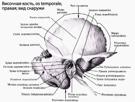
Temporal bone (OS temporale) steam room, is part of the base and side wall of the skull between the wedge-shaped bone of the front and the occipital bone from behind. It accommodates hearing and equilibrium organs. The composition of temporal bones distinguish the pyramid, drum and scaly parts.
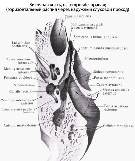
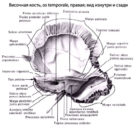
Pyramid, or stony part (Pars Petrosa), has a triangular shape, is located as a horizontal plane. The top of the pyramid is directed forward and medially, and the base is back and laterally. At the top of the pyramid is the inner hole of the sleepy channel (Canalis Caroticus). Nearby and laterally there is a muscular-tube channel (Canalis Musculotubarius), which partition is divided into two half-cups: half-chained the hearing pipe (Semicanalis Tubae Auditivae) and a semicanalis muscle straining (Semicanalis Musculi Tensoris Tympani).
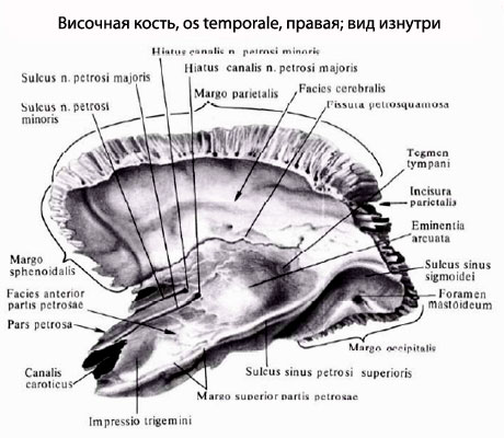
The pyramids allocate three surfaces: front, rear and lower. Front surfacethe pyramids are drawn up and forth. Near the top on this surface there is a small trigemini (Impressio Trigemini). Two holes are noticeable laterally. More than they are called a split (HIATUS CANALIS NERVI PETROSI MAJORIS), from which a narrow single-name groove is media. Kepened and lancerly located the cleft of a small rocky nerve (Hiatus Canalis Nervi Petrosi Minoris), turning into the furrow of this nerve. On the front surface of the pyramid there is a flattened section - the roof of the drum cavity (Tegmen Thympani), which is top wall. Along the upper edge of the pyramid is the groove of the top stony sinus (Sulcus Sinus Petrosi Superioris).
![]()
Rear surface of the pyramidframes for the post and medial. In the middle of this surface there is an internal auditory (Porus Acusticus Internus). It leads to an internal hearing pass (Medtus Acusticus Internus). Lateral and somewhat higher than this hole is a subdug fossa (FOSSA subarcuata), below and the laterally there is a little noticeable outdoor aperture (hole) of the runway (Apertura Externa Aqueductus Vestibuli). Along the rear edge of the pyramid passes the groove of the lower stony sinus (Sulcus Sinus Petrosi Inferioris). At the lateral end of this groove, next door to the jugular hole, there is a deepening, at the bottom of which the outer aperture of the Chanalculi Cochleae operates (Apertura Externa Canaliculi Cochleae).
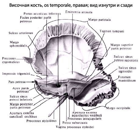
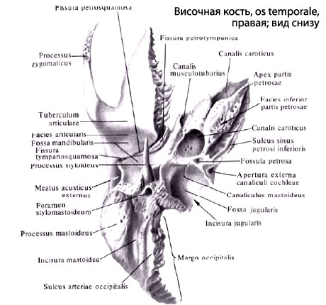
Lower surface of the pyramidit has a difficult relief. Next to the base of the pyramid is a deep jugular yam (Fossa Jugularis). Kepened from it is the rounded outdoor hole of the sleepy channel, inside of which, in its wall, there are 2-3 holes of the sleepy-drum tubuine connecting the sleepy channel with the drum cavity. On the scallop between the jugular straw and the outer hole of the sleepy canal there is a small lobe (Fossula Petrosa). The lateral and long-shaped ellone (Processus Styloideus) is directed laterally. Behind the process is a velocity vehicle (Formen Stylomastoideum), and behind this hole is directed down wide, easily tested through the skin of a mastoid process (Processus Mastoideus).

In the thick of the mining process there are air filled cells. The most coupling cell is a cottage cave (Antrum Mastoideum) communicates with the drum cavity. MEDIAL PREVICE PROCESS is limited to deep-like mastoidea (Incisure Mastoidea). MEDIALLY THIS CLEANS Located a groove of the occipital artery (Sulcus Ariae Occipitalis). At the base of the maternity process, there is sometimes a mining hole (Foramen Mastoideum).
The drum part (Pars tympanica) is formed by a curved narrow bone plate, which in front, from the bottom and rear limits the outer auditory (Porus Acusticus externus) leading to the outer hearing pass (Meatus Acusticus externus). Between the drum and the maternity process, there is a narrow drum-bed-like slot (Fissure Tympanomastoidea). Ahead of the outdoor auditory hole is the drum-scaled gap (Fissure Tympanosquamosa). In this slit, from the inside there is a narrow bone plate - the edge of the drum cavity. As a result, the drum-scaled gap is divided into the lying Khanged-Fissura Petrosquamosa (Fissura PetrotyMpanica, the Glut Glider), through which the branch of the facial nerve comes from the drum cavity - the drum string.
The scaly part (Pars Squamosa) is a convex duck with a plate having a bevelled free top edge for connecting with a dark bone and a large wing of a wedge-shaped bone. The outer temporal surface of the scales is smooth. On the inner cerebral surface of the scales there are brain elevations, finger-shaped pressure and arterial furrows. From scales, above and kaperi from an outdoor auditory passage, the process zygomaticus begins (Processus Zygomaticus). Connecting with the temporal process of zilly bone, it forms a zilly arc. Behind the zhilagogo process, at its base, there is a mandibular yam (Fossa MandiBularis) for articulation with a tiny process lower jaw For the formation of the temporomandibular joint.
Channels of temporal bone. Through the pyramid passes several channels of temporal bone for cranial nerves and blood vessels.
The canalis Cardticus sleep canal) begins on the lower surface of the pyramid with an outdoor sleepy hole, goes up, bends almost at right angles, then directly and forward. The channel ends with the inner sleepy hole at the top of the pyramid of the temporal bone. Through this channel in the cavity of the skull pass internal carotid artery and the nerves of the sleeping plexus.

Sleepy drum tubules (Canaliculi CaroticotyMpanic!), Number 2-3, depart from the sleepy canal and are sent to the drum cavity. Artery and nerves are located in these tubules.
The muscular-tube canal (Canalis Musculotubarius) begins on the top of the pyramid of the temporal bone, goes back and laterally and opens into the drum cavity. The horizontal partition divides it into two parts. Above is the half-channel muscle, straining eardrum (Semicanalis Musculi Tensoris Tympani) containing the muscle of the same name. Below is a hearing pipe (Semicanalis Tubae Auditivae).
The front channel (Canalis Facialis) begins in the inner hearing aisle. It comes first across in relation to the long axis of the pyramid to the level of cleft channel of a large stony nerve. Having reached the cleft, the channel forms the knee, then heads at right angles back and laterally. Having passed along the medial wall of the drum cavity, the channel rotates vertically down and ends with a veil vehicle. In this channel passes the face nerve.
Chanaliculus Chordae Tympani (Canaliculus Chordae Tympani) comes from the wall of the facial channel in the final department and opens into the drum cavity. In this channel passes the nerve - drum string.
Drum channel (Canaliculus Tympanicus) begins on the bottom of the rocky snaps, goes up, breaks the wall of the drum cavity. Next, the channel passes through its medial wall and ends in the area of \u200b\u200bthe cleft channel of a small stony nerve. The drum nerve passes in this canal.
CANALICULUS MASTOIDEUS (Canaliculus Mastoideus) begins in a yammer and ends in a drum-aparticle. In this canal, the ear branch of the vagus nerve is held.
Temple Bone, OS TemporaleThe steam bone, has a complex structure, since it performs all 3 functions of the skeleton and not only forms a part of the side wall and the base of the skull, but also contains hearing and gravity organs. It is a product of merging several bones (mixed bone), self-existing in some animals, and therefore consists of three parts:
- scaly Part, Pars Squamosa;
- drum part, Pars Tympanica and
- stony part, Pars Petrosa.
During the 1st year of life, they merge into a single bone, a closing outer hearing pass, Meatus Acusticus externus, in such a way that the scaly part lies above it, the stony part of Knuts from him, and the drum back, from the bottom and in front. Footprints separate parts The temporal bones are preserved for life in the form of intermediate seams and cracks, namely: on the border of Pars Squamosa and Pars Petrosa, on the front surface of the last - Fissura Petrosquamosa; In the depths of the mandibular fossa - Fissura Tympanosquamosa, which is divided by the process of the stony part on Fissura Petrosquamosa and Fissura PetrotyMpanica (through it the nerve Chorda Tympani).

Scaly Part, Pars Squamosa, participates in the formation of the side walls of the skull. It belongs to the coating bones, that is, it fables on the basis of the connective tissue and has a relatively simple structure in the form of a vertically standing plate with a rounded edge, superimposed on the respective edge of the dread bone, Margo Squamosa, in the form of fish scales, from where its name happened.
On the brain Surface of Her Facies Cerebralis, marrow traces are noticeable, finger pressure, Impressiones Digitatae, and ascending booster from a. Meningea Media. The outer surface of the scales is smooth, participates in the formation of temporal pits and therefore called Facies Temporalis. From it, there is a zygomatic process, Processus Zygomaticus, which goes ahead to the connection with the zilly bone. At its beginning, the zyloma process has two roots: the front and rear, between which there is a hole for articulation with the lower jaw, Fossa MandiBularis.
On the lower surface of the front root is placed on the articular tubercle, Tuberculum Articulare, which prevents the dislocation of the head of the lower jaw forward with a significant opening of the mouth.
Drum part, Pars Tympanica, temporal bone Forms the front, the lower and part of the rear edge of the external auditory passage, turns the endless and, like all the coating bones, has the form of a plate, only sharply curved. An external hearing pass, Meatus Acusticus externus, is a short channel heading inside and ahead and leading to the drum cavity. The top edge of its outdoor opening, POMS Acusticus externus, and a portion of the rear edge are formed with scales of temporal bones, and on the rest of the way the drum part.
The newborn, the outer hearing pass is not yet formed, since the drum portion is an incomplete ring (Annulus Tympanicus), tightened by the drumpot. Due to such a close location of the drumpoint of the dapper in newborns and children early age More often observed diseases of the drum cavity. The stony part, Pars Petrosa, is named so for the strength of its bone substance, due to the fact that this part of the bone is involved in the base of the skull, and is a bone container of hearing and gravity organs that have a very subtle structure and in need of strong protection against damage. It develops on the basis of cartilage. The second name of this part is the pyramid, given by its form of a three-grained pyramid, the base of which is drawn by the dust, and the top - forward and inside to the wedge-shaped bone.
The pyramid has three surfaces:front, rear and bottom. The front surface is part of the bottom of the middle skull; The rear surface is drawn back and media and forms a part of the front wall of the rear cranial fossa; The lower surface is drawn down and is visible only on the outer surface of the base of the skull. The external relief of the pyramid is complicated and due to the structure of it as extension for the average (drum cavity) and inner ear (A bone maze consisting of snail and semicircular channels), as well as the passage of nerves and vessels. On the front surface of the pyramid, near its top, noticeably a small pressure, Impressio Trigemini, from the trigeminal nerve unit (N. Trigemini). The duck from it is two thin grooves, medial - sulcus n. Petrbsi Majoris, and lateral - Sulcus N. Petrosi minoris. They lead to two co-holes: medial, HIATUS Canalis N. Petrosi Majoris, and lateral, Hiatus Canalis N. Petrosi Minoris. The duck from these holes is noticeable arcuate elevation, Eminentia Arcuata, sampled due to the absorption of a rapidly developing maze, in particular the upper semicircular channel.
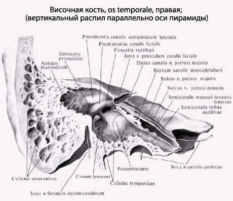
The surface of the bone between Eminentia Arcuata and Squama Temporalis forms the roof of the drum cavity, Tegmen Tympani. Approximately in the middle of the rear surface of the pyramid there is an inner hearing aid, Porus Acusticus Internus, which leads to an internal hearing pass, Meatus Acusticus Internus, where the facial and hearing nerves pass, as well as the arteries and veins of the maze. From the lower surface of the pyramid facing the base of the skull, a thin pointed cylinder process, Processus Styloideus, which serves as an anatomical bouquet muscle attachment site (MM. Styloglossus, Stylohyoideus, Stylopharyngeus), as well as ligaments - Ligg. Stylohyoideum and StylomandiBular. The breadless process represents a part of the temporal bone of gill origin. Together with Lig. Stylohyoideum He is the residue of the sub-speaking arc. Between the host and maternity process is a velocity vehicle, the ForaMen Stylomastoideum, through which n. Facialis and includes small artery. Mediality from a host-shaped process is a deep jugular fossa, Fossa Jugularis. Kepened from Fossa Jugulalis, separated from it with a sharp ridge, is the outer hole of the sleepy canal, ForaMen Caroticum Externum.

The pyramid has three edges: front, rear and top. The short front edge forms a sharp corner with scales. In this corner, the hole of the muscular tube canal, Canalis Musculotubarius leading to the drum cavity. The channel is divided into two departments: upper and lower. Upper, smaller, half-cup, semicanalis m. Tensoris Tympani, accommodates this muscle, and the lower, greater, Semicanalis Tubae Auditivae is a bone part of the hearing pipe that serves for air from the pharynx to the drum cavity. By upper edge The pyramids separating the front and rear surfaces passes a well-visible groove, Sulcus Sinus Petrosi Superiors, - the trail of the venous sinus of the same name. The rear edge of the Pyramid Kepende from Fossa Jugularis connects with the basilar part of the occipital bone and forms with this bone Sulcus Sinus Petrosi Inferioris - the trail of the lower rocky venous sinus.

The outer surface of the base of the pyramid is the place of attachment of the muscles than and is due to its outdoor relief (process, cutting, roughness). The book she is pulling into a mastoid process, Processus Mastoideus. It is attached to the breast-curable-bed-like muscle, which supports the head in the equilibrium, which is necessary in the vertical position of the body. Therefore, the maternity process is absent from four-legged and even man-like monkeys and develops only in a person due to its shine. On the medial side of the mastoid proof, there is a deep mastoid clipping, Incisura Mastoidea, - the place of attachment M. DigaStricus; Even more knutrice is a small groove, Sulcus a. Occipitalis, - trail of the artery of the same name. On the outer surface of the base of the mastoid process, a smooth triangle is distinguished, which is a place for prompt access to the cells of the mastoid process when filling them in gently.
Inside the mastoid process and contains these cellulae mastoideae cells, which are aircraft separated by bone crossbars that receive air from the drum cavity with which they communicate through Antrum Mastoideum. On the brain surface of the base of the pyramid passes a deep furrow, Sulcus Sinus Sigmoidei, where the venous sinus is the same name. Channels of temporal bone. The largest channel is Canalis Caroticus, through which internal carotid artery passes. Beginning by its outer opening on the lower surface of the pyramid, it rises upwards, then bent at right angles and opens with its inner hole at the top of the pyramid medial from Canalis Musculotubarius.
The front channel, Canalis Facialis, begins in the depths of Porus Acusticus Internus, from where the channel first goes ahead and laterally to the slots (HIATUS) on the front surface of the pyramid; These holes canal, remaining horizontal, turns at the right angle laterally and backward, forming the bend - the knee, geniculum canalis facialis, and then down and ends with the Formen Stylomastoideum, located on the lower surface of the temporal bone pyramid.
What doctors to apply for inspection of temporal bone:
Traumatologist
What diseases are associated with temporal bone:
What tests and diagnostics need to be held for temporal bones:
CT skull
MRI Telep
X-ray tight
Does something bothers you? Do you want to know more detailed information about the temporal bone or do you need an inspection? You can make an appointment to the doctor - Clinic Euro.lab always at your service! Top doctors will consider you, they will advise required help And make a diagnosis. you also can call a doctor. Clinic Euro.lab Opened for you around the clock.
How to contact the clinic:
Phone of our clinic in Kiev: (+38 044) 206-20-00 (multichannel). The secretary of the clinic will select you a convenient day and an hour of visiting to the doctor. Our coordinates and travel scheme are indicated. Look more detailed about all clinic services on it.
If you have previously been performed any research, be sure to take their results for a consultation to the doctor. If the studies have not been fulfilled, we will do everything you need in our clinic or our colleagues in other clinics.
It is necessary to carefully approach your health as a whole. There are many diseases that at the beginning do not show themselves in our body, but in the end it turns out that, unfortunately, they are already treated too late. For this you just need several times a year take a survey from a doctorso as not only to prevent terrible disease, but also support healthy Spirit In the body and the body as a whole.
If you want to ask a question to a doctor - use the online consultation section, you may find answers to your questions and read tips for care. If you are interested in reviews about clinics and doctors - try finding the information you need. Also register on medical portal Euro.labto be constantly in the know latest news and updates of information about temporal bone on the site that will be automatically sent to you by mail.
Others anatomical terms On the letter "B":
| Upper esophageal sphincter |
| Progress Golden |
| Vagina |
| Hair |