Temporal bone. The temporal bone pyramid. Temporal bone pyramid elements
Name
Anatomy
Traces of the brain in the form of impressions (impressiones digitatae) are visible on the cerebral surface. The zygomatic process (processus zygomaticus) departs from it, which is directed forward at the connection with the zygomatic bone. In the lower part there is a glenoid fossa for articulation with the lower jaw (fossa mandibularis).
The tympanic part (pars tympanica) is fused with the mastoid process (processus mastoideus) and the scaly part (pars squamosa), it is a thin plate that limits the front, back and bottom of the external auditory opening (porus acusticus externus) and the external auditory canal (meatus acusticus externus) ...
The stony part (pars petrosa) has the shape of a three-sided pyramid, the apex of which faces anteriorly and medially, and the base, passing into the mastoid process (processus mastoideus), to the posterior and lateral.
There are three surfaces: front, back and bottom, as well as three edges: front, back and top.
The front surface (facies anterior) is part of the bottom of the middle cranial fossa; the posterior (facies posterior) is directed backward and medially, forms part of the anterior wall of the posterior cranial fossa; the lower (facies inferior) faces downward and is visible only on the outer surface of the skull base.
The external relief of the pyramid is due to its structure as a receptacle for the middle and inner ear, as well as for the passage of blood vessels and nerves.
A thin pointed styloid process (processus styloideus) departs from the lower surface of the pyramid, serving as a place of muscle attachment. The relief of the outer surface of the pyramid is the place of attachment of the muscles; downwards, it is pulled into the mastoid process, to which the sternocleidomastoid muscle is attached.
On the mastoid process (on its anterior smooth surface) of the temporal bone, the Shipo triangle is distinguished, which is the place of operative access to the cells of the mastoid process. On the radiograph of the temporal bones, the so-called synodural angle (Chitelli angle) is distinguished. Inside, the mastoid process contains cells (cellulae mastoideae), which are air cavities that communicate with the tympanic cavity (middle ear) through the mastoid cave (antrum mastoideum).
Temporal bone connected to the occipital, parietal and sphenoid bones. Participates in the formation of the jugular foramen.
Temporal bone canals
- sleepy canal, canalis caroticus, in which the internal carotid artery lies. It begins on the lower surface of the pyramid, with an external carotid opening (foramen caroticum externum), directed vertically upward, bending at a right angle, directed forward and medially. The canal opens into the cranial cavity with an internal sleepy opening (foramen caroticum internum).
- drum string tubule, canaliculus chordae tympani, starts from the canal of the facial nerve, slightly above the styloid foramen (foramen stylomastoideum), goes forward and opens into the tympanic cavity. In this tubule, a branch of the facial nerve passes - the tympanic string, which then exits the tympanic cavity through the stony tympanic fissure (fissura petrotympanica).
- facial canal, canalis facialis, in which the facial nerve passes, it begins at the bottom of the internal auditory canal, then goes horizontally from back to front. Having reached the level of the cleft of the canal of the large stony nerve, the canal goes back and laterally, at a right angle, forming a bend, or knee of the facial canal. Further, the channel is directed backward, follows horizontally along the axis of the pyramid. Then it turns vertically downward, bending around the tympanic cavity, and ends with a styloid opening on the lower surface of the pyramid.
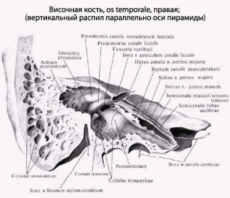
- musculocutaneous canal, canalis musculotubaris, has a common wall with the carotid canal. It starts in the corner formed by the anterior edge of the pyramid and the scales of the temporal bone, goes posteriorly and laterally, parallel to the anterior edge of the pyramid. The musculocutaneous canal is divided by a longitudinal horizontal partition into two semichannels. The upper semi-canal is occupied by a muscle that strains the eardrum, and the lower one is the bony part of the auditory tube. Both channels open into the tympanic cavity on its front wall.
- mastoid tubule, canaliculus mastoideus, originates at the bottom of the jugular fossa and ends in the tympanic-mastoid fissure. A branch of the vagus nerve passes through this tubule.
- tympanic tubule, canaliculus tympanicus, arises in a stony dimple (fossula petrosa) with an opening through which a branch of the glossopharyngeal nerve enters - the tympanic nerve. Having passed through the tympanic cavity, this nerve, called the lesser petrous nerve, exits through the cleft of the same name on the anterior surface of the pyramid.
- carotid tubules, canaliculi caroticotympanici, pass in the canal wall of the internal carotid artery near its external opening and open into the tympanic cavity. They serve for the passage of the vessels and nerves of the same name.
- vestibule water supply, aqueductus vestibuli, a canal in the pyramid of the temporal bone, connecting the vestibule of the bone labyrinth (an expanded part of the bone labyrinth between the cochlea inner ear and bony semicircular canals) with the cranial cavity (posterior cranial fossa). It opens with a slit on the posterior surface of the temporal bone pyramid, behind the opening of the internal auditory canal. In the canal, a vein of the aqueduct of the vestibule and ductus endolymphaticus passes, which ends in a blind sac (saccus endolymphaticus), on the posterior surface of the temporal bone pyramid, between the opening of the internal auditory canal and the sigmoid sinus.
- snail plumbing, aqueductus cochleae, about 10 mm long, connects the vestibule of the inner ear and the posterior surface of the temporal bone pyramid, opening at its lower edge, below the opening of the internal auditory canal. Its inner opening is located at the beginning of the cochlear drum ladder. The vein of the cochlear tubule passes through the canal.
Temporal bone, os temporale (Fig. 75 - 85), a steam room, is very complex in structure, since the organs of hearing and balance are enclosed in its thickness, and, in addition, bone permeated with a number of channels through which blood vessels and nerves pass. Temporal bone located in the lateral parts of the skull between the occipital, parietal and sphenoid bones, supplementing with one part the cranial vault, the other with the base of the skull. Temporal bone connected to the facial skull: with the help of the joint - with the lower jaw, and the suture - with the zygomatic bone.
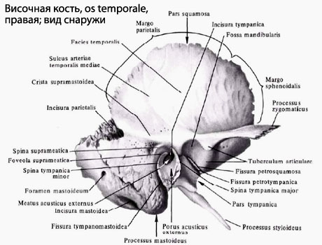
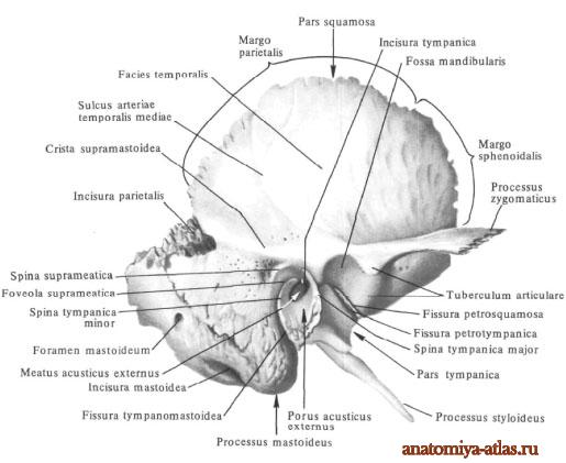
Temporal bone represents several accrete parts. When examining the temporal bone from the side of the external, temporal surface, at its lower edge, there is a large opening, which is called the external auditory opening, porus acusticus externus (Fig. 75, 79). The opening is surrounded by four component parts of the temporal bone: above and in front - a flat, with a pointed edge, the scales of the temporal bone, squama temporalis, in front and below - a small, in the form of a groove, the plate - the tympanic part, pars tympanica, behind - a powerful bone protrusion - the mastoid part , pars mastoidea, from the inside - in the form of a pyramid, tapering in the direction from the mastoid part obliquely inward and anteriorly - a stony part or pyramid, pars petrosa s. pyramis. The scales of the temporal bone, squama temporalis, has the form of a semicircular bone plate, facing its smooth temporal surface, fades temporalis, outward and the inner, cerebral surface, fades cerebralis, into the cranial cavity. The semicircular-shaped edge bounding the scales is not the same everywhere; the anterior and posterior portions of the edge are more serrated and less sharpened from the inside than the upper portion. The anterior edge is connected to the scaly edge of the large wing of the main bone and is called the main edge, margo sphenoidalis; the upper posterior edge, connecting with the scaly edge of the parietal bone, is called the parietal edge, margo parietalis. The posterior-lower part of the scales passes into the mastoid part.
In children, at the junction of these parts, there is an obliquely directed scaly-mastoid suture, sutura squamomastoidea, directed from top to bottom and anteriorly. Remnants of this suture are sometimes preserved in adults (Fig. 75). Somewhat higher and along it goes temporal line whose front end approaches the root zygomatic process temporal bone, processus zygomaticus ossis temporalis. The zygomatic process departs with two roots: posterior and anterior. It runs horizontally, first outward and then at an angle anteriorly, and ends with a serrated end. The latter, it connects to the temporal process of the zygomatic bone, forming with it a zygomatic arch, arcus zygomaticus. Below the zygomatic process and in front of the external auditory foramen, the glenoid fossa is located lower jaw, fossa mandibularis. In the anterior sections, the fossa is limited by a clearly visible articular tubercle, tuberculum articulare; in the back - smaller, behind - the articular process, processus retroarticularis. The anterior part of the fossa and the articular tubercle are covered with cartilage. In the posterior part of the outer surface, fades temporalis, the scales of the temporal bone bear the groove of the middle temporal artery, sulcus arteriae temporalis mediae. This furrow rises upward and branches in the upper segment of the scales.
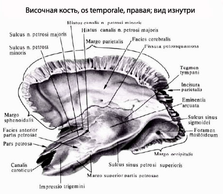
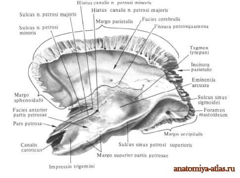
The cerebral surface, fades cerebralis, the bones are somewhat concave, has a well-defined, deep arterial groove in the anterior section, sulcus arteriosus (meningeus) (the place where the meningeal artery of the brain fits), traces of the depression of the brain convolutions - digital impressions, impressiones digitatae, and between the latter protrusions - cerebral eminences, juga cerebralia. Rocky part or pyramid, parspetrosa s. pyramis, has the appearance of a three-sided pyramid in a supine position, so that its base, basis pyramidis, is directed outward and connects with the mastoid and scaly parts of the temporal bone. At the place where the base of the pyramid adjoins to the scaly part in childhood there is a gap, flssura petrosquamosa (Fig. 83a), over the years it is filled with bone tissue, and thus the border between these two parts disappears (Fig. 77).
The top of the pyramid has an uneven edge. It is directed forward and inward, towards the lateral surface of the bodies of the sphenoid and occipital bones. The gap remaining between them on the whole skull is called torn hole, foramen lacerum (Fig. 124), filled with fibrous cartilage, fibrocartilago basilaris. Apex opens large sizes internal opening of the carotid canal, foramen caroticum intemum (Fig. 76 - 78). The upper corner of the pyramid, angulus superior pyramidis, protrudes freely into the cranial cavity at the border of the anterior and posterior surfaces of the pyramid, fades anterior and fades posterior pyramidis. The upper stony groove, sulcus petrosus superior, runs along the upper corner of the pyramid, a trace of the venous sinus of the same name. The anterior corner of the pyramid, angulus anterior pyramidis, is located on the border of the anterior and lower surfaces of the pyramid, facies anterior and facies inferior pyramidis. An internal segment of the anterior angle connects to the edge of the large wing of the main bone with the help of cartilage, forming a basic-stony synchondrosis, synchondrosis sphenopetrosa. The outer segment of the anterior angle is connected to the scales of the temporal bone, forming a stony-scaly slit, fissura petrosquamosa (Fig. 77).
Near the medial end of the stony-scaly fissure, in the corner where the anterior corner of the pyramid converges with the anterior edge of the scales, one can see tubal opening, canalis musculotubarius (Fig. 77, 78). The latter, located obliquely outward and backward, is divided by a horizontally standing thin bone plate - the septum of the muscular-tubal canal, septum canalis musculotubarii, into two parts: the upper - the semicanal of the muscle straining the tympanic membrane, semicanalis musculi tensoris tympani, and the lower - the semicanal of the auditory ) pipes, semicanalis tubae auditivae Eustachii (puc. 80). Both half-channels lead to the middle ear cavity. The posterior angle of the pyramid, angulus posterior pyramidis (Fig. 76), is located on the border of its posterior and lower surfaces, facies posterior et facies inferior pyramidis. It is adjacent to the lateral edges of the partes basilaris and lateralis ossis occipitalis. The inner part of the posterior angle adjoins the pars basilaris ossis occipitalis, and here a stony-occipital fissure, fissura petrooccipitalis (Fig. 124), is formed, made by cartilage connecting both bones, - synchondrosis petrooccipitalis. On the cerebral surface of this part of the posterior angle, there is a lower stony groove, sulcus petrosus inferior. The latter, connecting with the groove of the same name on the adjacent part of the occipital bone, is the location of the temporal sinus (sinus petrosus inferior).
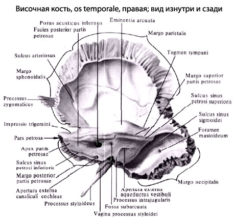
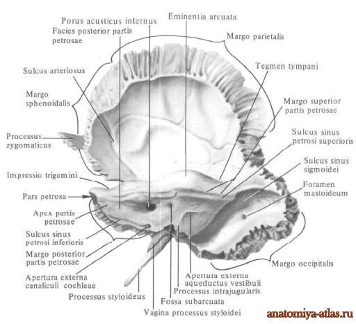
At the outer end of the groove, at the posterior corner of the pyramid, there is a small depression, at the bottom of which a small external opening of the cochlear canal opens, apertura externa canaliculi cochleae (Fig. 76). (Here v. Canaliculi cochleae and ductus perilymphaticus pass from the inner ear cavity). Side part the posterior corner of the pyramid belongs to the pars lateralis ossis occipitalis. There is a small jugular notch, incisurajugularis, which corresponds to the notch of the same name on the occipital bone and together with it on the whole skull forms a jugular foramen, foramen jugulare (Fig. 124).
At the indicated three corners of the pyramid, its three surfaces converge: front, back and bottom. The first two are directed into the cranial cavity, the latter is directed towards the outer surface of the skull base. The anterior surface of the pyramid, fades anterior pyramidis, is uneven, located obliquely anteriorly. Outside, it borders on scales, forming a stony-scaly fissure, fissura petrosquamosa (Fig. 124); from the inside, it borders on the body of the main bone, without reaching it and forming here uneven edge its apex is the ragged hole described above, foramen lacerum. The anterior-lower and posterior-upper boundaries are the corresponding corners or edges of the pyramid. On the front surface of the pyramid, near the apex, there is an impression of the trigeminal nerve, impressio nervi trigemini, - an imprint of the adjacent Gasser node of the trigeminal nerve (ganglion Gasseri).
A semicircular eminence, eminentia arcuata (Fig. 76, 77), is a relief of the upper semicircular canal slightly to the side of the middle of the anterior surface of the pyramid. The area of the anterior surface, located between the elevation and the stony-scaly fissure (fissura etrosquamosa), is the roof of the tympanic cavity, legmen tympani; which is a thin plate that forms the upper wall of the middle ear cavity. Tegmen tympani with its front edge enters the gap between pars tympanica from behind and pars squamosa in front, forming a ridge visible in the fossa mandibularis region, called processus inferior tegmenis tympani (s. Crista tegmcntalis) (see more about this in the description of pars tympanica).
A little inward and downward from eminentia arcuata, two holes are noticeable. One of them is located more medially and is the opening of the facial nerve canal, hiatus canalis facialis (Fig. 77, 80, 81). Through this opening, a branch of the facial nerve emerges - a large stony nerve, nervus petrosus superficialis major, which lies in the corresponding groove - sulcus nervi petrosi superficialis majoris, which runs longitudinally inward and anterior to the hiatus canalis facialis (Fig. 77, 80 - 82).
Another opening is located lateral and is the superior opening of the tympanic tubule, apertura superior canaliculi tympanici. Through this hole, a small stony nerve emerges - nervus petrosus superficialis minor, which lies in the groove of the same name - sulcus nervi petrosi superficialis minoris. This groove, heading inward and anterior to the pyramid, runs parallel and outward from the sulcus nervi petrosi superficialis majoris (Fig. 77, 80, 82). The posterior surface of the pyramid, fades anterior pyramidis, is located more vertically than the anterior, having, however, some slope backward and downward. Inwardly from the upper corner, closer to the middle of the posterior surface, there is a rather wide internal auditory opening, porus acusticus internus (Fig. 76). It opens into a channel leading into the rocky part. This channel is called internal auditory canal, meatus acusticus interims. (For its further course inside the rocky part, see "Ear".)
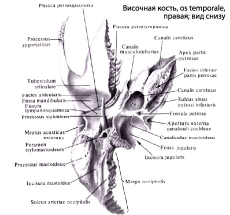
Outward and backward of porus acusticus internus, a small slit-shaped opening is visible, called external opening of the water supply-vestibule, apertura externa aquaeductus vestibuli (Fig. 76, 77), which is the exit site of the internal lymphatic duct, ductus endolymphaticus, from the inner ear cavity. Slightly above the water supply hole, at the upper corner of the pyramid, is sub-semicircular fossa, fossa subarcuata (Fig.83a), clearly visible in individuals young age... The lower surface of the pyramid, fades inferior pyramidis, is directed downward and facing the outer surface of the base of the skull; outside and somewhat in front, this surface is in contact with the tympanic part of the temporal bone. She carries a large number of holes, grooves and protrusions.
The central place on the lower surface of the pyramid is occupied by a large round shape the opening, which is the entrance to the carotid canal, the external opening of the carotid canal, foramen caroticum externum (Fig. 78). (The internal carotid artery and the nerve plexus enter through this hole.) Behind and outside of the foramen caroticum externum, separated from it by a crest, there is a wide jugular fossa, fossa jugularis, reaching the posterior edge of the lower surface of the stony part, where there is a jugular notch, incisura jugularis. It contains the bulb of the jugular vein. At the bottom of the jugular fossa, closer to its front edge, there is a groove of the mastoid canaliculus, sulcus canaliculi mastoidei, ending with the opening of the mastoid canaliculus, canaliculus mastoideus (Fig. 78).
On the ridge separating the fossa jugularis from the foramen caroticum externum, there is a barely noticeable stony dimple, fossula petrosa (Fig. 78), leading to the lower opening of the tympanic tubule, apertura inferior canaliculi tympanici. (Here pass a.tympanica inferior and n. Tympanicus - from the stony node.) At the very base of the pyramid, on the outer part of the lower surface, a styloid process, processus styloideus, which is semicircular in front of the bony sheath, vagina processus styloidei, formed by the tympanic part of the temporal bone.
Near the styloid process, on the border with the mastoid process, processus mastoideus, there is the styloid foramen, foramen stylomastoideum, the exit point of the facial nerve and blood vessels. In the pyramid of the temporal bone there are a number of channels through which the vessels and nerves pass, and the organ of hearing and the organ of balance of the body are laid , which is why the pyramid has such a complex structure. All these formations are visible on special preparations of cuts of the temporal bone, carried out in different directions.
1. Formations related to the structure of the organs of hearing and balance:
but). external auditory opening, porus acusticus externus, and its continuation into the external auditory canal, meatus acusticus externus, are the bony parts of the external ear;
b). tympanic lining, tegmen tympani, is top wall the cavity of the middle ear, where the canalis musculo-tubarius opens, lying on the outer edge of the anterior corner of the pyramid;
in). inner ear cavity(labyrinth) is indicated on the front surface of the pyramid by a semicircular elevation, eminentia arcuata, where the upper semicircular canal fits, and on the posterior surface by a fossa, fossa subarcuata.
Small holes on the back of the pyramid, apertura externa canaliculi cochleae and apertura externa aquaeductus vestibuli, lead to the inner ear; they contain vessels and lymphatic ducts through the porus acusticus internus passing through the auditory and facial nerves.
2. Canal of the facial nerve (fallopian canal), canalis facialis (Falloppii) (Fig. 80 - 82), inside the stony part of the temporal bone. It begins with the opening of the bottom of the internal auditory canal, in the area of its upper recess - the area facialis (see "Ear"), and continues the direction of the internal auditory meatus forward and outward under the anterior surface of the stony part. Here, to the front surface of the pyramid, a branch departs from it, ending in a hole - hiatus canalis facialis; the canal itself, turning outward and backward, forms the knee of the auditory nerve canal, geniculum canalis facialis, at the place of rotation (Fig. 80 - 82).
After the formation of the knee, the canal follows posteriorly and somewhat downward and, reaching the posterior part of the inner wall, cavum tympani, passes into the vertical part. Then it goes down and opens behind the base of the styloid and anterior to the mastoid processes - styloid opening, foramen stylomastoideum (Fig. 80, 81). The upper end of the vertical part of the canal forms the protrusion of the facial nerve canal, prominentia canalis facialis (Fig. 79), located in the posterior part of the medial wall of the inner ear. Slightly lower, the canal of the facial nerve gives a branch of the canadian drum string, canaliculus chordae tympani, through which the nerve passes - the drum string, chorda tympani, and which ends in fissura petrotympanica (Glaseri).
3. The tympanic tubule, canaliculus tympanicus (Fig. 80), passes the branch of the lingopharyngeal nerve. The canaliculus begins with the lower opening of the tympanic canaliculus at the bottom of the petrous fossa, fossula petrosa (from the side of the lower surface of the stony part), and, going arcuately posteriorly, upward and then forward, opens with the upper opening of the tympanic tubule, apertura superior canaliculi tympanici (Fig. 80) (on the front surface of the stony part). Canaliculus tympanicus communicates with canalis nervi facialis Falloppii in the area of his knee. 4. The canal of the carotid artery, canalis caroticus (Fig. 78), short, wide and curved. The internal carotid artery and its venous and nerve plexus pass through it. The channel begins with a hole located on the lower surface of the pyramid - foramen caroticum externum.
Further, the canal rises upwards, then forms a bend almost at a right angle and, going horizontally anteriorly and inside, opens with the internal opening of the carotid canal, foramen caroticum internum. Near the external opening, in the wall of the carotid canal, small openings of the carotid canaliculi open, canaliculi curaticotympanici. These short tubules go to the anterior wall of the cavum tympani, bypassing the wall of the carotid canal from above. Opening in the anterior wall of the cavum tympani, they pass the branches of the internal carotid artery and the superior and inferior carotid tympanic nerves.
Mastoid part, pars mastoidea (Fig. 75 - 83), located posterior to the external auditory canal. Outside, it smoothly turns into scales, and from the inside - into a rocky part. Downward, the mastoid part faces a free convex surface, posteriorly and outwardly - a rough surface. The posterior, occipital edge, margo occipitalis, is connected to the mastoid edge of the occipital bone, forming the occipital-mastoid suture, sutura occipitomastoidea (Fig. 123, 124).
Top edge, together with the posterior part of the parietal edge of the scales, forms the parietal notch, incisura parietalis. This notch is performed by the mastoid angle of the parietal bone, angulus mastoideus, which is connected to the mastoid part with the help of the mastoid-parietal suture, suturaparietomastoidea. In front, in the upper part, the mastoid part passes into scales, in the lower part it borders on the tympanic part, forming a drum-mastoid fissure with it, fissura tympanomastoidea. In the anterior section, which constitutes the upper-posterior part of the edge of the external auditory foramen, there is a small protrusion - the access spine, spina suprameatum, and near it posteriorly - the mastoid fossa, fossa mastoidea.
The rough antero-lower part of the outer surface and ends with a blunt and powerful mastoid process, processus mastoideus, which is directed obliquely anteriorly and downward and is easily felt through the skin, in adults it varies, the degree of its development in children of the first years of life is poorly expressed (Fig. 83 ). In the posterior-lower part of the outer surface of the process there is a mastoid opening, foramen mastoideum (Fig. 75, 76), belonging to the group of graduation openings, emissaria Santorini; it penetrates through the entire thickness of the bone and opens on the inner surface of the mastoid process. This hole is variable in size and position: sometimes it is one and located in the area of the sutura squamomastoidea, sometimes there are several of them.
WITH outside and below the mastoid process carries a deep mastoid notch, incisura mastoidea, - the place where the digastric muscle begins (m. digastricus). The groove of the occipital artery, sulcus arteriaeoccipitalis, runs medially and parallel to the notch (Fig. 78). On the inner, cerebral, surface of the mastoid there is an S-shaped groove, sulcus sigmoideus, - the place of occurrence of the venous sinus of the same name - sinus sigmoideus. Very often, the inlet of the above-mentioned foramen mastoideum opens into the same groove. Processus mastoideus belongs to the group of pneumatic bones. As can be seen from the figures (Fig. 79 - 82), depicting the cut of the mastoid process, it contains a large number of communicating cells, cellulae mastoideae, lined with a mucous membrane. The cells are filled with air coming here from the middle ear cavity. In the anteroposterior corner, inside the mastoid process, there is a large cell called the cave of the tympanic cavity, antrum tympanicum (Fig. 79 - 82, 85), communicating, on the one hand, with the middle ear cavity, and on the other, with the cells of the mastoid process ...
The number and size of cells may vary from individual to individual. The tympanic part, pars tympanica (Fig. 75), is laid in the period embryonic development in the form of a horseshoe-shaped half-ring - a tympanic ring, annuhis tympanicus (Fig. 83, 84, 84a), which forms the lower periphery of the external auditory canal. The ends of the semicircle: the anterior, large tympanic spine, spina tympanica major, and the posterior, minor tympanic spine, spina tympanica minor, limit the gap, called the tympanic notch, incisura tympanica (Rivini) (Fig. 84, 84a), above which (above both spines ) overhangs the lower edge of the scaly part of the temporal bone, thus closing the semicircle from above. The tympanic groove, sulcus tympanicus, is the site of attachment of the tympanic membrane along the circumference of the inner surface of the ring.
On the inner surface of spina tympanica major there is an obliquely passing spinous scallop, crista spinarum, the sharp ends of which are called: anterior - processus tympanicus anterior, and posterior - processus tympanicus posterior. A groove, sulcus mallei, runs along the ridge and below it. Due to the growth of bone substance from the outer surface of the semicircle, the latter takes the form of a grooved plate, which on the temporal bone of an adult forms the anterior, lower and part back wall external auditory opening, porus acusticus externus, and external auditory canal, meatus acusticus externus. With the lengthening of the bone groove of the tympanic part, the external auditory canal also lengthens with age: thus, the tympanic membrane, which lies more superficially in children, due to this, goes into depth.
The upper front edge of the tympanic part is separated from the scaly part over a large extent by the anterior edge of the stony part wedging between them - the lower process of the roof of the tympanic cavity, processus inferior tegmenis tympani (s. Crista tegmentalis) (Fig. 79). Between this process in front and pars tympanica behind, a stony-tympanic gap, fissura petrotympanica (Glaseri), is formed, through which they pass small vessels and the nerve is the drum string, chorda tympani. Between the process behind and the pars squamosa in front, another gap is formed - stony-scaly, fissura petrosquamosa, made by connective tissue.
The posterior lower edge of the tympanic part borders on the mastoid part of the temporal bone, forming at the point of contact a tympanic-mastoid fissure, fissura tympanomastoidea (Fig. 75), in the depth of which the outlet of the mastoid canaliculus, canaliculus mastoideus, starting in the fossa jugularis, opens. The edge is pointed and elongated downward in the form of a ridge, crista peirosa, part of which is most developed at the base of the processus styloideus, is called the styloid sheath, vagina processus styloidei (Fig. 76, 78). The lower surface of the tympanic part and the fossa at the root of the zygomatic process of the scaly part form the articular fossa of the lower jaw, fossa mandibularis, at the bottom of which are fissura petrotympanica (Glaseri) and fissura petrosquamosa. This fossa is divided by the glazed slit into two parts - anterior and posterior.
The front part, lined with articular cartilage, faces the cavity mandibular joint, she is called inside- or intracapsular part, pars intracapsularis; back - located outside the joint and is called outside- or extracapsular part, pars extracapsularis (see "Mandibular joint").
Temporal bone contains the organ of hearing and balance, serves as a support for the base of the skull and chewing apparatus. It consists of five parts - scaly, mastoid (mastoid). tympanic (tympanic), stony part and subulate complex. The base of the temporal bone is a pyramid, which has an apex directed towards the sphenoid bone, three faces and a base facing the mastoid process.
Upper inner face of the pyramid supports the middle cranial fossa. The cranial fossa itself is bounded in front by the small wings of the main bone, behind by the pyramid and partially by the back of the sella turcica. The main elements of the middle cranial fossa are the temporal lobes of the brain, the pituitary gland and the cavernous plexus.
Through a number of holes is carried out the connection between the middle cranial fossa, the pyramid and the cellular spaces of the face and neck. One of these openings is the optic nerve canal, where the optic nerve and the optic artery pass. Further, this is the superior orbital fissure, followed by the oculomotor, block and abducens nerves, as well as the ocular branch of the trigeminal nerve and the optic veins. The maxillary branch of the trigeminal nerve passes through the round opening; through the opening, the middle cranial fossa is connected to the pterygopalatine fossa. The carotid opening houses the canal of the internal carotid artery and the sympathetic carotid plexus. Through this hole, communication with the cellular tissue of the neck is carried out.
In the oval hole the mandibular branch of the trigeminal nerve passes through the opening, communication with the inter-pterygoid space is possible. Through the spinous opening, where the middle meningeal (meningeal) artery follows, communication with the temporo-pterygoid space is carried out.
TO upper inner face of the pyramid large nerves are related: oculomotor, block, trigeminal and abducens. On the upper part of the inner face of the pyramid, two anatomical elevations can be found. One eminence is formed by a gasser's node (trigeminal nerve node), the other by the superior semicircular canal. On the upper edge of the pyramid there are two cracks, in which stony nerves are located.
Back inner face of the pyramid creates support for the posterior cranial fossa. The posterior cranial fossa in front is formed by the pyramid of the temporal bone, in the back - by the cruciate elevation of the occipital bone. The main structures of the posterior fossa are the cerebellum, pons varoli and medulla oblongata.
Connection of the posterior cranial fossa with pyramid as well as the fiber of the face and neck can be carried through a series of holes.
Through large occipital foramen(in it pass: the medulla oblongata, accessory nerve, vertebral artery and spinal nerve) there is a communication with the spinal canal.
Through the jugular, a hole (through it follow: the internal jugular vein, the posterior sheath (meningeal) artery, glossopharyngeal, vagus and accessory nerves), anatomical contacts with the neck tissue are possible.
Through the hypoglossal nerve canal there is a communication with the tissue of the submandibular fossa. Through the emissary of the mastoid veins, the posterior cranial fossa communicates with the veins of the diploe, the veins of the integument of the skull and with the sigmoid sinus.
To the back of the pyramid the large cranial nerves are related: the branch of the trigeminal nerve, the facial nerve, the vestibular cochlear nerve, the glossopharyngeal nerve, the vagus. accessory, hypoglossal and intermediate nerves. Three sinuses run along the inner surface of the back face of the pyramid. By top edge the posterior inner face of the pyramid runs the upper stony sinus, along the lower surface of the pyramid - the lower stony sinus. They carry venous blood to the sigmoid sinus.
On the inner surface of the mastoid process there is a deep groove of the sigmoid sinus. The sigmoid sinus itself is located between the mastoid process and the cerebellum.
Transverse sine flows into the upper knee of the sigmoid sinus. The lower knee of the sigmoid sinus turns anteriorly and inward and passes into the bulb of the internal jugular vein, located under the bottom of the tympanic cavity. The sigmoid sinus directs its blood to the internal jugular vein.
On the the back inner face of the pyramid three main holes can be seen. This opening of the internal auditory canal (porus acusticus internus) with a diameter of 4-5 mm, behind it, at a distance of 5-6 mm horizontally, is the opening of the external aperture of the water supply of the vestibule. Downward from the opening of the internal auditory canal at a distance of 5-6 mm on the lower edge of the pyramid, the external aperture of the cochlear tubule (the aperture of the cochlear water supply) opens.
Table of contents of the subject "Hearing organ.":1. Pyramid of the temporal bone. Elements of the temporal bone pyramid.
Temporal bone, os temporale, a paired bone, has a complex structure, since it performs all 3 functions of the skeleton and not only forms part of the lateral wall and base of the skull, but also contains the organs of hearing and gravity. It is the product of the fusion of several bones (mixed bone) that exist independently in some animals, and therefore consists of three parts:
- scaly part, pars squamosa;
- drum part, pars tympanica and
- rocky part, pars petrosa.
During the 1st year of life, they merge into a single bone, closing the external auditory meatus, meatus acusticus externus, in such a way that the scaly part lies above it, the stony part is inward from it, and the tympanic part is behind, below and in front. Merge traces separate parts the temporal bone is preserved for life in the form of intermediate sutures and cracks, namely: on the border of pars squamosa and pars petrosa, on the anteroposterior surface of the latter - fissura petrosquamosa; deep in the mandibular fossa - fissura tympanosquamosa, which is divided by the process of the stony part into fissura petrosquamosa and fissura petrotympanica (the chorda tympani nerve leaves through it).
Scaly part, pars squamosa, participates in the formation of the lateral walls of the skull. It belongs to the integumentary bones, that is, it ossifies on the soil of connective tissue and has a relatively simple structure in the form of an upright plate with a rounded edge superimposed on the corresponding edge of the parietal bone, margo squamosa, in the form of fish scales, hence its name.
On the its cerebral surface, facies cerebralis, traces of the brain, digital impressions, impressiones digitatae, and an ascending groove from a. meningea media. The outer surface of the scales is smooth, participates in the formation of the temporal fossa and is therefore called facies temporalis. The zygomatic process, processus zygomaticus, departs from it, which goes forward to connect with the zygomatic bone. At its beginning, the zygomatic process has two roots: anterior and posterior, between which there is a fossa for articulation with the lower jaw, fossa mandibularis.
An articular tubercle, tuberculum articulare, is placed on the lower surface of the anterior root, which prevents the dislocation of the head of the lower jaw forward with a significant opening of the mouth.
Tympanic part, pars tympanica, temporal bone forms the anterior, lower and part of the posterior edge of the external auditory canal, ossifies endesmally and, like all integumentary bones, has the appearance of a plate, only sharply curved. The external auditory canal, meatus acusticus externus, is a short canal that goes inward and somewhat forward and leads into the tympanic cavity. The upper edge of its outer opening, poms acusticus externus, and part of the posterior edge are formed by the scales of the temporal bone, and the rest of the length - by the tympanic part.
In a newborn, the external auditory canal has not yet been formed, since the tympanic part is an incomplete ring (annulus tympanicus), tightened by the tympanic membrane. Due to such a close location of the tympanic membrane outward in newborns and children early age diseases of the tympanic cavity are more often observed. The stony part, pars petrosa, is so named for the strength of its bone substance, due to the fact that this part of the bone participates in the base of the skull, and is the bony receptacle of the organs of hearing and gravity, which have a very thin structure and need strong protection from damage. It develops on the basis of cartilage. The second name of this part is a pyramid, given by its shape of a triangular pyramid, the base of which is turned outward, and the top - forward and inward to the sphenoid bone.
The pyramid has three surfaces: front, back and bottom. The anterior surface is part of the bottom of the middle cranial fossa; the posterior surface faces posteriorly and medially and forms part of the anterior wall of the posterior cranial fossa; the lower surface faces downward and is visible only on the outer surface of the skull base. The external relief of the pyramid is complex and due to its structure as a receptacle for the middle (tympanic cavity) and inner ear (bony labyrinth, consisting of the cochlea and semicircular canals), as well as the passage of nerves and blood vessels. On the front surface of the pyramid, near its apex, there is a noticeable slight depression, impressio trigemini, from the node of the trigeminal nerve (n. Trigemini). Outwardly, two thin grooves pass from it, the medial one is sulcus n. petrbsi majoris, and lateral - sulcus n. petrosi minoris. They lead to two similar holes: medial, hiatus canalis n. petrosi majoris, and lateral, hiatus canalis n. petrosi minoris. Outside of these holes, an arcuate eminence, eminentia arcuata, is noticeable, formed due to the protrusion of a rapidly developing labyrinth, in particular the upper semicircular canal.
The surface of the bone between eminentia arcuata and squama temporalis forms the roof of the tympanic cavity, tegmen tympani. Approximately in the middle of the posterior surface of the pyramid is the internal auditory opening, porus acusticus internus, which leads to the internal auditory canal, meatus acusticus internus, where the facial and auditory nerves pass, as well as the artery and veins of the labyrinth. From the lower surface of the pyramid, facing the base of the skull, there is a thin pointed styloid process, processus styloideus, which serves as an attachment point for the muscles of the "anatomical bouquet" (mm. Styloglossus, stylohyoideus, stylopharyngeus), as well as ligaments - ligg. stylohyoideum and stylomandibular. The styloid process is a part of the temporal bone of the branchial origin. Together with lig. stylohyoideum it is the remainder of the hyoid arch. Between the styloid and mastoid processes there is a styloid opening, foramen stylomastoideum, through which n comes out. facialis and a small artery enters. A deep jugular fossa, fossa jugularis, is located medially from the styloid process. Anterior to the fossa jugulalis, separated from it by a sharp crest, is the outer opening of the carotid canal, foramen caroticum externum.

The pyramid has three edges: front, back and top. The short anterior margin forms an acute angle with scales. In this corner, the opening of the musculoskeletal canal, canalis musculotubarius, leading to the tympanic cavity is noticeable. This canal is divided by a partition into two sections: upper and lower. Upper, smaller, semi-channel, semicanalis m. tensoris tympani, contains this muscle, and the lower, larger, semicanalis tubae auditivae, is the bony part of the auditory tube, which serves to conduct air from the pharynx into the tympanic cavity. Along the upper edge of the pyramid, dividing the anterior and posterior surfaces, there is a clearly visible groove, sulcus sinus petrosi superiors, - the trace of the venous sinus of the same name. The posterior edge of the pyramid anterior to the fossa jugularis connects with the basilar part of the occipital bone and forms, together with this bone, sulcus sinus petrosi inferioris - the trace of the lower petrosal venous sinus.

The outer surface of the base of the pyramid serves as a place of muscle attachment, which determines its external relief (process, notches, roughness). Downward, it stretches into the mastoid process, processus mastoideus. Attached to it is the sternocleidomastoid muscle, which supports the head in the balance required when the body is upright. Therefore, the mastoid process is absent in tetrapods and even apes and develops only in humans in connection with his upright posture. On the medial side of the mastoid process there is a deep mastoid notch, incisura mastoidea, - the place of attachment of m. digastricus; even more inwards - a small furrow, sulcus a. occipitalis, - a trace of the artery of the same name. On the outer surface of the base of the mastoid process, a smooth triangle is distinguished, which is a place for prompt access to the cells of the mastoid process when they are filled with pus.
Inside, the mastoid process contains these cells, the cellulae mastoideae, which are air cavities separated by bone crossbars, receiving air from the tympanic cavity, with which they communicate through the antrum mastoideum. On the cerebral surface of the base of the pyramid there is a deep groove, sulcus sinus sigmoidei, where the venous sinus of the same name lies. Temporal bone canals. The largest canal is the canalis caroticus, through which the internal carotid artery passes. Starting with its outer opening on the lower surface of the pyramid, it rises upward, then bends at a right angle and opens with its inner opening at the apex of the pyramid medially from canalis musculotubarius.
The facial canal, canalis facialis, begins deep in the porus acusticus internus, from where the canal first goes forward and laterally to the cracks (hiatus) on the front surface of the pyramid; at these holes, the canal, while remaining horizontal, turns at a right angle laterally and backward, forming a bend - a knee, geniculum canalis facialis, and then downward and ends through the foramen stylomastoideum, located on the lower surface of the temporal bone pyramid.
Which doctors should I contact for examining the Temporal bone:
Traumatologist
What diseases are associated with the temporal bone:
What tests and diagnostics need to be done for the Temporal bone:
CT scan of the skull
MRI of the skull
X-ray of the skull
Are you worried about something? Do you want to know more detailed information about the Temporal bone or do you need an examination? You can make an appointment with the doctor- clinic Eurolab always at your service! Top Doctors will examine you, advise you, provide help needed and diagnose. you also can call a doctor at home... Clinic Eurolab open for you around the clock.
How to contact the clinic:
The phone number of our clinic in Kiev: (+38 044) 206-20-00 (multichannel). The clinic secretary will select a convenient day and hour for you to visit the doctor. Our coordinates and directions are indicated. Look in more detail about all the services of the clinic on her.
If you have previously performed any research, be sure to take their results for a consultation with your doctor. If the research has not been performed, we will do everything necessary in our clinic or with our colleagues in other clinics.
You need to be very careful about your overall health. There are many diseases that at first do not manifest themselves in our body, but in the end it turns out that, unfortunately, it is too late to treat them. To do this, you just need to several times a year. be examined by a doctor, in order not only to prevent a terrible disease, but also to maintain healthy mind in the body and the body as a whole.
If you want to ask a question to the doctor, use the section of the online consultation, perhaps you will find answers to your questions there and read self-care tips... If you are interested in reviews of clinics and doctors, try to find the information you need on. Also register on medical portal Eurolab to be constantly updated latest news and updates of information about the Temporal bone on the site, which will be automatically sent to your mail.
Other anatomical terms with the letter "B":
| Upper esophageal sphincter |
| Larynx protrusion |
| Vagina |
| Hair |
Os temporale, steam room, participates in the formation of the base of the skull and the lateral wall of its vault. It contains the organ of hearing and balance. It articulates with and is the support of the chewing apparatus.
On the outer surface of the bone there is an external auditory opening, porus acusticus externus, around which there are three parts of the temporal bone; above - a scaly part, inwardly and behind - a stony part, or a pyramid, in front and below - a drum part.
The scaly part, pars squamosa, has the shape of a plate and is located almost in the sagittal direction. The outer temporal surface, facies temporalis, of the scaly part is slightly rough and slightly convex. In the back section, it passes in vertical direction sulcus of the middle temporal artery, sulcus arteriae temporalis mediae (trace of the adhesion of the artery of the same name).
In the posterior lower part of the scaly part, an arcuate line passes, which continues into the lower temporal line, linea temporalis inferior,.
From the scaly part, above and somewhat anterior to the external auditory opening, the zygomatic process, processus zygomaticus, departs in a horizontal direction. It is, as it were, a continuation of the supra-mastoid ridge, crista supramastoidea, located horizontally along the lower edge of the outer surface of the scaly part. Starting with a broad root, the zygomatic process then narrows. It has an inner and outer surface and two edges - a longer top and bottom, shorter. The anterior end of the zygomatic process is serrated. The zygomatic process of the temporal bone and the temporal process, processus temporalis, the zygomatic bone are connected using the temporomandibular suture, sutura temporozygomatica forming a zygomatic arch, arcus zygomaticus.
On the lower surface of the root of the zygomatic process is a transverse-oval mandibular fossa, fossa mandibularis. The anterior half of the fossa, up to the stony-scaly fissure, is the articular surface, fades articularis, of the temporomandibular joint. In front, the mandibular fossa is limited by the articular tubercle, tuberculum articulare.
The outer surface of the scaly part is involved in the formation of the temporal fossa,
fossa temporalis (beams begin here, m. temporalis).
The inner brain surface, facies cerebralis, is slightly concave. It has finger-like indentations, impressiones digitatae, as well as an arterial groove, sulcus arteriosus (it contains the middle meningeal artery, a.meningea media).
The scaly part of the temporal bone has two free edges- wedge-shaped and parietal.
The antero-inferior wedge-shaped edge, margo sphenoidalis, wide, serrated, connects to the scaly edge of the large wing of the sphenoid bone and forms a wedge-scaly suture, sutura sphenosquamosa.
The superior posterior parietal margin, margo parietalis, is pointed, longer than the previous one, connected to the scaly edge of the parietal bone.
The pyramid (stony part), pars petrosa, of the temporal bone consists of the posterolateral and anteromedial sections.
The posterolateral part of the petrous part of the temporal bone is the mastoid process, processus mastoideus, which is located posterior to the external auditory opening. It distinguishes between external and inner surface... The outer surface is convex, rough and is the site of muscle attachment. Downward, the mastoid process turns into a cone-shaped protrusion, which is well felt through the skin,
On the inside, the process is limited by a deep mastoid notch, incisura mastoidea (from which the posterior abdomen of the digastric muscle originates, venter posterior m. Digastrici). Parallel to the notch and somewhat posteriorly is the groove of the occipital artery, sulcus arteriae occipitalis (a trace of the adhesion of the artery of the same name).
On the inner, cerebral, surface of the mastoid process, there is a wide S-shaped groove of the sigmoid sinus, sulcus sinus sigmoidei, passing at the top into the groove of the parietal bone of the same name and further into the groove of the transverse sinus of the occipital bone (the venous sinus, sinus transversa lies in it). Downward, the sigmoid sinus groove continues as the eponymous groove of the occipital bone.
The posterior border of the mastoid process is the serrated occipital margin, margo occipitalis, which, connecting with the mastoid margin of the occipital bone, forms the occipital-mastoid suture, sutura occipitomastoidea. In the middle of the length of the suture or in the occipital edge there is a mastoid opening, foramen mastoideum (sometimes there are several of them), which is the location of the mastoid veins, vv. emissariae mastoidea, connecting the saphenous veins of the head with the sigmoid venous sinus, as well as the mastoid branch of the occipital artery, ramus mastoideus a. occipitalis.
Above, the mastoid process is bounded by the parietal edge, which, on the border with the same edge of the scaly part of the temporal bone, forms a parietal notch, incisura parietalis; it includes the mastoid angle of the parietal bone, forming the parieto-mastoid suture, sutura parietomastoidea.
At the transition point of the outer surface of the mastoid process to the outer surface of the scaly part, you can see the remnants of the scaly-mastoid suture, sutura squamosomastoidea, which is well pronounced on the skull of children.
On the cut of the mastoid process, the bony air cavities inside it are visible - mastoid cells, cellulae mastoideae. These cells are separated from one another by the bony mastoid walls, paries mastoideus. The permanent cavity is the mastoid cave, antrum mastoideum, in the central part of the process; mastoid cells open into it, it connects to the tympanic cavity, cavitas tympanica. The mastoid cells and the mastoid cavity are lined with mucous membranes.
The anteromedial part of the stony part lies medially from the scaly part and the mastoid process. It has the shape of a triangular pyramid, the long axis of which is directed from the outside and back to front and medially. The base of the stony part faces outwards and backwards; the top of the pyramid, apex partis petrosae, is directed inward and anteriorly.
In the stony part, three surfaces are distinguished: anterior, posterior and lower, and three edges: upper, posterior and anterior.
The front surface of the pyramid, facies anterior partis petrosae, is smooth and wide, facing the cranial cavity, directed obliquely from top to bottom and in front and passes into the brain surface of the scaly part. It is sometimes separated from the latter by a stony-scaly gap, fissura petrosquamosa. Almost in the middle of the anterior surface there is an arcuate eminence, eminentia arcuata, which is formed by the anterior semicircular canal of the labyrinth lying beneath it. Between the elevation and the stony-scaly fissure, there is a small platform - the roof of the tympanic cavity, tegmen tympani, under which is the tympanic cavity, cavum tympani. On the front surface, near the apex of the stony part, there is a small trigeminal depression, impressio trigemini (the place where the trigeminal node fits, ganglion trigeminale).
Lateral to the impression is the cleft of the canal of the large stony nerve, hiatus canalis n. petrosi majoris, from which a narrow groove of the large stony nerve, sulcus n. petrosi majoris. Anteriorly and somewhat laterally from the indicated opening, there is a small cleft of the canal of the small stony nerve, hiatus canalis n. petrosi minoris, from which the groove of the small stony nerve, sulcus n. petrosi minoris.
The posterior surface of the pyramid, facies posterior partis petrosae, as well as the front, faces the cranial cavity, but goes up and posteriorly, where it passes into the mastoid process. Almost in the middle of it there is a round internal auditory opening, porus acusticus internus, which leads to the internal auditory canal, meatus acusticus internus (it contains the facial, intermediate, vestibular cochlear nerves, nn.facialis, intermedius, vestibulocochlearis, as well as an artery and the vein of the labyrinth, a. et v. labirinthi). Slightly higher and laterally from the internal auditory opening there is a well-defined, small depth subarc fossa in newborns, fossa subarcuata (it includes the process of the dura mater of the brain). Even more lateral lies the slit-like outer aperture of the vestibule aqueduct, apertura externa aqueductus vestibuli, opening into the vestibule aqueduct, aqueductus vestibuli. Through the aperture, the endolymphatic duct emerges from the inner ear cavity.
The lower surface of the pyramid, facies inferior partis petrosae, rough and uneven, forms part of the lower surface of the base of the skull. On it is a rounded or oval jugular fossa, fossa jugularis (the place where the upper bulb of the internal jugular vein fits).
You will be interested in this read: