The length of the femur does not correspond to the gestational age. The size and increase of the fetus by weeks. The simplest algorithm for reading an ultrasound report
For many women during pregnancy, the issue of internal growth and development of the child is very important. Such data can only be provided by ultrasound early dates. The size of the fetus is calculated in conjunction with other measurements, as the fetus is in a comfortable position for him with his legs tucked in.
There are average norms for the size of a child on ultrasound, but it is worth knowing that this is individual and depends on many factors, including the genetics of the parents.
The length of the skull-caudal has an accuracy of ¾ in 95% of cases. When calculating time using this method, it may vary slightly compared to that determined last period. With the same purpose and mode, with obstetric ultrasound, it is possible to determine the bimetallic diameter. This measurement, calculated between the twelfth and sixteenth weeks, provides a similar accuracy with regard to the length of the cough.
How to Read a Pregnancy Ultrasound
In obstetric ultrasound reports, we find several signals associated with numbers that we often do not understand: how to read a pregnancy ultrasound. In the reports of obstetric surgeons, we find various numbers associated with numbers that we often do not understand. But we're also curious because it's by far the most emotional and engaging exam for expectant mothers.
Fetal size by week of pregnancy
3 weeks.
On the initial stage development of pregnancy, the embryo is a small germinal vesicle. On ultrasound, its size in diameter is about 0.2 mm.
4 weeks.
The size of the embryo reaches 0.5 mm in diameter.
5 weeks.
The length of the embryo is from 1 to 1.5 mm. The fertilized egg has an internal diameter of 18 mm.
The coccyx-parietal size (KTP) is 3 mm.
So, here's how to read a pregnancy ultrasound and a glossary of the most common ultrasound episodes. This is the length of the fetus from the top of the skull to the sacral bone, estimated in the first trimester for exact date pregnancy. In addition to the first trimester, it is no longer possible to accurately estimate the length of the fetus, since this measure will not be accurate enough.
Thus, from the 14th week, measurements of the head, abdomen and long bone. If you want to have an approximate measurement from head to heel, it is multiplied by 7 times over size femur. And this is the measurement of the distance between the two ears. This measure is evaluated starting from the 12th week. This is usually not important, for example, dolicocephaly is very common in sucking fetuses, that is, the head takes on a more “crushed” shape.
6 weeks.
The length of the embryo is about 4 mm. Inner diameter gestational sac 22 mm.
KTR reaches 6 mm.
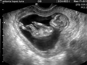
PHOTO: 6 weeks pregnant: fetal size by ultrasound
7 weeks.
The length increases to 1.5 cm. The embryo has a disproportionate big head. The inner diameter of the fetal egg is 24 mm.
KTR is 10 mm.
PHOTO: 7th week of pregnancy: fetal size by ultrasound
The control value is up to 10 mm. . The intracranial part, located at the back of the skull, at the base of the brain, in the posterior skull, below and behind the cerebellum. Measured in the lobby "not maid" should be 10 mm higher. This allows early relief of some indirect signs fetal chromosomal disease.
Initial values depend on gestational age and used growth curves. If you are wondering if this is possible from the acronyms or measures indicated on the ultrasound, the answer is no, as other parameters seen on the ultrasound images need to be analyzed.
8 weeks.
It is from this time that ultrasound includes indicators of the weight of the fetus, which at week 8 is from 1 to 1.5 g.
The length of the embryo from the coccyx to the parietal region can reach 2.2 cm. It is the measurement of the parietal-coccygeal length that continues throughout pregnancy, since even after the fetus is fully formed, it cannot be measured from the crown to the heels (the natural position of the embryo is bent legs).
The inner diameter of the egg has an average size of 30 mm.
KTR is 16 mm.
Biparental head size (BDP) - 6 mm.
PHOTO: 8th week of pregnancy: fetal size by ultrasound
Pregnancy for pregnant women. The information contained on this site is not intended to, and should in no way be a substitute for a direct relationship between healthcare professionals and the reader. This is the complete or partial absence of the white matter structure that connects the two hemispheres of the brain, forming the interepidermal forest floor and the third roof of the ventricle and the lateral ventricular frontal horns.
The following parts are distinguished: the trunk, which forms the main part, the knee, which forms the folded front part, the rostrum, in front of the knee and the spleen, behind the trunk. Top surface of the heartless body is connected to the lower edge of the large vasculature of the brain and is limited by the heartless circumcision via a solitary pathway from mandible cerebral arteries.
9 weeks.
Weight approximately 2 g. Length from the parietal region to the sacrum from 13 to 17 cm.
The fertilized egg has an internal diameter of 33 mm.
KTP - 23 mm.
BPR - 8.5 mm.
PHOTO: 9th week of pregnancy: fetal size by ultrasound
10 week.
The lower surface of the heartless body attaches middle part transparent septum and forms part of the lateral ventricles, from which the fornix is formed. black body represents the main neocortical commissure of the cerebral hemispheres. The corresponding areas of the cortex of the two hemispheres, with the exception of the frontal parts of the temporal lobes, are connected to each other by fibers that extend into the heartless body.
Among the most important functions is access to information stored in the cortex of the hemisphere, also for the corresponding area of the cortex of the opposite hemisphere. The two halves of the brain have independent abilities of consciousness, awareness, communication, and control. motor activity. Acute bodily function is necessary for their functional cooperation.
Weight no more than 4 g.
The distance from the crown to the waist is from 27 to 35 mm.
The average size of the internal diameter of the fetal egg is 39 mm.
KTP - 31 mm.
BPR - 11 mm.
PHOTO: 10th week of pregnancy: fetal size by ultrasound
The tumor body appears late in the obstruction of the brain. This comes from the fusion of the lateral fur fields that separate the primitive encephalic vesicles. The first part to grow is the rostrum, followed by the knee, body, and spleen. Complete agenesis of the corpus callosum may result from primary process before the 12th week or secondly before the destructive process of the insensible body, which is already formed after the 20th week of pregnancy.
It can be the result of both a malformative event that usually occurs, tardiness, and a disruptive event that occurs at any time during pregnancy. The incidence of body sputum affect is 0.7-1%. The data are certainly underestimated for the presence of asymptomatic individuals, especially in partial form.
11 weeks.
The length of the fetus from the crown to the sacrum is about 55 mm. Weight reaches 7 g.
The inner diameter of the fetal egg is 47 mm.
KTP - 41 mm.
BPR - 15 mm.
From 11 weeks with the help of ultrasound is calculated approximate height and fetal weight. This requires measurements of the length of the thigh and the diameter of the chest.
Fruit size:
Fetal growth - 6.8 cm.
Thigh length - 6.5 mm.
Rib cage in diameter 20 mm.
PHOTO: 11th week of pregnancy: fetal size by ultrasound
The karyotype is abnormal in 20% of cases, with an association with a syndrome of aneuploidy and chromosomal translocation. Family repetition suggests genetic heterogeneity with autosomal dominant, autosomal recessive, and X-linked recessive inheritance. Among the teratogens involved, alcohol, valproate, cocaine, toxoplasma, and influenza virus have been documented.
The mechanism by which damage occurs is uncertain, predominantly due to developmental anomalies middle line, which fluctuate when commissural lamination fails. Another way the disease occurs is the appearance of a heart attack in the distribution of the anterior cerebral artery, as a rule, as a result of intrauterine insults during various etiologies.
12 weeks.
The length of the fetus from the crown to the sacrum is from 70 to 90 mm.
Weight reaches 14-15 g.
The average value of the internal diameter of the fetal egg is 56 mm.
KTP - 53 mm.
BPR - 20 mm.
Fruit size:
Fetal growth - 8.2 cm.
Thigh length - 9 mm.
PHOTO: 12th week of pregnancy: fetal size by ultrasound
The frequency of relapses depends on the etiology in a particular case. 1% recurrence if sporadic or chromosomal. 25% recurrence if transmission is an autosomal recessive type. An anecogenic structure of a semi-solid form is placed between the hypoecogenicity of the saline tract from the pericaltic artery, in the upper part, and the subcutaneous cable, below.
From the notes of introductory anatomy and embryology, from which we observe the close structural relationships between the nuchal body, the septum pellucidum, and the lateral ventricles, we understand how complete anagenesis of the vermicular body is usually associated with significant changes in the intracranial anatomy, which, however, are more nuanced into partial forms.
13 weeks.
The length of the fetus from the crown to the sacrum is 10 cm. The weight reaches 25 g.
The average value of the inner diameter of the fetal egg is 65 mm.
KTP - 65 mm.
BPR - 24 mm.
Fruit size:
Fetal growth - 10 cm.
Thigh length - 12 mm.
The chest is 24 mm in diameter.
PHOTO: 13th week of pregnancy: fetal size by ultrasound
Sometimes the absence of a body causes an increased separation of the hemispheres with a prominent intervertebral suture. The lateral ventricles tend to be wider than usual, especially with occipital auras and horns. This anomaly is responsible for the "lacrimation" of the configuration of the lateral ventricles.
Usually anterior horns standard size but more distant from the midline. The likely mechanism by which this dilatation occurs is the absence of the posterior part of the heartless body, resulting in a distortion of the occipital whiteness protein tract that leads to caudal dilatation of the lateral ventricles.
14 weeks.
The length of the fetus from the crown to the sacrum is 11 cm. The weight can reach 43 g.
Baby head size:
BPR - 26 mm.
The perimeter of the skull is 80 mm.
Skull area - 510 mm2.
Fruit size:
Fetal growth - 12 cm.
Thigh length - 16 mm.
The chest in diameter is 26 mm.
PHOTO: 14th week of pregnancy: fetal size by ultrasound
The third ventricle is dislocated high up, reaching the area occupied by the callous body. Arterial vascularization modifications are essential for antenatal diagnosis. An in-depth analysis with color power Doppler in the mediosagittal scans for the loss of the classic semicircular arrangement of the pericelial artery and with the relief of the radiating arrangement of the branches of the anterior cerebral artery.
The differential diagnosis must be set with. Fundamental, every time you document the absence of an insensible body, you must rule out the presence of an anomaly associated with it. From the observation that the callous body is a structure that appears late in the phylogeny, it is clear that its absence is incompatible with life.
15 weeks.
The length of the fetus from the crown to the sacrum is 11 cm. The weight can reach 70 g.
Baby head size:
BPR - 32 mm.
The perimeter of the skull is 90 mm.
Skull area - 675 mm2.
Fruit size:
Fetal growth - 14.2 cm.
Thigh length - 19 mm.
The chest is 28 mm in diameter.
PHOTO: 15th week of pregnancy: fetal size by ultrasound
Wonderful confusion in clinical manifestations stems from the fact that in many cases corpus callosum agenesis is not distinguished by patterns in which this lesion is associated with other syndromic, congenital metabolic errors or other associated anatomical abnormalities.
If extrapolated cases are available from the case history where casein agenesis is an isolated event, it is noted that this disease is compatible with postnatal borderline development in most cases, and neurodevelopmental delay does not exceed 20% of subjects.
16 weeks.
The length of the fetus from the crown to the sacrum is 16 cm. The weight can reach 85 g.
Baby head size:
BPR - 35 mm.
The perimeter of the skull is 102 mm.
Skull area - 860 mm2.
Fruit size:
Fetal growth - 16.2 cm.
Thigh length - 22 mm.
The chest is 34 mm in diameter.
PHOTO: 16th week of pregnancy: fetal size by ultrasound
These children may have sensory integration problems, deficiencies in non-verbal learning, difficulty adequately managing the changes that occur in their routine, difficulty with abstract reasoning such as math, irony or word games, difficulty understanding good suggestions social nature, difficulty in focusing on argumentation.
There is no evidence that dislocation of the superior third ventricle, the degree of ventricular ligation, and significant dilatation of the interepidermal fissure is more associated with aica, where the outcome is worse, provide predictive value for these ultrasound procedures.
17 weeks.
The length of the fetus from the crown to the sacrum is 17 cm. Weight is approximately 142 g.
Baby head size:
BPR - 39 mm.
The perimeter of the skull is 120 mm.
Skull area - 1080 mm2.
Fruit size:
Fetal growth - 18 cm.
Thigh length - 24 mm.
The chest is 38 mm in diameter.
PHOTO: 17th week of pregnancy: fetal size by ultrasound
If this is confirmed to be an isolated agenesis of a heartless body, the couple can be reassured that the cognitive and behavioral defect is often modest, and the absence of a calloson body necessarily implies that the individual will not be capable of a complex intellectual life.
Standard obstetric care will not be changed unless the indication, if necessary, is recommended by a pediatric neurologist. Ultrasonic waves pass through the body and are reflected from individual organs or from transitions between tissues with different acoustic impedances. This is a painless study that has an irreplaceable place in medicine. Unwanted Effects ultrasound not yet demonstrated, it has only very low energy and biological activity.
18 weeks.
The length of the fetus from the crown to the sacrum is 20 cm. The weight can reach 200 g.
Baby head size:
BPR - 42 mm.
The perimeter of the skull is 126 mm.
Skull area - 1320 mm2.
Fruit size:
Fetal growth - 20 cm.
Thigh length - 28 mm.
The chest is 41 mm in diameter.
PHOTO: 18th week of pregnancy: fetal size by ultrasound
Pregnant women go through a lot ultrasound examinations throughout your pregnancy. At each stage of pregnancy, other important values determined and measured by ultrasound. Only in this way can fetal developmental defects be recognized in a timely manner, as well as prevent possible complications birth itself.
Most important research in the current prenatal ultrasound diagnosis in the week of pregnancy. It should always be carefully planned and carefully executed. Although it is still relatively small, it is now possible to find the first signs of the disease and find out the most serious morphological anomalies. If significant developmental defects are detected during the week of pregnancy, parents are advised to terminate the pregnancy. Some developmental defects that are compatible with life are eliminated after childbirth. In limited cases, you can also use the possibilities intrauterine treatment.
19 weeks.
The length of the fetus from the crown to the sacrum is 20-22 cm. The weight can reach 230 g.
Baby head size:
BPR - 44 mm.
The perimeter of the skull is 138 mm.
Skull area - 1450 mm2.
Fruit size:
Fetal growth - 22 cm.
Thigh length - 31 mm.
The chest is 44 mm in diameter.
PHOTO: 19th week of pregnancy: fetal size by ultrasound
20 weeks.
The length of the fetus from the crown to the sacrum is 25 cm. The weight can reach 280 g.
Baby head size:
BPR - 47 mm.
The perimeter of the skull is 144 mm.
Skull area - 1730 mm2.
Fruit size:
Fetal growth - 24 cm.
Thigh length - 34 mm.
The chest is 48 mm in diameter.
PHOTO: 20th week of pregnancy: fetal size by ultrasound
21 weeks.
The length of the fetus from the crown to the sacrum is 25 cm. Weight is from 360 to 370 g.
Baby head size:
BPR - 51 mm.
The perimeter of the skull is 151 mm.
Skull area - 1870 mm2.
Fruit size:
Fetal growth - 26 cm.
Thigh length - 37 mm.
The chest is 50 mm in diameter.
PHOTO: 21 weeks of pregnancy: the size of the fetus by ultrasound
22 weeks.
The length of the fetus from the crown to the sacrum is 27 cm. Weight is from 425 to 430 g.
Baby head size:
BPR - 54 mm.
The perimeter of the skull is 162 mm.
Skull area - 2190 mm2.
Fruit size:
Fetal growth - 28 cm.
Thigh length - 40 mm.
The chest is 53 mm in diameter.
PHOTO: 22nd week of pregnancy: fetal size by ultrasound
23 weeks.
The length of the fetus from the crown to the sacrum is 30 cm. Weight 500 g.
Baby head size:
BPR - 58 mm.
The perimeter of the skull is 170 mm.
Skull area - 2520 mm2.
Fruit size:
Fetal growth - 30 cm.
Thigh length - 43 mm.
The chest is 56 mm in diameter.
PHOTO: 23rd week of pregnancy: fetal size by ultrasound
24 weeks.
The length of the fetus from the crown to the sacrum is 30 cm. Weight 590 g.
Baby head size:
BPR - 61 mm.
The perimeter of the skull is 183 mm.
Skull area - 2710 mm2.
Fruit size:
Fetal growth - 31 cm.
Thigh length - 46 mm.
The chest is 59 mm in diameter.
PHOTO: 24th week of pregnancy: fetal size by ultrasound
25 weeks.
The length of the fetus from the crown to the sacrum is approximately 31 cm.
Weight from 700 g.
Baby head size:
BPR - 64 mm.
The perimeter of the skull is 194 mm.
Skull area -3072 mm2.
Fruit size:
Fetal growth - 32 cm.
Thigh length - 48 mm.
The chest in diameter is 62 mm.
PHOTO: 25th week of pregnancy: fetal size by ultrasound
26 weeks.
The length of the fetus from the crown to the sacrum is approximately 32-33 cm. Weight - 800 g.
Baby head size:
BPR - 67 mm.
The perimeter of the skull is 199 mm.
Skull area - 3260 mm2.
Fruit size:
Fetal growth - 33 cm.
Thigh length - 51 mm.
The chest in diameter is 64 mm.
PHOTO: 26th week of pregnancy: fetal size by ultrasound
27 weeks.
The length of the fetus from the crown to the sacrum is approximately 34 cm. Weight - 900 g.
Baby head size:
BPR - 68 mm.
The perimeter of the skull is 215 mm.
Skull area - 3675 mm2.
Fruit size:
Fetal growth - 35.5 cm.
Thigh length - 53 mm.
The chest in diameter is 69 mm.
PHOTO: 27th week of pregnancy: fetal size by ultrasound
28 weeks.
The length of the fetus from the crown to the sacrum is approximately 35 cm. Weight - 1000 g.
Baby head size:
BPR - 72 mm.
The perimeter of the skull is 218 mm.
Area - 3880 mm2.
Fruit size:
Fetal growth - 37 cm.
Thigh length - 55 mm.
The chest in diameter is 73 mm.
PHOTO: 28th week of pregnancy: fetal size by ultrasound
29 weeks.
The length of the fetus from the crown to the sacrum is approximately 37 cm.
Weight - 1150 g.
Baby head size:
BPR - 75 mm.
The perimeter of the skull is 225 mm.
Area - 4100 mm2.
Fruit size:
Fetal growth - 39 cm.
Thigh length - 57 mm.
The chest in diameter is 76 mm.
PHOTO: 29th week of pregnancy: fetal size by ultrasound
30 weeks.
The length of the fetus from the crown to the sacrum is approximately 37.5 cm.
Weight can reach 1400 g.
Baby head size:
BPR - 78 mm.
The perimeter of the skull is 234 mm.
Skull area - 4563 mm2.
Fruit size:
Fetal growth - 40 cm.
Thigh length - 59 mm.
The chest in diameter is 79 mm.
PHOTO: 30th week of pregnancy: fetal size by ultrasound
31 weeks.
The length of the fetus from the crown to the sacrum is 38-39 cm. The weight can reach 1500 g.
Baby head size:
BPR - 80 mm.
The perimeter of the skull is 240 mm.
Skull area - 4810 mm2.
Fruit size:
Fetal growth - 41 cm.
Thigh length - 61 mm.
The chest in diameter is 81 mm.
PHOTO: 31 weeks of pregnancy: the size of the fetus by ultrasound
32 weeks.
The length of the fetus from the crown to the sacrum is approximately 40 cm. Weight - 1700 g.
Baby head size:
BPR - 82 mm.
The perimeter of the skull is 246 mm.
Skull area - 5040 mm2.
Fruit size:
Fetal growth - 42 cm.
Thigh length - 63 mm.
The chest in diameter is 83 mm.
PHOTO: 32 weeks pregnant: fetal size by ultrasound
33 weeks.
The length of the fetus from the crown to the sacrum is 42 cm. Weight is 1800 g.
Baby head size:
BPR - 84 mm.
The perimeter of the skull is 255 mm.
Skull area - 5290 mm2.
Fruit size:
Fetal growth - 43.5 cm.
Thigh length - 65 mm.
The chest in diameter is 85 mm.
PHOTO: 33rd week of pregnancy: fetal size by ultrasound
34 weeks.
The length of the fetus from the crown to the sacrum is 42 cm. Weight is 2000.
Baby head size:
BPR - 86 mm.
The perimeter of the skull is 265 mm.
Skull area - 5547 mm2.
Fruit size:
Fetal growth - 44.5 cm.
Thigh length - 66 mm.
The chest in diameter is 88 mm.
PHOTO: 34 weeks of pregnancy: the size of the fetus by ultrasound
35 weeks.
The length of the fetus from the crown to the sacrum is 45 cm. Weight is 2200 g.
Baby head size:
BPR - 88 mm.
The perimeter of the skull is 270 mm.
Skull area - 5810 mm2.
Fruit size:
Fetal growth - 45.5 cm.
Thigh length - 67 mm.
The chest in diameter is 91 mm.
36 weeks.
The length of the fetus from the crown to the sacrum is 46 cm. Weight is 2300 g.
Baby head size:
BPR - 90 mm.
The perimeter of the skull is 272 mm.
Skull area - 6075 mm2.
Fruit size:
Fetal growth - 46.5 cm.
Thigh length - 69 mm.
The chest in diameter is 94 mm.
37 weeks.
The length of the fetus from the crown to the sacrum is 48 cm. Weight is 2800 g.
Baby head size:
BPR - 91 mm.
The perimeter of the skull is 276 mm.
Skull area - 6348 mm2.
Fruit size:
Fetal growth - 48 cm.
Thigh length - 71 mm.
The chest in diameter is 97 mm.
38 weeks.
The length of the fetus from the crown to the sacrum is 50 cm. Weight is 2900 g.
Baby head size:
BPR - 92 mm.
The perimeter of the skull is 282 mm.
Skull area - 6620 mm2.
Fruit size:
Fetal growth - 48 cm.
Thigh length - 71 mm.
The chest in diameter is 98 mm.
39 weeks.
The length of the fetus from the crown to the sacrum is 50 cm. Weight is 3000 g.
Baby head size:
BPR - 94 mm.
The perimeter of the skull is 285 mm.
Skull area - 6680 mm2.
Fruit size:
Fetal growth - 49 cm.
Thigh length - 73 mm.
The chest in diameter is 99 mm.
40 weeks.
The length of the fetus from the crown to the sacrum is 51 cm.
Weight - 3000 g.
Baby head size:
BPR - 95 mm.
The perimeter of the skull is 290 mm.
Skull area - 6770 mm2.
Fruit size:
Fetal growth - 50 cm.
Thigh length - 75 mm.
The chest is 100 mm in diameter.
Do not forget about the error in the average values, as well as the correctly set gestational age, which can lead to an error in calculating the size of the fetus. At multiple pregnancy values have different parameters and some lag in size.
Material prepared specifically for the site
For all nine months of bearing a child future mom she is very worried about how well her baby develops.
Regular visits to the doctor, compliance with his recommendations and mandatory examinations will help you monitor the course of pregnancy.
Most best method diagnostics is considered to be visited by pregnant women several times. It is this study that will help track the growth and development of the child literally by weeks and even days.
Significant deviations from the norm will indicate any violations or other problems, the timely detection of which will make it possible to correct the situation and help the mother and baby.
This concept denotes an assessment tool that is given by experts in all indicators: the growth rate of the baby, the symmetry of its development, and also the correspondence of these data to your gestational age.
Based on the obtained figures, the doctor will be able to draw conclusions and predict further development and child development.
What will be measured?
 To assess the condition, the doctor will record the indicators of many parameters.
They are different for every period. In the first trimester (from 8 to the end of 13 weeks), only a few possible ones are measured:
- the most important indicator is the coccygeal-parietal size (CTE). At this time, it is impossible to measure the bent legs of the child, so the figure is taken from the head (temechka) to the coccyx;
- as part of the screening study, the thickness is measured collar space(TVP) and the size of the nasal bone (these indicators are markers by which doctors may suggest the development birth defects and developmental anomalies);
- also evaluated, its weight, the presence and development of various internal organs.
KTP is measured up to a maximum of 14 weeks. The value of the indicators depends on the gestational age and allows you to determine it to the nearest day, because the child grows in height every day.
However, already at the second planned ultrasound, other indicators appear that become more important for the study:
- biparental size (BDP) - the distance that is measured between the two widest points of the baby's head (this indicator can be observed in the early stages, but then it does not matter as much as the KTR);
- fronto-occipital size (LZR) or longitudinal line;
- values of the circumferences of the head and tummy, the diameter of the abdomen and chest girth;
- limb length and tubular bones(bones of the lower leg, forearm, femur and humerus and others).
The symmetry of all sizes of the child, his weight, compliance of indicators specific date. In addition, other indicators also matter: the quality and size, and the place of attachment of the placenta, the assessment of the umbilical cord and cervix.
For more later dates doctors look and additionally determine the position and presentation of the baby in order to be able to predict the method of delivery.
Norms of the main dimensions of the fetus
 There are special tables that indicate the average parameters of all indicators.
Keep in mind that absolutely all babies develop in different ways, and even if some figures for the size of your child do not match the tabular norms, this is not a reason to panic: perhaps just lagging behind the average parameters is due to the usual individual features organism.
The same goes for exceeding standards. The doctor will definitely take repeated measurements after some time in order to track the dynamics of growth.
An alarming signal will only be a significant difference up or down in all parameters, recorded several times in a row.
Biometric indicators are measured at each ultrasound (the calculation is in percentiles: from 10 to 95 - fluctuations within the normal range, 50th percentile - its average indicator). As a rule, doctors take the average figure as a basis, but focus on the minimum and maximum size parameters.
Table. Fetal size by week of pregnancy
| week of pregnancy | Length cm | KTR, mm | Fronto-occipital size (LZR), mm | BPR, mm | Abdominal circumference, mm | Head circumference, mm | Femur, mm | Lower leg bones, mm | Forearm bone length, mm | Humerus length, mm |
| 8 | 1,6 | 10-18 | — | — | — | — | — | — | — | — |
| 9 | 2,3 | 16-27 | — | — | — | — | — | — | — | — |
| 10 | 3,1 | 24-38 | — | — | — | — | — | — | — | — |
| 11 | 4,1 | 34-50 | — | 13-21 | 40-62 | 53-73 | 3,4-7,8 | — | — | — |
| 12 | 5,4 | 42-59 | — | 18-24 | 50-72 | 58-84 | 4,0-10,8 | — | — | — |
| 13 | 7,4 | 51-75 | — | 20-28 | 58-80 | 73-96 | 7,0-11,8 | — | — | — |
| 14 | 8,7 | 63-89 | — | 23-31 | 66-90 | 84-110 | 9,0-15,8 | — | — | — |
| 15 | 10,1 | — | — | 27-35 | 90 | 110 | 16,2 | — | — | — |
| 16 | 11,6 | — | 41-49 | 31-37 | 88-116 | 112-136 | 17-23 | 15-21 | 12-18 | 15-21 |
| 17 | 13 | — | 46-54 | 34-43 | 93-131 | 121-149 | 20-28 | 17-25 | 15-21 | 17-25 |
| 18 | 14,2 | — | 49-59 | 37-47 | 104-144 | 131-161 | 23-31 | 20-28 | 17-23 | 20-28 |
| 19 | 15,3 | — | 53-63 | 41-49 | 114-154 | 142-174 | 26-34 | 23-31 | 20-26 | 23-31 |
| 20 | 16,4 | — | 56-68 | 43-53 | 124-164 | 154-186 | 29-37 | 26-34 | 22-29 | 26-34 |
| 21 | 26,7 | — | 60-72 | 46-56 | 137-177 | 166-200 | 32-40 | 29-37 | 24-32 | 29-37 |
| 22 | 27,8 | — | 64-76 | 48-60 | 148-190 | 178-212 | 35-43 | 31-39 | 26-34 | 31-39 |
| 23 | 28,9 | — | 67-81 | 52-64 | 160-202 | 190-224 | 37-45 | 34-42 | 29-37 | 34-42 |
| 24 | 30 | — | 71-85 | 55-67 | 172-224 | 201-237 | 40-48 | 36-44 | 31-39 | 36-44 |
| 25 | 34,6 | — | 73-89 | 58-70 | 183-229 | 214-250 | 42-50 | 38-46 | 33-41 | 39-47 |
| 26 | 35,6 | — | 77-93 | 61-73 | 194-240 | 224-262 | 45-53 | 41-49 | 35-43 | 41-49 |
| 27 | 36,6 | — | 80-96 | 64-76 | 205-253 | 235-273 | 47-55 | 43-51 | 37-45 | 43-51 |
| 28 | 37,6 | — | 83-99 | 67-79 | 217-265 | 245-285 | 49-57 | 45-53 | 39-47 | 45-53 |
| 29 | 38,6 | — | 86-102 | 70-82 | 228-278 | 255-295 | 50-60 | 48-55 | 40-48 | 47-55 |
| 30 | 39,9 | — | 89-105 | 71-85 | 238-290 | 265-305 | 52-62 | 49-57 | 42-50 | 49-57 |
| 31 | 41,1 | — | 93-109 | 73-87 | 247-301 | 273-315 | 54-64 | 50-60 | 44-52 | 51-59 |
| 32 | 42,4 | — | 95-113 | 75-89 | 258-314 | 283-325 | 56-66 | 51-61 | 45-53 | 52-59 |
| 33 | 43,7 | — | 98-116 | 77-91 | 267-325 | 289-333 | 58-68 | 53-63 | 46-54 | 54-62 |
| 34 | 45 | — | 101-119 | 79-93 | 276-336 | 295-339 | 60-70 | 55-65 | 48-56 | 55-63 |
| 35 | 46,2 | — | 103-121 | 81-95 | 285-345 | 299-345 | 62-72 | 56-66 | 49-57 | 57-65 |
| 36 | 47,4 | — | 104-124 | 83-97 | 292-354 | 303-349 | 64-74 | 57-67 | 50-58 | 58-66 |
| 37 | 48,6 | — | 106-126 | 85-98 | 299-361 | 307-353 | 66-76 | 59-69 | 51-59 | 59-67 |
| 38 | 49,8 | — | 108-128 | 86-100 | 304-368 | 309-357 | 68-78 | 60-70 | 52-60 | 60-68 |
| 39 | 50,7 | — | 109-129 | 88-102 | 310-374 | 311-359 | 69-80 | 61-71 | 53-61 | 60-70 |
| 40 | 51,2 | — | 110-130 | 89-103 | 313-381 | 312-362 | 70-80 | 62-72 | 54-62 | 61-71 |
Conclusion
The scorecard contains average data that helps doctors determine how symmetrical your child is developing. The measurement of the baby's parameters is supplemented by other studies - to observe the full picture and track the dynamics.
Be sure to see a doctor: regular examinations will make it possible to record all stages of development and growth of the child.