The vertebral column. The number of vertebrae in different species of animals and humans
Vertebral column ( columna vertebralis) forms the main longitudinal axis of the body. The spine is built from 50 to 55 separate bones - the vertebrae.
Vertebra (vertebra) is a structural element of the spinal column and consists of a body ( corpus vertebrae) and arcs ( arcus vertebrae). There is a bulge at the cranial end of the body - the head of the vertebra ( caput vertebrae), at the caudal end - concavity - the fossa of the vertebra ( fossa vertebrae). On the ventral surface of the body is the ventral ridge ( crista ventralis).
A vertebral foramen ( foramen vertebrae). All vertebral openings together form the spinal canal ( canalis vertebralis), in which the spinal cord lies. At the base of the cranial edge of the arch is the cranial vertebral notch ( incisura vertebralis cranialis), and at the base of the caudal margin - the caudal vertebral notch ( incisura vertebralis caudalis). These notches of two adjacent vertebrae form the intervertebral foramen ( foramen intervertebrale), through which the vessels enter and the nerves exit.
Cranial and caudal articular processes protrude along the edges of the arches, which serve to connect the vertebrae to each other. On the sides of the vertebral body, there are transverse or transverse costal for the attachment of muscles and ribs. From the middle of the arch, the spinous process rises dorsally ( processus spinosus) - for muscles.
Spine sections of the dog
The vertebral column is subdivided into cervical, thoracic, lumbar, sacral and tail departments.
Cervical vertebrae
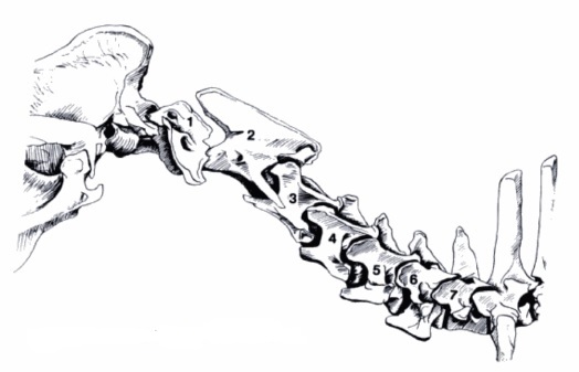
Cervical vertebrae
Cervical vertebrae ( vertebrae cervicales) are highly mobile in different directions (well-developed and widely spaced articular processes) and have a large surface for muscle attachment.
Dogs, like most mammals, have 7 cervical vertebrae, among them there are:
atypical: 1 (atlas), 2 (epistrophy), 6, 7
and typical: 3, 4, 5.
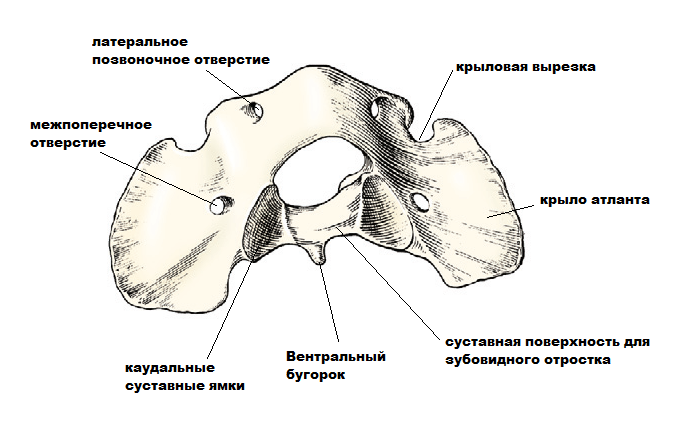
Atlant of the dog
First cervical vertebra - atlant (atlas) - the widest, formed by a wider dorsal and narrower ventral arches, which are connected in lateral (lateral) masses. On the dorsal arch, a dorsal tubercle is placed in the form of a small irregularity, on the ventral arch - a ventral tubercle, represented by a small, backward-directed protrusion for attaching muscles that provide flexion and extension of the head. The transverse processes formed the horizontal, thin, long, straight wings of the Atlas. At the base of each wing there is a transverse opening, which extends caudally to the surface of the wing in the dog. On the cranial edge of the wing, there is a clearly visible wing notch. Next to it, the lateral vertebral foramen opens, through which the first cervical nerve passes. The ventral surface of the wings is flat and bears a flat pterygoid fossa. The transverse opening is well pronounced. The cranial glenoid fossa is fairly deep, while the caudal glenoid fossa is flatter, triangular in shape, and their surfaces are directed caudo-medially. They pass into a flat facet - a tooth fossa located on the dorsal surface of the ventral arch of the atlas to connect with the tooth of the II cervical vertebra.
Second cervical vertebra - epistrophy (epistropheus) - the longest cervical vertebra, at the front end, instead of the head of the vertebra, it has a dentate process with a day-to-day surface for articulation with the atlas. In dogs, it has a thin vertebral crest strongly pushed forward, the intervertebral foramen are well developed.
Typical vertebrae. The middle cervical vertebrae are the most typical in their structure: flat and obliquely set head and fossa of the vertebra, the presence of a ventral crest at the caudal ends of the body and mastoid processes at the caudal articular processes; each of them has its own structural features. So, in vertebra 3, the ventral crest, costal process (the anterior part of the transverse costal process) is well developed, the rounded spinous process is absent. In vertebra 4, the ventral crest is less developed than in vertebra 3; the costal process is pointed. In the 5th vertebra, the head and fossa are well developed, the directed cranial spinous process is high and powerful (in decorative rocks poorly developed), the ventral crest is practically absent.
Sixth and seventh cervical vertebrae differ in structure from typical cervical vertebrae. The sixth cervical vertebra has a plate of the transverse costal process, the ventral ridge is absent. The seventh cervical vertebra does not have an intervertebral foramen, and the caudal costal fossa is poorly developed.
Thoracic vertebrae
Thoracic vertebrae ( vertebrae thoracales) together with the ribs and sternum form chest... Dogs usually have 13 thoracic vertebrae. But sometimes there are 12 of them, less often 14. All of them also have spinous processes. There are no ridges on the vertebral bodies. The length of the vertebral bodies decreases from the 1st to the 9th, and then increases to the last. In dogs, the 11 thoracic vertebra is diaphragmatic.
In the thoracic region, the ribs are connected to the vertebrae ( costae), for which there are articular surfaces on the body and transverse process of the thoracic vertebra - costal fossa (cranial, caudal and transverse).
Lumbar vertebrae
Lumbar vertebrae ( vertebrae lumbales) have more oval shape and are characterized by the presence of long, flat, ribbon-like transverse costal processes and well-developed articular processes. The number of vertebrae is usually 7. In very rare cases, there may be 6. In dogs, the spinous processes of the lumbar vertebrae are tilted forward; the transverse costal processes are directed forward downward and laterally; their length increases up to 5 vertebra, and then sharply decreases. The articular surfaces are in the sagittal plane. On the cranial articular processes, the mastoid processes are well developed for the attachment of muscles, under the caudal articular processes there are additional processes also for the attachment of muscles.
Sacral vertebrae
Sacral vertebrae ( vertebrae sacralis), of which there are 3 (less often 4) in dogs, merged into one sacrum bone ( os sacrum). The spinal column is firmly connected to the girdle of the pelvic limb, while experiencing static and dynamic loads. The final fusion occurs at the age of two years. In females, the sacrum is relatively longer, wider, and more curved ventrally than in males.
At the sacral bone, the spinous processes merged into the sacral crest ( crista sacralis medialis), but often the process of the first vertebra remains isolated. There are no intercutaneous holes. The intervertebral notches form the dorsal sacral foramen for nerves and blood vessels. The transverse costal processes merged into the lateral parts - for the attachment of muscles and ligaments. In dogs, the wings of the sacrum are located in the lateral sagittal plane.
Tail vertebrae
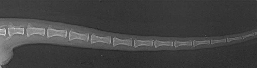
X-ray of the tail spine
Caudal vertebrae ( vertebra caudales, coccygeae) - different breeds of dogs may have a different number of 20-23 (less often 15-25). Of these, only the first two or four are still well developed, having all the characteristic anatomical structures for a typical vertebra. The rest undergo reduction and are the site of attachment of the muscles that set the tail in motion. The vertebrae get longer and the processes are gradually reduced. Starting from the X-XII vertebrae, their bodies are shortened again, and the vertebrae are elongated cylinders. On the V-XV vertebrae from the ventral surface there are hemal processes ( proc. hemalis), which on the V-VIII vertebrae form closed hemal arches ( arcus hemalis), forming a channel for the passage of the main tail vessel.
Sources of
Arlene Coulson Atlas of Interpretative Radiographic Anatomy of the Dog and Cat, Blackwell Science Ltd, 2002.
Volmerhaus B., Frewein J. et al. Anatomy of a dog and a cat. M .: "Aquarium Buk", 2003.
Sloths differ from other mammals primarily in that their neck contains the largest number of vertebrae among the representatives of this group. And in manatees, the situation is different - the number of their cervical vertebrae is minimal among mammals. It is curious that both of these adaptations are associated with the slowness of these animals.
In all mammals, the number of cervical vertebrae is the same: that in a giraffe, in a mouse, in a person - all have exactly seven vertebrae. However, there are no rules without exceptions. One representative of the fauna - sloth - has more cervical vertebrae than other mammals.
Birds, reptiles and amphibians can have different numbers of vertebrae. A swan, for example, has 22 to 25. Mammals have a different story: additional vertebrae that suddenly "grew" at the embryonic stage increase the risk of stillbirth, and if the animal is born alive, it is at risk of cancer or problems with nervous system.
But sloths have a great life with the "wrong" number of vertebrae. Moreover, each family of these animals has a different number: two-toed sloths (Choloepus) have cervical vertebrae from five to seven, and three-toed sloths (Bradypus) have eight or nine. Some specimens from this family even have 10 cervical vertebrae. And nothing - they live, they don't complain.
During formation, the mammalian spine goes through several stages: the vertebrae ossify, first in the thoracic region, and then in the cervical region. In the case of sloths, ossification begins immediately in the thoracic region, and several adjacent noncostal vertebrae, which are usually referred to as cervical, although, based on the characteristics of their formation, it would be more correct to consider them thoracic. In addition, rudimentary ribs are preserved on one or two of the last cervical vertebrae, although they do not reach the sternum.
Why this happened is a long-standing mystery. Even before the publication of Charles Darwin's famous theory of evolution, there was a heated debate among zoologists on this topic. However, even such a brilliant specialist in comparative anatomy as Georges Cuvier could not explain this fact. To tell the truth, scientists still don't know exactly why sloths' necks suddenly lengthened abnormally during evolution. However, there are some hypotheses trying to explain this phenomenon.
According to one version, the increase in the number of cervical vertebrae could be caused by an arbitrary mutation of homeotic genes (also called Hox genes) that control early development organism and are responsible for the differentiation of tissues and the laying of organs in the embryo. True, in this case, the changes should concern not only the spine, but also other organs. And, in addition, it is logical to assume that since this mutation was supported by natural selection, then, consequently, the mutant received some benefit from it. But which one?
That the change has affected many internal organs, is brilliantly confirmed by the data of biologists who have studied internal structure sloths. Bradypus is known to have rib asymmetry, tracheal curvature, vertebral fusion, and pelvic ossification. Undoubtedly, all this is a consequence of the increase in the number of vertebrae. It is much more difficult to answer the question - why did sloths need to mutilate themselves like this? Apparently, this is due to some peculiarities of their lifestyle.
Dr. Galis, a spokesman for the Netherlands Center for Biodiversity, says that the only thing that saves sloths from all the unpleasant consequences of eight or nine vertebrae is their slow metabolism. Indeed, from the point of view of physiology, these funny creatures are more reptiles than mammals. Their body temperature can fluctuate from 24 to 33-35 ° C, that is, by almost 10 ° C, which is usual for reptiles, but not for their warm-blooded descendants. That is why it can often take sloths about a month to digest a portion of eaten leaves, and they can go to the toilet only once every two weeks.
True, a leisurely metabolism and low temperature protects these "reptilian" mammals from a number of diseases - such as cancer, for example. However, they also cause a lot of inconvenience - in particular, when the temperature is low, the supply of energy to the muscles slows down, so it becomes very difficult for them to move.
This is where it helps abnormally long neck - it allows these sluggish animals to turn their heads 270 degrees, partly compensating for their limited ability to move: hanging on a tree, the sloth twists its neck to get to the fresh foliage, while remaining in place. To work cervical muscles you don't need to spend a lot of energy.
It is fair to add that there is one more "exception to the vertebral rule" in the mammalian kingdom. This is a manatee (Trichechus), a huge aquatic animal of the order of sirens (Sirenia). Manatees have only six cervical vertebrae, which, in addition, are fused and shortened. Interestingly, the reason is most likely also the way of life of this creature.
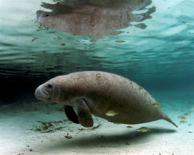
Let me remind you that, unlike seals and walruses, manatees are vegetarians. These slow and good-natured creatures leisurely graze in the algal meadows like land cows. Manatees have practically no enemies in the world of animals - few people can cope with an animal with such impressive dimensions (up to 5 meters in length and weighing half a ton), and large sharks that could do this rarely visit shallow waters where these bumpers.
Because of this lifestyle, the mobility of the neck for these animals has become not particularly relevant. And reducing its length, on the contrary, is beneficial - as a result of such a restructuring, manatees were able to bring their head closer to the body, which had a positive effect on the general buoyancy of the body (the body became close to an oval in shape, and this shape is most beneficial for those who soar in the water column ).
As you can see, for some, as a result of slowness, the neck increases, and for some it shortens. Needless to say, evolution is sometimes paradoxical.
Cervical vertebrae - vertebrae cervicales - seven. Their long bodies (the longest is the 2nd vertebra) are gradually shortened posteriorly. The vertebrae from the 3rd to the 5th are typical and close in structure, while the 6th and 7th, and especially the 2nd and 1st, have their own characteristic differences.
The head and fossa - caput et fossa vertebrae (Fig. 47-c, m) - are very pronounced; the head is almost hemispherical. The ventral crest (n) is highly developed, as a result of which the vertebral body acquires a prismatic shape.
The transcostal processes - processus costotransversarii (b, l) - bifurcate at the ends, with one part directed cranially (rib homologue), and the other - caudally. At the base of the transverse costal processes there is a significant intertransverse opening - foramen transversarium (k) - stretching along the vertebral bodies. The combination of these holes forms an intertransverse canal - canalis transversarius - in which the vessels and nerve pass. The articular processes are widely spaced, flat and voluminous. Of these, the cranial ones - processus articulares craniales (e) - face the articular surfaces dorso-medially, and the caudal ones - processus articulares caudales (h) - ventro-lateral. The cranial and caudal articular processes are connected to each other by ridges, which, together with the transcostal processes, gives the cervical vertebrae a typical tetrahedral shape. The neural arches are highly developed. Intermediate holes are insignificant. Intervertebral notches - incisurae intervertebrales (d) - deep.
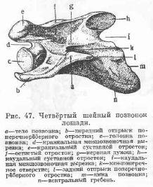
The spinous processes - processus spinosi - are present only in the form of weak rough scallops (f).
The sixth cervical vertebra in its shape is close to those described, but differs from them in a shorter length, a wider intertransverse foramen, a more strongly developed spinous process, and most importantly - a special shape of the transcostal process, which has a ventrally directed bony plate connecting with the anterior part of the process ...
The seventh cervical vertebra, the shortest of all, has a pronounced spinous process and an unbranched transcostal vertebra. There is no transverse hole.
At the caudal end of the body, there is a pair of glenoid fossae for articulation with the heads of the first pair of ribs.
The second cervical vertebra - epistrophy - epistropheus (Fig. 48-A) - the longest. Instead of a head, the cranial end of his body has a semi-cylindrical odontoid process - dens epistrophei (b). The ventral rounded side of this process bears an articular surface for connection with the body of the Atlantean. From the odontoid process, it extends back into the adjacent, almost segmented, articular processes separated by the notch, which correspond to the cranial articular processes. Thus, a device for the rotation of the Atlantean is created.
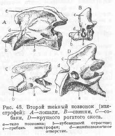
The inner surface of the odontoid process is rough and serves as a place of attachment of the ligament of the odontoid process. The transverse processes are not bifurcated, poorly expressed and directed backward with the apex.
There is an inter-transverse hole, but it is small in diameter. Instead of the cranial intervertebral notch, there is an independent intervertebral foramen, separated from the intervertebral space by a small bone bridge. At the site of the spinous process, the crest - crista epistrophei (c) - is strongly developed, which bifurcates posteriorly and carries the caudal articular processes - processus articulares caudales. The ventral crest is developed.
The first cervical vertebra, or atlas, - atlas (Fig. 49 - A) - represents wide ringcomposed of dorsal and ventral arches - arcus dorsalis et ventralis (a, 6); the latter is equipped on the outer surface with a significantly protruding ventral tubercle - tuberculum ventrale - and on the inner surface - with a pit-shaped inner articular surface for articulation with the odontoid process of the 2nd vertebra. This surface continues back and to the sides, to the wings of the atlas, where it merges with the caudal articular processes, which stand almost in the segmental plane.
In the direction of the occipital bone on the atlas there are cranial glenoid fossa-foveae articulares craniales. The dorsal arch is more strongly convex and on outside instead of the spinous process, it has a rough, slightly noticeable thickening, called the dorsal tubercle - tuberculum dorsale.
![]()
The transverse processes are extremely wide, lamellar, with rounded, slightly thickened edges. They wear special name wings of the atlant - alae atlantis (c) - and directed to the lateral sides and slightly bent downward, as a result of which an atlas fossa is formed on each wing on its ventral surface, into the bottom of which an opening of the pterygoid fossa leads from the spinal canal. Posteriorly, the base of the wing has an intertransverse foramen - foramen transversarium (e), - and in front - an intervertebral foramen - foramen intervertebrale (d). It is separated by a bone plate from the occipital-atlas joint and, on the dorsal surface, continues laterally into a short wide trough, at the lateral end of which there is a wing opening - foramen alare - leading to the ventral surface of the wing (d).
Cervical in all mammals consists of seven vertebrae... The cervical vertebrae of mammals are distinguished by a long body, a spherical shape of the head, a deep fossa of the vertebra, a bifurcated transverse process (the transverse process connected to the rudiment of the rib), the presence of a transverse opening at the base of the transverse costal process, well-defined 22 anterior and posterior articular processes connected to each other, with extensive articular areas, which provides greater mobility of this part of the spinal column. The spinous processes are small in size.
The first two vertebrae are built differently from the other vertebrae. The first cervical vertebra is called atlas, and the second is called epistrophy, or axial.
Atlant in animals it is ring-shaped and has two arches: upper and lower. The transverse processes are extensive and are called the wings of the Atlantean. At the front end of the atlas, there are glenoid fossae for articulation with the skull, and on the posterior end there are convex articular surfaces for connection with the second cervical vertebra. The Atlanta has a series of holes: the vertebral, which forms the beginning of the spinal canal; winged; intervertebral and transverse for vessels and nerves.
The second cervical vertebra, or epistrophy in animals, is the longest; its head has turned into an epistropheus tooth, and its massive spinous process has turned into a crest. The transverse costal processes are not bifurcated.
In cattle, the cervical vertebrae have a short body, a well-defined spinous process, the head and fossa of the vertebra. The atlas is devoid of a transverse opening; the epistrophy has a semi-cylindrical tooth. In pigs, the body of the cervical vertebrae is short, the heads are flat and the fossa is well defined; the shape of the transverse costal processes is lamellar; the transverse foramen on the atlas is located at the posterior end of the vertebra next to the articular surfaces; the epistrophy has a blunted odontoid process; the epistrophy crest is narrow and raised behind. In a horse, the cervical vertebrae, with the exception of the seventh, do not have spinous processes, their bodies are longer. The atlas has an intervertebral, wing and transverse foramen; epistropheus tooth convex below and flat above; the epistrophy crest is powerful, bifurcates behind and carries the posterior articular processes.
Thoracic vertebrae in animals, they have a high spinous process, anterior and posterior articular costal fossa, small transverse processes, on which there are facets for the costal tubercle and mastoid processes.
The thoracic vertebra of cattle is distinguished by a posterior intervertebral foramen instead of a notch, a flat and wide spinous process. In pigs, an additional opening is located on the thoracic vertebra at the base of the transverse process. In the horse, the thoracic vertebra has a prismatic body due to the presence of a ventral ridge. The spinous processes at the free ends are strongly thickened.
29. The structure of the lumbar and sacral vertebrae.
The lumbar vertebrae in animals are distinguished by long transverse-costal processes, which originated from the fusion of transverse processes with rib rudiments, flat fossae and a head, well-defined articular processes.
In a cattle cattle six lumbar vertebrae; the anterior articular processes have a grooved shape, and the posterior ones are cylindrical. The width of the spinous processes is greater than the height. The pig has seven lumbar vertebrae. The articular processes are built in the same way as in cattle. There is an opening or notch on the transverse costal process. The horse has six lumbar vertebrae. Articular processes with flat articular surfaces. On the last two vertebrae, the transverse costal processes bear articular surfaces for connection with each other and the sacrum. The spinous processes are higher than wide.
Sacral vertebrae fused into the sacrum, which is the vault of the pelvic cavity.
On the sacrum bone in animals, wings (the first pair of transverse processes), anterior articular processes, lateral parts, spinous processes, upper and lower sacral foramen are distinguished. The sacrum bone is connected to the pelvic bone with the help of wings.
In cattle, the sacrum is formed from five vertebrae. It differs in the quadrangular shape of the wings with one articular surface for connection with the pelvic bone, grooved anterior articular processes, accrete spinous processes. In a pig, the sacrum is formed from four vertebrae. The spinous processes are absent. A horse has five (six) vertebrae. The wings are triangular in shape with two articular surfaces, the anterior articular processes are flat, the spinous processes are fused only at the base.
Tail vertebrae mostly reduced, and only the first of them have elements inherent in the vertebrae of other departments.
In cattle, there are 18-20 vertebrae, in a pig - 20-23, in a horse - 18.
All vertebrae form the vertebral column, and the vertebral foramen form the vertebral canal in which the spinal cord is located.
Spine section: Cervical - (number of vertebrae) 7
Pectoral -13
Lumbar – 6
Sacral – 5
Tail – 18–20
Total – 49–51
The rib cage is formed by the ribs and sternum. Ribs - paired arcuate bones, movably attached to the right and left to the vertebrae thoracic spinal column. They are less mobile in the front of the chest, where the scapula is attached to them. In this regard, the anterior lobes of the lungs are more often affected by lung diseases. All ribs make up a fairly voluminous conical chest, in which the heart and lungs are located.
The peripheral skeleton, or skeleton of the limbs, is represented by 2 thoracic (front) and 2 pelvic (hind) limbs.
The thoracic limb includes: a scapula attached to the body in the area of \u200b\u200bthe first ribs; shoulder consisting of humerus; forearm, represented by the radius and ulna; a hand (Fig. 4), consisting of the wrist (6 bones), the metacarpus (2 fused bones) and the phalanges of the fingers (2 fingers having 3 phalanges, the third phalanx being called the coffin bone).
Fig. 4. The skeleton of an autopodium (hand) of a cow:
1 - radius bone; 2 - the ulna; 3 - carpal bones; 4 - metacarpal bones; 5 - phalanges
The pelvic limb consists of a pelvis (Fig. 5), each half of which is formed by a nameless bone, at the top is the ilium, below the pubic and ischial bones; thighs presented femur and a patella that slides over the femur block; tibia, consisting of the tibia and fibula; the foot, represented by the tarsus (6 bones), metatarsal (2 fused bones) and phalanges of the fingers (2 fingers having 3 phalanges, with the third phalanx called the coffin bone).
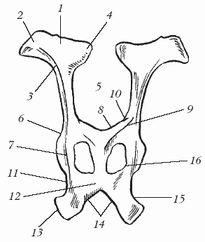
Fig. 5. Bones pelvic girdle (pelvis) cows:
1 - wing ilium; 2 - maklokovy hillock; 3 - the body of the ilium; 4 - sacral tubercle; 5 - large ischial notch; 6 - glenoid cavity; 7 - ischial spine; 8 - cavity of the pubic bone; 9 - suture branch of the pubic bone; 10 - ilio-pubic eminence; 11 - the hollow branch of the ischium; 12 - plate of the ischium; 13 - ischial tubercle; 14 - sciatic arch; 15 - small ischial notch; 16 - locked hole
It must be remembered that the maturity of the skeleton occurs later than the maturity of the body or puberty, and deprivation of animals motor activity leads to the birth of calves with an underdeveloped skeleton. IN embryonic period is happening fast growth peripheral skeleton, since after birth the calves must move independently and reach the nipples of the mother, who feeds them while standing. After birth, the ribs, spine, sternum and pelvic bones... The increase in body size in cattle ends at 5-6 years. The aging process begins in the skeleton from the caudal vertebrae and the last ribs. All this affects the mineralization of bones, which must be taken into account when developing the diet of animals for different stages development.
Ligaments - These are bundles of collagen fibers that connect bones or cartilage to each other. They experience the same body weight load as the bones, but by connecting the bones to each other, the ligaments impart the necessary buffering to the skeleton, significantly increasing the resistance to the loads on the bone junctions as supporting structures.
There are 2 types of bone connection:
›Continuous. This type of connection has great elasticity, strength and very limited mobility;
. Discontinuous (synovial) type of connection, or joints. It provides more range of motion and is built more complexly. The joint has an articular capsule, consisting of 2 layers of the outer (fused with the periosteum of the bone) and the inner (synovial, which secretes synovium into the joint cavity, due to which the bones do not rub against each other). Most of the joints, except for the capsule, are fixed with a different number of ligaments. In case of ruptures and severe sprains of the ligaments, the bones are separated from each other and the joint is dislocated.
Among diseases of the organs of the apparatus of movement in animals, there are more pathological processes at the junction of bones, especially the joints of the limbs. Pathology at the junction of the bones is dangerous with consequences such as loss of mobility, which is accompanied by the loss of the ability to move normally and significant pain.
Muscle possesses important property: it contracts, causing movement (dynamic work), and provides the tone of the muscles themselves, strengthening the joints at a certain angle of combination with a stationary body (static work), while maintaining a certain posture. Only the work (training) of the muscles contributes to the increase in their mass, both by increasing the diameter of the muscle fibers (hypertrophy) and by increasing their number (hyperplasia).
Muscle tissue is of 3 types, depending on the location of muscle fibers: smooth (vascular walls), striated (skeletal muscles), cardiac striated (in the heart). By the nature of their activity and the work performed, they are divided into flexion and extension, adduction and abduction, locking (sphincters), rotating, etc.
The work of the muscular apparatus is built on the principle of antagonism. In total, the body contains up to 200–250 paired muscles and several unpaired ones.
Muscle mass in cattle is approximately 42–47% of the total body weight. Each muscle has a supporting part (connective tissue stroma) and a working part (muscle parenchyma). The greater the static load a muscle performs, the more developed the stroma in it.
Skin covering
The body of cattle is covered with hairy skin and organs, or derivatives skin... Their appearance, consistency, temperature and sensitivity reflect the state of metabolism and functions of a number of organ systems.
Leather protects the body from external influences through a variety of nerve endings, acts as a receptor link in the skin analyzer external environment (tactile, pain, temperature sensitivity). Through a lot of sweats and sebaceous glands a number of metabolic products are secreted through the skin; through the mouths of hair sacs and skin glands, the surface of the skin can absorb a small amount of solutions. The blood vessels of the skin can hold up to 10% of the blood of the animal's body. Reduction and dilation of blood vessels have essential in the regulation of body temperature. The skin contains provitamins. Under the influence of ultraviolet light, vitamin D is formed.
In cattle, the skin makes up 3–8% of the total weight of the animal. In a bull, the mass of the skin can be in the range of 60–80 kg, its thickness ranges from 2 to 6 mm.
In the skin covered with hair, the following layers are distinguished:
›Cuticle (epidermis) - the outer layer. It determines the color of the skin, and keratinized cells slough off, thereby removing dirt, microorganisms, etc. from the skin surface. Hair grows on the epidermis;
›Dermis (skin itself) formed by:
a) the pilar layer, which contains the sebaceous and sweat glands, hair roots in hair follicles, muscles - hair lifters, many blood and lymphatic vessels and nerve endings;
b) a mesh layer consisting of a plexus of collagen and insignificant amount elastic fibers.
subcutaneous base ( subcutaneous layer), represented by loose connective and adipose tissue. This layer is attached to the superficial fascia covering the body of the cattle (Fig. 6). It stores spare nutrients in the form of fat. Skin with hair and subcutaneous tissueremoved from the body of an animal is called the skin.
Fig. 6. Scheme of the structure of the skin with hair (according to Techver):
1 - epidermis; 2 - dermis; 3 - subcutaneous layer; 4 - sebaceous glands; 5 - sweat glands; 6 - hair shaft; 7 - hair root; 8 - hair follicle; 9 - hair papilla; 10 - hair bag
Derivatives of the skin include sweat, sebaceous, mammary glands, hooves, crumbs, horns, hair, nasolabial speculum.
Sebaceous glands are located at the base of the skin, and their ducts open into the orifices of the hair follicles. The sebaceous glands secrete a sebaceous secretion, which, by lubricating the skin and hair, gives them softness and elasticity, protects against fragility, and protects the body from moisture penetration.
Sweat glands located in the reticular layer of the skin. Their excretory ducts open to the surface of the epidermis, through which a liquid secretion - sweat - is released. The secretion of sweat helps to cool the animal, that is, the sweat glands are involved in thermoregulation. In cattle, a large number of them are located on the head.
Breast cattle is called the udder. It consists of four quarters, or lobes, formed by the fusion of two pairs of glands. Inside the udder there are alveoli lined with secretory epithelium from the inside. The alveoli pass into the milk ducts. The latter, merging, form a milk cistern, passing into the nipple canal. Each part of the udder has a teat for milk withdrawal (fig. 7). The upper udder is covered with elastic skin. The more productive the animal is, the softer and more elastic this skin is.
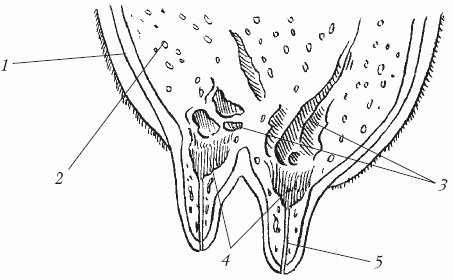
Fig. 7. The structure of the cow's mammary gland:
1 - leather; 2 - alveoli; 3 - milk ducts; 4 - milk tank; 5 - nipple canal
Hoof - this is the hard dermal tip of the third phalanx of the fingers (3 and 4) of artiodactyls. It is represented by a skin area, the epidermis of which, in certain places of the hoof, forms horny layers of various structures and consistencies. According to the location and nature of the produced stratum corneum on the hoof, 4 parts are distinguished: the border, the corolla, the wall and the sole (Fig. 8).

Fig. 8. The structure of the hoof:
a - border; b - a whisk; в - wall; d - sole: 1 - epidermis; 2 - skin base; 3 - subcutaneous layer; 4 - tendon of the common digital extensor; 5 - subcutaneous layer of the border; 6 - the basis of the skin of the border; 7 - epidermis of the border; 8 - corolla epidermis; 9 - wall glaze; 10 - tubular horn; 11 - leaf horn; 12 - lamellar layer of the base of the skin; thirteen - white line; 14 - the epidermis of the sole; 15 - base of sole leather; 16 - periosteum; 17 - the epidermis of the digital crumb; 18 - crumb skin base; 19 - crumb cushion epidermis; 20 - crumb cushion skin base; 21 - subcutaneous layer of the crumb cushion
Myakishi - these are the supporting areas of the limbs. They are rich in nerve endings, due to which they serve as an organ of touch. In cattle, only modified digital crumbs remained, which became mainly shock absorbers of the horny capsules of the hoof.
Horns - These are solid formations in the head region of cattle, located on the horny processes of the frontal bones. Outside, they are covered with a horny capsule formed by the horn epidermis. The growth of the horn depends on the metabolism of the whole organism, which is expressed in the appearance of rings. Changes in metabolism during pregnancy retard horn growth.
The growth of two horn buds in young animals is terminated by cauterization or excision. To dehydrate adult animals, it is necessary to squeeze the wax or rim of the horn (soft horn at the border of the base of the horn with the skin) with rubber rings, which contributes to the cessation of blood supply and innervation of the horn, leading to its death.
Hair.The entire body of cattle is covered with wool. For 1 cm2 of skin, these animals can have up to 2500 or more hairs. Hair is spindle-shaped filaments of stratified keratinized and keratinized epithelium. The part of the hair that rises above the surface of the skin is called the shaft, which is inside the skin - the root, which is surrounded by blood capillaries. The root passes into the bulb, and inside the bulb is the hair papilla. Each hair has its own muscles that allow it to straighten, as well as sebaceous glands.
By structure, 4 main types of hair are distinguished: guard (short integumentary hair of the body and long hair at the end of the tail), downy (hair around the guard and covered by them), transitional, vibrissae, or sensitive hair (hair on the skin around the lips, nostrils, chin and eyelids).
In cattle, like in other animals, there is a change in the integument of the body, or molt. In this case, the hair or coat is completely or partially replaced (except for tactile hairs). When molting, the skin thickens, becomes looser, often the stratum corneum of the epidermis is renewed. Distinguish between physiological and pathological molting. The physiological change in the coat is divided into 3 types: age, seasonal and compensatory.
Nervous system
The structural and functional unit of the nervous system is a nerve cell - neurocyte - together with gliocytes. The latter dress the nerve cells and provide them with a support-trophic and barrier function... Nerve cells have several processes - sensitive tree-like branching dendrites, which conduct excitation to the body of the neuron that occurs on their sensitive nerve endings located in the organs, and one motor axon, through which a nerve impulse is transmitted from a neuron to a working organ or another neuron. Neurons come into contact with each other with the help of the endings of the processes and form reflex circuits along which nerve impulses are transmitted (propagated).
The processes of nerve cells in combination with neuroglia cells form nerve fibers... These fibers in the brain and spinal cord make up the bulk of the white matter. From the processes of nerve cells, bundles are formed, from a group of bundles, dressed with a common sheath, are formed nervesin the form of cord-like formations.
Anatomically, the nervous system is divided into the central, including the brain and spinal cord with spinal ganglia, and the peripheral, consisting of the cranial and spinal nerves connecting the central nervous system with receptors and effector apparatus various bodies... This includes the nerves of the skeletal muscles and skin (the somatic part of the nervous system), as well as the vessels (the parasympathetic part). These last two parts are united by the concept of "autonomous, or autonomic, nervous system."
Brain - this is head part the central part of the nervous system, located in the cranial cavity. The brain consists of 2 hemispheres separated by a groove. The hemispheres have convolutions and are covered with cortex, or cortex.
The following sections are distinguished in the brain: the large brain, the telencephalon (olfactory brain and cloak), diencephalon (optic hillocks (thalamus), epithalamus (epithalamus), hypothalamus (hypothalamus), perihumor (metathalamus), midbrain (legs of the large brain and quadruple), rhomboid brain, hindbrain (cerebellum and pons) and medulla oblongata.
The brain is dressed in 3 membranes: hard, arachnoid and soft. Between the hard and arachnoid membranes there is a subdural space filled with cerebrospinal fluid (its outflow is possible into the venous system and into the lymph circulation organs), and between the arachnoid and soft - the subarachnoid space. The brain is composed of white and gray matter. The gray matter in it is located on the periphery of the cerebral cortex, and white matter in the center.
The brain is the highest part of the nervous system, which controls the activity of the whole organism, unites and coordinates the functions of all internal organs and systems. In pathology (trauma, swelling, inflammation), there is a violation of the functions of the entire brain.
The absolute weight of the brain of cattle varies widely from 410 to 550 g, and the relative is inversely proportional to the weight of the animal and is 1 / 600-1 / 770.
Spinal cord Is part of the central nervous system. It is a cord of brain tissue with remnants of the cerebral cavity. The spinal cord is located in the vertebral canal and starts from the medulla oblongata and ends in the region of the 7th lumbar vertebra. The spinal cord is conditionally subdivided without visible boundaries into the cervical, thoracic and lumbosacral regions, consisting of gray and white medulla. In the gray matter, there are a number of somatic nerve centers that carry out various unconditioned reflexes, for example, at the level of the lumbar segments, there are centers that innervate the pelvic limbs and the abdominal wall. The gray matter is located in the center spinal cord and in section it looks like the letter "H", and the white matter is located around the gray.
The spinal cord is covered with three membranes: hard, arachnoid and soft, between which there are gaps filled with cerebrospinal fluid.
In cattle, the length of the spinal cord is on average 160–180 cm. The mass of the spinal cord is 220–260 g, which is on average 47% of the mass of the brain.
Peripheral nervous system - a topographically separated part of the unified nervous system. This section is located outside the brain and spinal cord. It includes the cranial and spinal nerves with their roots, as well as plexuses, ganglia and nerve endings embedded in organs and tissues. So, 31 pairs of peripheral nerves depart from the spinal cord, and 12 pairs from the brain.
In the peripheral nervous system, it is customary to distinguish 3 parts - somatic (connecting centers with skeletal muscles), sympathetic (associated with smooth muscles of the vessels of the body and internal organs), visceral, or parasympathetic (associated with smooth muscles and glands of internal organs), and trophic (innervating connective tissue).
Autonomic nervous system has special centers in the spinal cord and brain, as well as a number of nerve nodes located outside the spinal cord and brain. This part of the nervous system is subdivided into:
›Sympathetic (innervation of vascular smooth muscles, internal organs, glands), the centers of which are located in the thoracolumbar spinal cord;
. Parasympathetic (innervation of the pupil, salivary and lacrimal glands, respiratory organs, as well as organs located in the pelvic cavity), whose centers are located in the brain.
A feature of these 2 parts is their antagonistic nature in providing them with internal organs, that is, where the sympathetic nervous system acts excitingly, the parasympathetic one has a depressing effect.
The central nervous system and cerebral cortex regulate the entire higher nervous activity through reflexes. There are genetically fixed reactions of the central nervous system to external and internal stimuli - food, sexual, defensive, orienting, sucking reaction in newborns, the appearance of saliva at the sight of food. These reactions are called innate, or unconditioned, reflexes. They are provided by the activity of the brain, the spinal cord stem and the autonomic nervous system. Conditioned reflexes - acquired individual adaptive reactions of animals, arising on the basis of the formation of a temporary connection between the stimulus and the unconditional reflex act. An example of such reflexes is the milking of cows in certain time... If the clock is adjusted, milk yield may decrease.