Assessment of the nasal bone in the first trimester of pregnancy: how, where, when and why we do it.
Hello!
I really need to know your opinion! I would be grateful for any information!
I am 29 years old, I have three children and I am expecting a 4th child.
Here's what happened to me:
On ultrasound at 12 weeks in all respects, the norm. I did not donate blood, tk. in the Republic of Belarus, not everyone is given such an analysis. (Mogilev regional diagnostic and treatment center, department of medical genetics - ultrasound was performed here)
Since this is a screening test and not diagnostic test, measurements obtained by morphological ultrasonography and biochemical analysis are not final to say that the fetus has some chromosomal syndrome- they only offer a probability, which can be high or low. Abnormal results should be investigated with other types of tests.
The duration of the morphological ultrasound of the first quarter always depends on a number of factors, the main of which is the position of the child, as the occipital translucency measurement should be obtained on the basis of strict criteria. The position of the fetus can also make it easier or more difficult to assess the anatomy.
On October 15, 2014, at 20.3 weeks (the date of the ultrasound scan performed on that day), hypoplasia of the nasal bone of the fetus was revealed at the Mother and Child Republican Scientific and Practical Center. Right - 2.4, left - 4.7. Everything else is normal (rechecked several times). We looked at two doctors: Minaicheva OP, and then called the head. branch prenatal diagnosis N.A. Venchikova. (The ultrasound device is not indicated in the report, lying on the couch I saw the inscriptions, I remember .... MATRIX, but in the photo, which was given the inscription Philips).
There are no special recommendations after morphological ultrasound of the first quarter. The pregnant woman can return to normal activities. The first trimester ultrasound is usually done once during pregnancy. Every woman should have this examination, therefore, at least once a pregnancy.
Since this is a non-invasive test, the morphological ultrasound is the first quarter without harm to the mother or baby in any way. After the completion of an ultrasound scan in the first trimester of pregnancy and the dosage of biochemical markers from the maternal blood, a calculation is made. chromosomal risks under a specific program enrolled in fetal medicine London. At the same time, you can also calculate the risk of developing preeclampsia in pregnancy.
Geneticist L.A. Savenko advised (Republican Scientific and Practical Center "Mother and Child"). On the same day immediately after the ultrasound. (I was not ready for a conversation, I was just sitting in shock, and now there were questions that I would like to ask ...) She said that the nasal bone important indicator at 12 weeks, and at 20 is not so informative. They look at this marker at the first ultrasound screening and therefore the risk (taking into account the norm at the first ultrasound) is 0.3%. (I've already started to calm down.)
Here are the indicators, based on which they put the risk of 0.3%:
CTE: 55 mm. Thickness collar space(NT): 1.1 mm. Imaging of the nasal bone: +
Features of the anatomy of the fetus: no. The risk of Down's syndrome is 1: 4841 (further it is probably not so important, therefore I am not giving the text, but everything is in order).
Malformations visible during an ultrasound examination at a given gestational age: not identified.
The deadline was set at 11.6 by ultrasound, and by menstruation. 12.1. BUT there is a mistake here. For menstruation actually 11.6 and in the document in the field "date last menstruation”Is indicated correctly, but in the conclusion for some reason they got it wrong. I assumed that I just swapped the numbers and it doesn't matter. Obstetrician-gynecologist: T.M. Kravets. Mogilev Regional Diagnostic and Treatment Center, Department of Medical Genetics. (VOLUSON E8 apparatus - indicated in the document)
The report is drawn up in writing and accompanied by photographic documentation of the images obtained. The report contains all the parameters assessed in the test and what possible risks calculated from the collection of this data. The variables analyzed in the first quarter of the morphological study are.
Sonographic evaluation of the fetus Dosage of maternal biochemical markers measures maternal blood pressure. These data are entered into the program to calculate the risk of chromosomal abnormalities and preeclampsia. All of these results are included in the report that is delivered to pregnant women. In some laboratories, the patient has the opportunity to take the result in writing on the same day, after a few minutes. There are cases when the report will be ready for several hours or days.
I came home after an ultrasound scan and a geneticist consultation and started reading - Internet sources say the opposite ... the indicator is important even at 20 weeks. It got even worse. I am sure that I had a consultation with an excellent doctor, but I would like to know one more opinion, statistics on such cases.
Also, I started looking for something in my family (before I said that as far as I know, there was nothing), and it turned out that two Native sister her husband had a CHD. And his own sister died at the age of 8 months. According to another sister (my mother is dead, we cannot ask any more, and the father does not yet know about our situation), the girl was absolutely healthy, calm child... Death came as a surprise. Parents left on their older brother, went away on business, and she began to cry and scream, could not calm down, and then died. I don’t know if an autopsy was done, but according to my sister, an ambulance arrived, it was not a violent death, I don’t know exactly what and how, but she remembers that my mother said “something childish”.
If there are no changes in fetal growth and blood flow from the umbilical cord and artery maternal uterus are as expected during the period, the patient receives a written report with images of the fetus and information collected during the study, such as.
Any questions can be clarified in the future medical consultation with an obstetrician or during an exam. Values above the 95th percentile by fetal size tend to carry higher risks. Faced with this situation, the patient and your obstetrician discuss the results and decide which the best way... More echographic and biochemical markers are included in the tracking, the more accurate becomes the final calculation of the risk of chromosomal abnormalities.
I do not have time to do Amniocentesis on time, because Uzi is Wednesday, and on Saturday it is already 21 weeks (according to menstruation). As the geneticist explained to me, it is necessary to donate blood, urine, smear and good results do this procedure. It turns out on Thursday (tomorrow after the ultrasound) to pass, find out the results and come on Friday (the day after tomorrow for the procedure).
I would fly at all times and did everything, but the doctor (gynecologist-geneticist) did not offer.
Geneticist L.A. Savenko (in the Republican Scientific and Practical Center “Mother and Child”) set the probability of 0.3% only on the basis of the 1st ultrasound scan, since there the nasal bone "+" is in custody, and I did not donate blood (otherwise I would not have worried so much, because it would be: ultrasound + blood + words genetics). It seems that you can and calm down, tk. There are population risks according to the 1st ultrasound, but we have girls on the parent forum "Child.vu" who, through the entire pregnancy, did not fall into the risk group by ultrasound and only after childbirth learned the diagnosis. In the articles (or on the forums), I came across information that only 1-1.5% of children with hypoplasia of the nasal bone are born healthy. But there is no clear division over how long the diagnosis of NK hypoplasia was made, most of them at the first ultrasound screening.
And I am also worried about what arguments she gave me when L.A. Savenko explained everything. (geneticist), why shouldn't amniocentesis be done at 21 weeks (I only have time to do all the tests for amniocentesis at 21 weeks). (Other tests and procedures were not offered to me)
According to her, it turns out that a miscarriage as a consequence of amniocentesis (the risk is not great) - up to 21 weeks, and after 21 weeks - premature birth is already considered (risk 30% - according to the geneticist) and the child will be tried to nurture, so that you can eventually get a disabled person.
But more she pointed out that exactly no one and nothing will do in case of confirmation of the presumptive diagnosis, tk. the period by the time the amniocentesis result is obtained will be 24-26 weeks.
Therefore, in the conclusion, she wrote: “Due to the late pregnancy, by the time of amniocentesis (22 weeks), amniocentesis is not performed according to this indication". (Savenko L.A., geneticist, Republican Scientific and Practical Center "Mother and Child")
I asked about additional ultrasound when to do it and whether it is necessary to control the size of the nasal bone. She replied that it was not necessary. All ultrasound scans are to be done on time. Not why once again disturb the child.
V recent times he was also designed to screen for preeclampsia, which can be performed in the same first-quarter morphological study. Tracking preeclampsia in the first quarter consists of evaluating factors such as. As with any screening, the ultrasound result does not say whether the patient will or will not develop a high blood pressure during pregnancy, but if he has a higher or lower risk of developing symptoms, thereby directing this patient's prenatal care to a higher focus on early signs development of this state.
Naturally, I could not stand it and the next day I went to a private medical center to do an ultrasound scan ( ultrasound machine: VOLUSON E8, more than 512 digital channels - indicated in the document).
Dr. Schroeder S.A. confirmed nasal hypoplasia. All other indicators are normal, the term was set at 21.1 weeks. (on the eve of “Mother and Child” they put 20.3) About the fact that O.P. Minaicheva and Venchikova N.A. in the Republican Scientific and Practical Center “Mother and Child” they intended a 2.4 mm bone on the right, and 4.7 mm on the left said that it was not clear what they were measuring there at all. there should be a clearly visible, bright line, the same as the skull bone. In our case, it turns out that the nose has a barely visible line (most likely cartilaginous tissue, as I understand it). Schroeder S.A. He explained everything to me and showed that if you measure a barely visible line, then you can even intend the norm, BUT there must be a bone and that's it! No fine lines are measured, there is bone tissue - they are measured, no means no! Therefore, in the document, in the parameter plate we have it written: the assessment of the nasal bone: "-". When asked whether it is necessary to consult another geneticist or constantly do an ultrasound scan and see how things are going, he replied that only an analysis can give exact result.
In all cases, the nasal bone was measured transabdominal. In a recent study, the inclusion of nasal bone assessment with occipital translucency and maternal biochemical serum test showed a 90% detection rate for trisomy 21, however it reduced false positives from 5 to 2.5.
Therefore, the ethnic factor for using the length of the nasal bone must be adjusted when screening for trisomy 21 in the first trimester of pregnancy. There are no studies providing reference values for the length of the nasal bone in the first trimester of pregnancy for the Brazilian population, in one study in the second trimester of pregnancy.
Now I am trying to understand or decide whether to do (insist) amniocentesis or another procedure, because I think that it will be calmer for me to know for sure, even at that time when nothing is being done in the Republic of Bashkortostan. And if it turns out that everything is in order, then calmly wait for the birth of the baby and prepare with joy.
I read a bunch of information, I'm worried, I don't know what to think about.
I can’t collect my thoughts ... I don’t know where to go, go, run and what to do. It seems that hearing the opinion of several experts and at least some examples or statistics will become easier or clearer ...
METHODS: We evaluated 171 pregnant women by ultrasound screening for first trimester chromosomal abnormalities, which are considered to be of low risk. Exclusion criteria were abortion or fetal death during prenatal care. A sagittal incision with a probe angle of 45 ° to 135 ° relative to the facial plane was used to obtain a fetal profile image for the measurement of the nasal bone. The magnification of the image was used so that the head and top part chest occupied 75% of the screen.
The nasal bone and frontonasal synostosis, which manifests as an anechoic region in the glabella region, have been identified, as well as two other linear, parallel and echogenic images that correspond to the skin boundaries just above the nasal bone. The caliber has been adjusted so that with each movement it changes by 0.1 mm. Then a new image was prepared and a new measurement of the nasal bone was taken. The final measure was obtained by the arithmetic mean of two measurements.
What else do I need to do, where should I go or can I calm down and wait for the birth?
I really look forward to your reply.
Task prenatal screening- identification of pregnant women of the group high risk by birth of children with chromosomal diseases and congenital malformations with the aim of a more detailed analysis of the condition of the fetus using special methods.
All examinations were performed by three ultrasound scans with experience in fetal morphological evaluation in the first trimester, who were certified by the Fetal Medicine Foundation. Polynomial regression analysis was performed to determine the curves that the best way correspond to the mean and standard deviations depending on gestational age... Model fit was assessed using residual analysis.
Evaluate the correlation of nasal bone measurement using embryonic anthropometric parameters? Bilateral diameter, cranial circumference, circumference abdominal cavity, length femur, length humerus and the length of the head and buttocks? Spearman's correlation coefficient was used with a confidence interval of 95%.
Since the end of the last century, the calculation of the individual combined risk, in which the central place is occupied by ultrasonic and biochemical screening in the first trimester (11-14 weeks of pregnancy). Were created computer programs risk calculation, taking into account age, ultrasound marker of the first trimester (collar space thickness - TVP) and biochemical blood markers (β-hCG and PAPP-A) of a pregnant woman.
All 171 pregnant women chose inclusion and exclusion criteria and were assigned to the final statistical analysis. The nasal bone was visualized and measured in an assay of 171 fetuses. The behavior of the measurement of the nasal bone is observed depending on the gestational age.
This shows that the measurement of the nasal bone was statistically correlated with all embryonic anthropometric parameters... Consistent with this, linear growth of the nasal bone measurement was observed as a function of gestational age, and the resulting linear correlation provided an explanation coefficient of 59.4%.
Over the past 10 years, this system has fully justified itself and received further development, by adding additional ultrasound markers to the risk calculation (assessment of the nasal bone, ductus venosus, tricuspid regurgitation, some marker congenital malformations development). Expansion of the examination protocol with the assessment of new ultrasound markers (assessment of the nasal bone, blood flow in venous duct and on the tricuspid valve) improves the sensitivity of combination screening by increasing the detection rate and decreasing the frequency false positives.
It should be noted that the maternal ethnicity affects the incidence of nasal bone loss. 992 fetuses were prospectively assessed and noted that the prevalence of nasal bone loss was 5.8% in African mothers, 3.4% in Asian mothers, and only 2.6% in white mothers.
The authors point out the need for racial correction for the use of the nasal bone in screening for trisomy 21 in the first trimester of pregnancy. In the present study, the nasal bone was visualized and measured in 100% of cases. Most studies show high rates assessments of the nasal bone in the first trimester.
However, their assessment requires appropriate in-depth training of the ultrasound doctor and obtaining a certificate of competence to conduct this type of study, since only after gaining access to a specific type of study, the risk calculation program will take these data into account in its calculations.
The advantages of performing an ultrasound scan at 11-14 weeks apart from the installation exact date pregnancy are: early diagnosis many, assessment of markers of chromosomal abnormalities to identify high-risk pregnant women chromosomal abnormalities in the fetus, with multiple pregnancy exactly at early date it is possible to establish chorion, which is the most important factor determining the outcome of multiple pregnancies, the ability to identify women at high risk for the development of preeclampsia in late dates pregnancy.
These results lead to the conclusion that ethnic and racial factors are very important in assessing the nasal bone, therefore it is necessary to control these factors, including the consideration of the nasal bone when screening for trisomy 21 in the first trimester of pregnancy. Accordingly, in the Korean and Chinese populations. This finding proves that the length of the nasal bone is a parameter for assessing fetal growth.
Our preliminary results show that the measurements of the length of the nasal bone are very different from other ethnic groups, while at the same time, the Brazilian population is known to be of great ethnic and racial diversity. Therefore, new multicenter studies with more casuistry are needed to include the length of the nasal bone in screening for trisomy.
The fetus () for the first trimester screening should be within 45-84 mm. To assess the nasal bone in the first trimester of pregnancy, strict conditions must be observed. This is an adequate increase (the image should only show the head and upper part of the chest), a mid-sagittal scan (the echogenic tip of the nose, the palatine process should be visualized upper jaw, diencephalon), the nose is represented by three "K" (tip of the nose, skin, bone). Skin integument and the bones of the nose are visualized as an equal sign, the nose is parallel to the probe.
Absence of fetal nasal bone and aneuploidy on first trimester occipital transparency screening in unselected pregnancies. Thesis submitted to the Federal University of São Paulo - Escola Paulista de Medicina to obtain the title of Master of Science.
Luis Claudio de Silva Bussama. Graduate program in obstetrics. English Title: Reference Fetal nasal bone length range at 11-14 weeks gestation. Federal University of São Paulo Paulista School of Medicine Department of Obstetrics. Coordinator of the PhD program prof.
Rules such as fetal size, adequate enlargement, and mid-sagittal scan are identical to those when measuring TVP. Thus, when the correct scan is taken to measure TVP, which is mandatory when conducting an ultrasound study at 11-14 weeks of gestation, the assessment of the nasal bone is carried out in the same section, without requiring additional images.
Making progress in patient care. Certainly one of the most difficult tasks medical research, but this is also his nobility, and the only reason its existence. If your plans are for a year, plant some grain. If it takes ten years, plant a tree. If they go for a hundred years, educate people.
My wife Sonya and my daughter are nicer than Mariana and Lily. My brothers Isildinha, Lucigno, Monica and Luis Francisco for everything difficult minutes and the joy why we spent together. Antonio Fernandez Moron, my great friend, opened the doors of this university to a simple doctor from the inside, who only sought to "attend the meetings of fruit medicine" and for valuable advice.
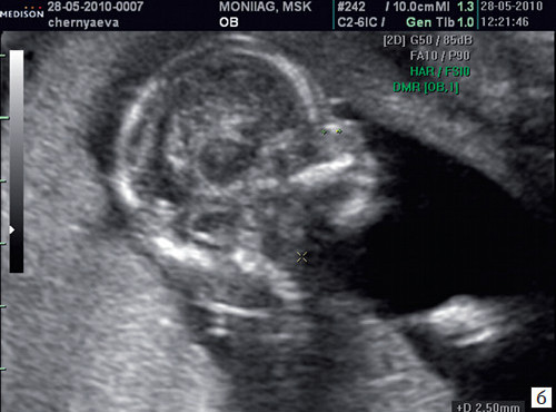
If all the criteria are met, then at the level of the fetal nose, three clearly distinguishable lines should be visible: the upper line represents the skin, down from it a thicker and more echogenic nasal bone than the skin is visualized. The third line, visualized anterior to the nasal bone and more high level than the skin is the tip of the nose (Fig. 1).
Luis Clauido Bussamra, without words, because they are not enough to express my gratitude. Rosian Mattar, for the patience and opportunity to work together and for the commitment to coordinate the postgraduate course in midwifery. Mary Uchiyama Nakamura, Head of the Midwifery Department, for the ease with which she runs this department.
Renato Martins Santana, Head of the Fetal Medicine Discipline, for the support, support and it has always given me. Luciano Nardozza, for the stimulating forces and partnerships at the Fridays. To all the professors of the Department of Obstetrics of Escola Paulista de Medicina, with whom I enjoyed sharing a healthy coexistence on Friday mornings.
Rice. 1. Normal nasal bone.
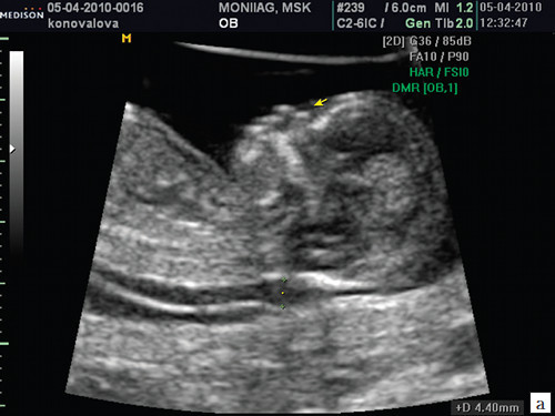
The nasal bone is considered normal when it is more echogenic in structure than the proper skin and pathological if it is not visible (aplasia) (Fig. 2) or its length is less than normal (hypoplasia) (Fig. 3). In the case of the same or less echogenicity of the nasal bone than the skin, the nasal bone is considered pathological (Fig. 4).
For friends of the third age, consisting of odd figures, such as: Sebastiano Saraiva, Guarachi Garcia. Postdoctoral Fellow in the Discipline of Obstetrics, Rosine Pereira Lima Gonsalvis, for her responsiveness and dedication to the guidelines. For the secretaries of obstetrics Natalia Diaz Dofa and fetal medicine, Lucy Alexander, for the organization, dedication.
Friend, Adriana Senudo, strange statistic, for patience and willingness when formulating statistics. To my "sister" Marcia Amalia, for her dedication to my data. My mother-in-law and my father-in-law Maria and Miro Manfredi have always treated me with love.
Rice. 2. Aplasia of the nasal bone.
a) The arrow indicates the echogenic skin of the fetus.
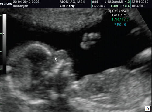
b) The arrow indicates the absence of the nasal bone.
Rice. 3. Hypoplasia of the nasal bone.
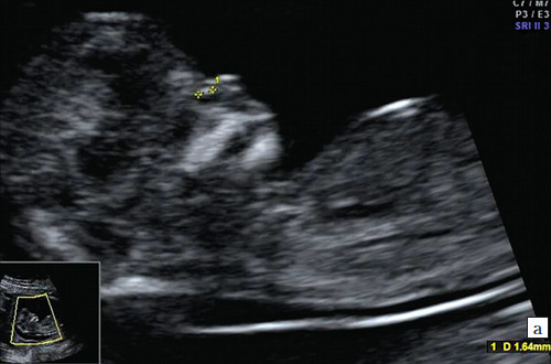
a) Nasal bone at 12 weeks and 2 days, 1.4 mm long (less than the lower limit of the norm).
b) Nasal bone 2.1 mm at 14 weeks in a fetus with Down syndrome.
Rice. 4. Nasal bone pathology
Rice. 4. Reduced echogenicity of the nasal bone.
So, the pathology of the nasal bone is considered:
- lack of nasal bone (aplasia);
- change in its length (hypoplasia);
- change in its echogenicity.
Considering that many studies of this important marker have been carried out in populations of different composition, the data on the frequency of the absence of the nasal bone in different authors differ. So, according to averaged data from multicenter FMF studies at 11-14 weeks, the nasal bone is absent in euploids (in the case of a normal karyotype) from 1 to 2.6% of fetuses, with chromosomal pathologies: in fetuses with trisomy 21 - 60%, with trisomy 18 - in 50%, in fruits with trisomy 13 - in 40%.
Numerous studies have been carried out on the measurement and assessment of the nasal bone at 11-14 weeks of gestation. Some authors suggest evaluating only its presence or absence (+/-). Some works, in addition to assessing the nasal bone, are devoted to its measurement, comparing the length with the standard for this period values.
The evolution of the development of the assessment of this marker and the opinion of specialists in this regard is perhaps one of the most controversial problems that have not been fully resolved in the screening of the first trimester of pregnancy. Most authors consider the assessment of the nasal bone in the first trimester one of the most difficult tasks among all other markers. And this opinion is not without foundation.
Of course, supporters of the theory that for each race (Asians, African-Americans, etc.) and population of peoples (Buryats, Kalmyks, peoples North Caucasus) there must be their own percentile standards for each KTR are right. However, these studies can only be carried out when, as part of a randomized screening for normal fruit multicenter studies with nasal bone measurements will be carried out.
In the program for calculating the risk of Astraia when assessing the nasal bone, there are 4 fields: norm, pathology (aplasia / hypoplasia), it is not clearly visible, it was not possible to evaluate, i.e. in order to diagnose "hypoplasia of the nasal bone", you need to make sure that it is actually less than the standard values for a given gestational age, and this can only be done by measuring it and comparing it with a known standard.
The method of assessing the nasal bone is only "yes / no" when it is suggested only to see nasal bone and compare its echogenicity with the skin is very "apparatus-dependent", i.e. very variable and depends on technical settings ultrasound scanner... When receiving a "hard" image, typical for some with specific factory presets (settings) for examining the fetus in the first trimester, the echogenicity of the skin will always be comparable, ie, the same as the echogenicity of the nasal bone. Thus, practical doctors who are unable to work on premium scanners have objective difficulties in assessing this important additional diagnostic marker.
As supporters of the method of measuring the nasal bone at 11-14 weeks, we present data for the Moscow region. The region is diverse in terms of its ethnic composition. In our work, we used the standard values for the length of the nasal bone published by J. Sonek et al. in 2003, taking the 5th percentile value as the lower limit of the norm (table).
Table. Standards for the length of the nasal bone (NK) in a period of 11-14 weeks.
Experts from the district offices of the Moscow region assessed not only the presence and absence of the nasal bone, but also its measurement in all pregnant women (about 150 thousand examined in 3.5 years of screening). All 31 experts of the Moscow region have a valid FMF certificate of competence both for the TVP and for the assessment of the nasal bone. The analysis of the detection of pathology (aplasia / hypoplasia) of the nasal bone in fetuses with chromosomal abnormalities showed that of the prenatally detected 266 cases of Down syndrome in the fetus in the first trimester, the nasal bone was pathological in 248 cases, which is 93.2%.
This high incidence of nasal bone pathology in Down's syndrome testifies to the correctly chosen algorithm for assessing the nasal bone, which we never intend to abandon, receiving such highly sensitive results, especially with regard to the diagnosis of Down syndrome. In cases of detection of other chromasome abnormalities, the frequency of detection of nasal bone pathology was comparable to the literature data. In Edwards syndrome, the nasal bone is pathological in 78 fetuses, which is 71%, in Patau's syndrome - in 24 (59%) fetuses, with monosomy X - in 24 (42%) cases, with triploidy - in 22 (49%) fetuses.
I would especially like to emphasize that in our study there were 10 pregnant women of Korean nationality who were at risk for chromosomal pathology. Four of them were diagnosed with fetal nasal bone pathology. It could be expected that this is an ethnic feature, however, all these fetuses during prenatal karyotyping had chromosomal pathology(trisomy 21). And, conversely, in 6 fetuses with a normal karyotype, both in length and in echogenicity of the nasal bone were within the normative values for a given period.
In the works of some authors, it was found that with trisomy 21 in the first trimester of pregnancy, only 25% of the fetuses had no nasal bone, at a higher frequency it was hypoplastic (36%).
Since in normal fetuses, the absence of a nasal bone is more common at 11 weeks of gestation than at 13 weeks, FMF gives practical recommendation that if during this period (11 - beginning of 12 weeks) the fetus has no nasal bone, provided normal performance other markers (ultrasound and biochemical) should not take this indicator into account when calculating the individual risk. In the future, it is recommended to conduct an additional ultrasound examination after one week. In the event that the nasal bone remains pathological, this fact must be taken into account when recalculating the value of the individual risk for chromosomal abnormalities.
Assessment of the nasal bone improves the results of combination screening. The detection rate of pathology increases from 90 to 93%. The false positive rate is reduced from 3.0% to 2.5%.
Thus, our own data allow us to recommend evaluating the nasal bone within 11-14 weeks by two parameters: echogenicity and length, taking its absence, hypoplasia, and a decrease in echogenicity for the pathology of the nasal bone.
Literature
- Baranov V.S., Kuznetsova T.V., Kashcheeva T.K. and others. Modern algorithms and new possibilities for prenatal diagnosis of hereditary and congenital diseases. Guidelines... St. Petersburg, 2013.S. 23-46.
- Nicolaides K.H. Screening for fetal aneuploidies at 11-13 weeks // Prenatal diagnosis. 2011, 31: 7-15.
- Nicolaides K.H. Per. from English Mikhailova A., Nekrasova E. Ultrasound procedure at 11-13 + 6 weeks of gestation. St. Petersburg, 2007. Publishing House "Petropolis", 142 p.
- Kagan K.O., Cicero S., Staboulidou I., Wright D., Nicolaides K.H. Fetal nasal bone in screening for trisomies 21, 18 and 13 and Turner syndrome at 11-13 weeks of gestation // Ultrasound Obstet Gynecol. 2009; 33: 259-264.
- Kagan K.O., Staboulidou I., Cruz J., Wright D., Nicoladides K.H. Two-stage first-trimester screening for trisomy 21 by ultrasound assessment and biochemical testing // Ultrasound Obstet Gynecol. 2010. V. 36. N 5. P. 542-547.
- Cicero S., Curcio P., Papageorghiou A., Sonek J., Nicolaides K. Absence of nasal bone in fetuses with trisomy 21 at 11-14 weeks of gestation: an observational study // Lancet 2001; 358: 1665-1667.
- Sonek J.D., Mckenna D., Webb D., Croom C., Nicolaides K. Nasal bone length throughout gestation: normal ranges based on 3537 fetal ultrasound measurements // Ultrasound in Obstetrics & Gynecology. 2003. V. 21. No. 2. P. 152-155.
- Kanellopoulos V., Katsetos C., Economides D.L. Examination of fetal nasal bone and repeatability of measurement in early pregnancy // Ultrasound Obstet Gynecol. 2003 Aug; 22 (2): 131-4.
- Cicero S., Bindra R., Rembouskos G., Tripsanas C., Nicolaides K.H. Fetal nasal bone length in chromosomally normal and abnormal fetuses at 11-14 weeks of gestation // Matern Fetal Neonatal Med. 2002; 11: 400-402.
- Keeling J. W., Hansen B. F., Kjaer I. Pattern of malformations in the axial skeleton in human trisomy 21 fetuses // Am J Med Genet. 1997; 68: 466-471.