Is it possible to put multiple sclerosis on mri. Treatment of multiple sclerosis, drugs used in the treatment. MRI as one of the most effective diagnostic methods
Currently, the most informative instrumental method for the diagnosis of multiple sclerosis is magnetic resonance imaging (MRI). The method allows high efficiency visualize pathological foci in the CNS. To speak unequivocally about the diagnosis, to distinguish a number of pathological conditions occurring under the mask of multiple sclerosis, systematized tomographic signs help. It was magnetic resonance imaging that made it possible to establish the multifocal lesion of the central nervous system with multiple sclerosis. The principle of its operation is based on the existence of a natural difference in the content of hydrogen atoms (protons) in water and lipids. The intensity of the signal depends on the density of protons in the tissue, the proton relaxation time (T1 and T2), the pulse repetition time, and the echo occurrence time. Image contrast is achieved due to the different content of water and lipids in the tissues and their areas. In addition to studying the proton density, it is possible to adjust the device in order to emphasize the T1 or T2 relaxation time, which makes it possible to enhance the image contrast - T1 and T2 modes. In T1 mode, the normal white matter of the brain has a light signal, in T2 mode it is dark. Demyelination foci due to the increased water content have a signal of reduced intensity on T1- and increased on T2-weighted images.
Camelia Babes was diagnosed with multiple sclerosis at the age of 20 when she was a happy student climbing mountains with her backpack. After 15 years of isolation, but after he got up with this disease, he got out of bed, graduated from his faculty and even made a master. Now they are fighting for people with multiple sclerosis to get medicines, just like European patients, to open more centers so that patients do not cross the country from one end to the other. for advice and treatment.
Tests for viral infections
For years, Camellia didn't want to leave the house, see her friends, or talk to the world. She felt the need to remain isolated from her illness and her family, her husband's parents and daughter. The disease first affected her leg, then a few months later and the second.
Typical localization of demyelination foci in the brain is the periventricular zones, more often in the angle between the caudate and corpus callosum, in areas adjacent to the upper lateral angle of the lateral ventricles, in the white matter of the semioval center, temporal lobes, as well as in the brain stem and cerebellum. At the border of gray and white matter or in the gray matter there is a small proportion of foci (5-10%). Their size is from 0.2 cm to 3.0 cm, more often oval or rounded. In the spinal cord, lesions are usually oblong in shape and located along the axis spinal cord, reaching a size of 2.0 cm. The total number of foci In each patient, the total number of foci varies significantly. Large lesions may result from the emergence of an active new lesion with a significant area of edema or fusion of individual plaques. As a result, on late stages diseases, the formation of very large foci (up to 8 cm) is possible, which may need to be differentiated from brain tumors.
Being very ambitious, it was terrible to stop doing nothing, not to go alone, not to depend on others. In fact, the hardest thing is to admit my illness. He graduated from his faculty, made a master and devoted himself exclusively to helping the sick. So he came to Bucharest as president of the Craiova Multiple Sclerosis Society to discuss with the Minister of Health Florian Bodog and other authorities about the needs of patients. They met at round table organized by the Multiple Sclerosis Society.
These patients have many problems. Agglomerated centers, multiple sclerosis drug list not updated for two years, patients in the country treated in Bucharest who cannot be transferred to proximity centers are just some of the problems of those who suffer from this disease.
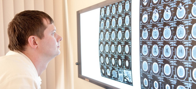
The use of MRI in T1 and T2 modes visualizes these foci and makes it possible to prove dissemination of the process. On T1-weighted images, demyelinating lesions appear darker than normal white matter (“black holes”). These are chronic lesions reflecting the loss of axons. In T2 mode, the plaques appear bright white. In this mode, the assessment of the focus volume is more informative. To identify foci, it is advisable to conduct both sagittal and axial sections. The most revealing is the parasagittal scan at the level of the lateral ventricles.
The multiple sclerosis treatment program is not run by the Ministry of Health, but Florian Bodog promised them help and told them that every new treatment center established or already operating in Romania should provide safe care for patients with multiple sclerosis.
Patients from other counties who can benefit from treatment travel hundreds of kilometers, although most of them have a social budget. The second problem is the transfer to new centers, because after the authorization commissions for the treatment dossier, which was in the Ministry of Health until winter, the clinics now had some reserve for transferring to other centers, from administrative fear, says Laurentiu Lazar.
Often, studies of proton density in both modes are not informative enough to make a diagnosis of multiple sclerosis. In this regard, contrast agents have been introduced into practice. The use of contrasting helps to identify small foci of demyelination that are not visualized with non-contrast MRI. In addition, the accumulation of contrast allows you to determine the degree of activity pathological process. The appearance of a white ring around the old focus in T2-weighted mode due to the accumulation of contrast indicates an exacerbation of the process. Thus, the use of contrast agents allows for differential diagnosis between foci of active inflammation, foci in the stage of fading exacerbation, and chronic inactive foci. It should be noted that dynamic control does not always make it possible to unambiguously assess the activity of the disease due to the observed motley picture: some plaques disappear, others appear, the affected areas increase and decrease in size at the same time.
Patients with multiple sclerosis receive free treatment but only have access to 7 drugs, while in other EU countries those who suffer from this disease have 13 drugs. Therapy for a patient with multiple sclerosis costs about $000.
Role of contrast enhancement
Currently, 930 patients are enrolled in the National Treatment Program in 13 authorized clinics, out of a total of over 1000 patients evaluated in Romania. Multiple sclerosis is a chronic neurological disease, the mechanisms of which are not fully known. It is known to be an autoimmune disease that causes the body to attack its own cells. It affects the central nervous system with physical and emotional consequences.
Demyelination, as the next stage in the development of foci in multiple sclerosis, can also be determined on MRI (loss of a short echo of the T2 signal). On the this stage it is expedient to use magnetic resonance spectroscopy (MrS), with the help of which it is possible to determine the activity of the process in the early stages and, in addition, to evaluate the biochemical and neurophysiological manifestations of demyelination. Proton spectroscopy is most often used. This technique allows you to evaluate the functional and biochemical processes in the CNS based on the content of certain metabolites in it, such as: N-acetylaspartate (NAA), creatine (Cr), choline-containing compounds (Cho), inositol (Ins), taurine (Tau) and, in some conditions, lipids. In multiple sclerosis, there is an increase in the content of choline in the foci, which reflects the degree of destruction of myelin, since choline-containing compounds are necessary component myelin membrane. Another symptom characteristic of multiple sclerosis is a decrease in the intensity of NAA signals relative to Cr and Cho signals. Low level index NAA/Cr is the most characteristic change in the foci of multiple sclerosis.
Multiple sclerosis is by no means a mental, fatal, hereditary or contagious disease. The disease cannot be cured. However, Romania has a national program offering free medicines for those suffering from multiple sclerosis. This treatment does not stop the disease, but improves the patient's condition.
Diagnosis is difficult. Certain symptoms and imaging of the brain are important in diagnosing the disease. There are no tests that can be used to say yes or no. None of the studies that help doctors diagnose multiple sclerosis is 100%. reliable. Various Methods have been used to help doctors diagnose the disease as early as possible. Due to improved diagnostic methods for early stage disease doctors can start treatment for multiple sclerosis.
In patients with multiple sclerosis, a pattern was revealed between changes on MRI and variants of the clinical course of the disease:
- at the debut of the disease, as a rule, there is at least 1 large focus (1.5-2 cm) with fuzzy contours. On average, the size of the foci is 0.5-0.8 cm. There is a clear tendency to merge. The plaques are oval or round shape and oriented parallel to the subependymal veins. MRI control in dynamics shows the reversibility of acute demyelination;
- with relapsing course of multiple sclerosis (up to 1 year) - a large number of foci and the presence of one large lesion (up to 2.5 cm);
- with a relapsing course of multiple sclerosis (more than 5 years) - a large number of medium-sized foci that increase in size or reappear with each exacerbation of the disease;
- in the primary progressive course of multiple sclerosis - a small number of foci of predominantly subepindemic localization in the region of the horns of the ventricles. The defeat of the corpus callosum is not necessary;
- in the secondary progressive course of multiple sclerosis - initially, as in the case of the primary progressive course, then - the appearance of chronic periventricular confluent foci.
It should be noted that the MRI picture, taken in isolation, is not an absolute criterion in the diagnosis of multiple sclerosis.
When examining a patient, doctors look for as much evidence as possible to confirm the disease. For this purpose, neurological studies, magnetic resonance imaging, laboratory tests, lumbar puncture and induced potential studies that determine the electrical conductivity of the brain and spinal cord or motor pathways.
How does it work, is multiple sclerosis visible on an MRI?
A neurological examination determines the functioning of the nervous system. Study of changes in vision, hearing, speech and sensory disturbances. Later, the reflexes of the elbows, wrists, knees, ankles and shoulders are examined to determine the damage. Examination of the stomach and leg may reveal ataxia or loss of sensation, indicating damage to the spinal cord or brain. The doctor may also examine hearing, linguistic changes, and facial abnormalities. The optic nerve, which travels through nerve impulses to the brain, is a common site that is damaged.
In 2001, the diagnostic criteria for multiple sclerosis recommended by the International Expert Group were adopted, supplemented in 2005. Changes diagnostic criteria adopted taking into account the special role of MRI in the diagnosis of multiple sclerosis in patients with different start diseases.
Multiple sclerosis is very dangerous disease neurological type, characterized by a gradual loss of human control over their movements, impaired speech, breathing, deterioration intellectual abilities and attention. As a result of all this, a person can remain an invalid, bedridden for the rest of his life.
It is examined by examining vision, visual acuity, and eye examinations. This is the only visual examination used in the diagnosis of the disease, but its results cannot be considered definitive. To do this, use contrast enhancement. This study, along with the medical history and neurological examination, is very important in determining multiple sclerosis.
Visualized potentials are useful not only for diagnosis, but also for assessing the course of the disease. In this study, damage to the optic nerve is determined. This is why a patient with normal vision can help diagnose multiple sclerosis. Diagnosis of multiple sclerosis is not direct, so laboratory testing can be helpful. extra help. One of the studies is the removal of fluid from the spinal cord.
But in the case of multiple sclerosis, it all starts with the central nervous system, or to be more precise, then. The point is that in this body an atypical situation occurs - healthy cells begin to perceive their "relatives" as foreign substances.
Thus, the body, including the mechanism of protection from external influences, begins to produce antibodies that are designed to fight those cells that cause mistrust, although in fact they can be absolutely healthy.
A lumbar puncture is used to confirm or reject a diagnosis of a disease. Several studies can be done from liquid samples that indicate disease activity. When examining the liver, most people found signs of the disease. Diagnosis cannot be based on responses from a single study. All of these tests help determine the final diagnosis, but each should be evaluated by a responsible, an experienced specialist. Diagnosis should determine the type of path of the disease.
Using different modes of MRI diagnostics
Four are known different kind multiple sclerosis. Recurrent remission is the most common, starting at 80%. with multiple sclerosis. This form is present only at the beginning of the disease, and then passes into secondary progressive multiple sclerosis. On the initial stage there may be no symptoms for many years, but flare-ups are unpredictable and can occur at any time. New or earlier existing symptoms may come on suddenly, last for days or weeks, and then go away again.
Based on this, it is classified as an autoimmune disease - ailments in which a person's immunity fights with its own healthy cells. This serious violation can be caused by various reasons.
Often a sick person at first does not even suspect that such a restructuring of immunity is taking place in his body. Signs of the disease begin to appear later. Therefore, in order to fight multiple sclerosis as successfully as possible, it would be useful to know its possible root causes, the factors that cause it:
Up to 40% of patients with relapsing multiple sclerosis develop into secondary progressive MS. After exacerbations and remissions, the secondary progressive form of the disease can develop continuously with or without significant relapses, less periods of remission or stabilization. Primary progression is very rare and affects only 10%. patients with multiple sclerosis. From the very beginning of the disease there is a constant progression. Symptoms develop and disability gradually increases. There will be no flare-ups or remissions, only occasional periods of stabilization or temporary minor improvements.
- genetic factor. If there are relatives in the family of a person who suffer from multiple sclerosis, then he is at risk
- Hormonal changes in the body
- Transmitted viruses and infections
- Various brain lesions - injuries, tumors, cysts
Multiple sclerosis doesn't just affect the elderly. At risk are also young people and people in adulthood. This fact indicates that one should not neglect one's health and one should monitor various external symptoms.
Beneficial manifests itself with a slight exacerbation and possible additional relapse. The patient is then healed up to possible second exacerbation, which may be absent for 20 years, and the disease may progress slightly. In the next issue of the healthy person, read about life with multiple sclerosis.
Multiple sclerosis is an inflammatory demyelinating disease of the central nervous system, usually beginning on its own. early age, is characterized by recurring or continuous progressive neurological symptoms of dysfunction that cause progressive disability. This is a fundamental neurological disease. It is hard to find another disease of similar importance that will receive so much attention and leave so many unanswered questions.
Symptoms of multiple sclerosis
The disease is very dangerous, first of all, because its signs for a very long time at its early stage do not manifest themselves outwardly. Moreover, even with a diagnosis that includes a clinical general examination, the specialist will not reveal any deviations from the norm.
Thorlakur tells the story of a Viking woman whose blindness and disorientation returned after several days of prayer. The prevalence of the disease is highly dependent on geographical factors and the composition of the ethnic population. With multiple sclerosis, women are 1.6-2 times more likely than men.
Exacerbation activate T cell adhesion molecule is promoted, adhere to the venules and penetrate the endothelium, such as through the blood-brain barrier, enter the finesse of the brain. It accumulates at the site of injury and monocytes, increased pro-inflammatory cytokines: interleukin-2, interleukin-12, interleukin-15, interferon-gamma, tumor necrosis factor alpha production. Important and humoral immunity, since activated B-lymphocytes and antibodies attack surface myelin antigens and oligodendrocytes, which are responsible for remylinization.
In this case, there are two options left - either wait for the first symptoms to appear, or sign up for a magnetic resonance imaging procedure. Tomography also initially may not give the desired result.
Therefore, in his observations, the doctor often relies on the patient's description of his feelings. Multiple sclerosis can be recognized by the following symptoms:
The antibodies bind to myelin and activate complement and attract macrophages. The gland's asthma spreads when myelin and oligodendrocytes disappear. Plates are heterogeneous, consisting of lymphocytes, macrophages, demyelinated axons. Clinic Symptoms of multiple sclerosis are very diverse, and the disease can begin in a completely different way. However, it is indicated that the disease is also characterized by general fatigue, mood, cognitive impairment, paroxysmal symptoms and pain. These are central neuropathic pain, pain caused by demyelination, various discastheses due to pulmonary edema, inflammatory, visceral and orthopedic pain.
- deterioration of vision. With this symptom, a person usually suffers from one eye, which can simply stop seeing. In patients with multiple sclerosis, the eyes become very sensitive to bright light, they often water,
- headaches of various nature
- deterioration of intellectual abilities, attention, memory
- coordination. This symptom It is associated primarily with the appearance of abnormal reflexes in a person and with the partial inability to perform the movements that the patient conceived. The point is that at autoimmune diseases the connection between the brain and the neurons going from it to the limbs and back is broken or inhibited. Healthy man does not think about any action, performing it mainly on the machine with the help of the lightning speed of the response of the limbs to signals from the brain. In multiple sclerosis, this connection first slows down, and then slowly breaks down, while being observed with voluntary movements
- . One of the most severe symptoms, which may follow from the previous
- urinary incontinence, impotence
- frequent stressful and unstable emotional states
All these signs are interconnected, they characterize pathologies of an autoimmune nature, therefore, they can represent the development of multiple sclerosis. Wherein, this disease cannot be cured by getting rid of its original causes, it must be considered as a separate substance.
It is controlled in about one-fifth of patients and is diagnosed if, after exclusion of other causes, neurological symptoms progress for more than one year. If there are many fireplaces, they are relatively symmetrical, more periventricular. Preliminary potential studies are important for diagnosis as they may be clinically asymptomatic. Immunosuppression with cyclophosphamide and mitoxantrone and the less potent azathioprine and methotrexate are used to treat progressive, non-flaring forms.
For these products, hematologists or chemotherapists should be consulted.
It is controlled in about one-fifth of patients and is diagnosed if, after exclusion of other causes, neurological symptoms progress for more than one year. If there are many fireplaces, they are relatively symmetrical, more periventricular. Preliminary potential studies are important for diagnosis as they may be clinically asymptomatic. Immunosuppression with cyclophosphamide and mitoxantrone and the less potent azathioprine and methotrexate are used to treat progressive, non-flaring forms.
MRI - diagnosis of multiple sclerosis
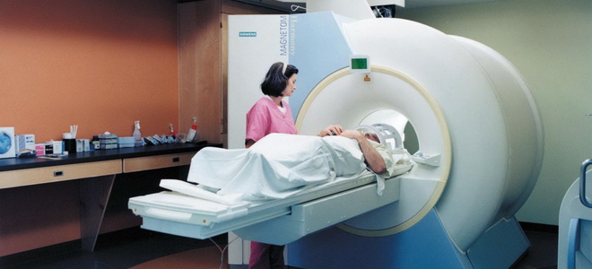
Most informative way diagnosis of multiple sclerosis - magnetic resonance. MRI, due to its safety and clarity of the obtained images, is used everywhere in this disease.
The fact is that with the help of tomography, it is possible not only to detect foci of brain damage, but also to exclude some other pathologies. Doctors, studying the development of foci of the disease, can predict its course, and therefore prescribe an effective treatment.
Of course, it should be noted that even when applying, specialists face certain difficulties when making a diagnosis. Multiple sclerosis is a specific disease that manifests itself at stages in different ways, so it is very difficult to predict and predict something about the symptoms and development of the disease.
MRI is a study, the basis of which is the effect of electromagnetic waves generated by the device. magnetic field on brain tissue. These waves find a response in the tissues, repelling them and giving an image to the monitor.
The amplitude of the reverse waves emanating from the very structures of the brain, their clarity, frequency can indicate the presence or absence of pathologies, because healthy and affected tissues react differently to electromagnetic impulses.
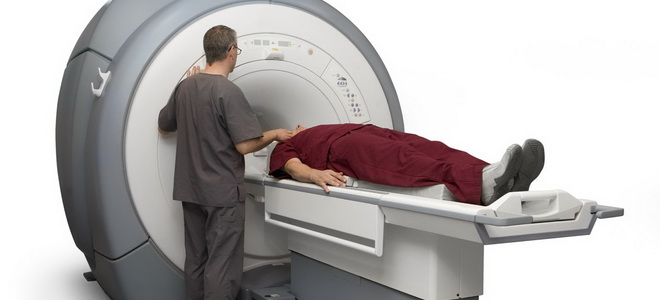
Of course, the MRI procedure for suspected multiple sclerosis is carried out on a high-field device that has the maximum power of the electromagnetic field, therefore, creating the most detailed image. Thus, the doctor in the pictures can see all the structures of the brain.

To exclude some other pathologies from the suspicions of specialists, a contrast agent containing iodine may be introduced into the body of the subject. The contrast, dissolving through the blood, provides clear information about the state of the blood vessels, showing in detail vascular network within the cranial cavity.
The contrast agent due to its chemical features tends to tend to places where pathology is born, accumulating there. According to the pictures obtained as a result of the survey, this is quite realistic to follow. Therefore, it is possible to exclude with the help of contrast and some types infectious diseases and degenerative disorders of the brain.
Signs of multiple sclerosis on MRI scans
When deciphering images, specialists should rely not only on comparisons of healthy and affected brain structures. It is also important to identify some parameters characteristic of multiple sclerosis. These signs are easy to see in the pictures, and if they occur, then we can talk about an emerging or already developing pathology.
- accumulation of contrast in inflamed and affected areas
- an increase in lesions during several MRI procedures separated by certain intervals of time. Multiple sclerosis is a disease that develops gradually. This can be seen both in the slow onset of symptoms and in the MRI series.
- swelling around the lesion
- lesions of the brainstem, cerebellum, spinal cord
- noticeable emergence of new lesions in various areas of the brain as a result of a series of several procedures
Among other things, tomography in multiple sclerosis can reveal disorders such as compression of blood vessels or nerve endings by the bones of the spine. This can be affected, for example, by osteochondrosis cervical spine, intervertebral hernia etc.
Compression and interference with the functioning of the spinal cord can cause the development of multiple sclerosis, because it is a kind of mediator in the relationship between the brain and the body.
MRI results in multiple sclerosis
Usually, experts are satisfied with the images obtained with MRI, because the procedure allows you to recreate images from several angles. Magnetic resonance imaging is indicated in the case of this disease not only for its diagnosis, but also for further monitoring.
Disease monitoring includes monitoring possible development disease and verify the correctness and effectiveness of treatment. Therefore, if a patient is diagnosed with multiple sclerosis, then he needs to be prepared for a series of MRI examinations, with the help of which control will be carried out.
These arguments are another positive moment in the use of MRI - a procedure for suspected multiple sclerosis.