Ultrasound of the intestines and abdominal organs. What is included in a comprehensive ultrasound of the internal organs of the abdominal cavity for a child and an adult
Hello!
Taking care of your health is important and necessary. Today's review will focus on ultrasound diagnostics of organs abdominal cavity... The procedure is necessary and useful, it can help identify abnormalities in the organs of the abdominal system.
I was sent for examination by an endocrinologist, after my appeal to her with a prolonged fever (one of the scheduled examinations).
The following organs are included in the abdominal cavity:
liver, pancreas, spleen and gallbladder. You can also do ultrasound obp (abdominal organs) with the kidneys (look at the kidneys and adrenal glands). I was prescribed just such an examination.
I underwent research at the Persona clinic. Along with the examination of the abdominal cavity on the same day with one specialist, I did ultrasound of the pelvic organs and the thyroid gland.
It is written on the door of the office that it is better to turn off the Cell phones since they have a bad effect on the ultrasound machine.
Preparing for a diagnostic examination:
- The study is carried out on an empty stomach, preferably in the morning. If the study is carried out after 15:00, you can have breakfast at 8-11 hours, after which food and water should not be consumed.
- On the eve of the study, it is necessary to exclude from the diet foods that increase gas production in the intestines (white cabbage, peas, beans, beans, fresh apples, grapes, tomatoes, kefir, fermented baked milk)
- It is not recommended to chew gum, suck lollipops, or smoke.
I signed up for 10:30. I haven't eaten or drank since morning. It felt as if I had not fully woken up. After all, when you eat the body starts up. But in general, it is tolerable without food. After the examination, I went to work and had a snack there.
The whole the necessary training I observed.
The doctor who performed the ultrasound was very pleasant. Before the study, the required area was smeared with a special gel. The survey began with thyroid gland, then there was the abdominal cavity and kidneys, and then the small pelvis. The abdominal cavity was examined the longest. Looked through the front abdominal wall and from both sides. Several times it was necessary to roll over and change position. We looked at the gallbladder with special attention, under the right hypochondrium. There were deviations.
Abdominal ultrasound result:
an ultrasound protocol is issued with the signature of the doctor and the seal of the organization.
It indicates the course of the study, the size and features of the organs. In conclusion, the ultrasound specialist wrote that I have echoscopically - diffuse changes in the parenchyma of the pancreas and chronic acalculous cholecystitis. Based on the results of the examination, the ultrasound specialist recommended contacting a gastroenterologist.
Price my examination was 1000 rubles. If they looked at the abdominal organs without kidneys, the cost would be 750 rubles.
By time ultrasound was carried out for about 15 minutes. After the examination, the doctor gives wipes to remove the remaining gel.
Outcome: doing ultrasound of the abdominal organs is not scary, painful and not expensive. In the process of carrying out, abnormalities in the abdominal organs are revealed. Preparation for the survey is not difficult, but due to the limitations that it carries, I reduce one point to the survey.
Abdominal ultrasound is the most painless and reliable method examinations of the intestines, liver, stomach, spleen and other organs. Diagnostics is based on the use of ultrasound. The procedure is completely safe for health, it helps to identify the development of diseases and any pathological changes.
Indications for the appointment of ultrasound of the abdominal cavity
IN preventive purposes An abdominal ultrasound is recommended annually. But since the procedure is paid and takes some time, most patients ignore the advice of doctors and do not undergo a timely examination, but seek help only when symptoms of diseases appear.
Diagnostic indications:
- excessive gas formation in the intestines;
- systematic pain in the abdomen;
- bitterness in the mouth;
- feeling of heaviness or pain after eating;
- abdominal trauma;
- suspicion of appendicitis;
- diseases of the genitourinary system;
- diagnostics of tumors, metastases;
- pregnancy.
Ultrasound diagnostics usually take about 25-60 minutes. All this time, the patient must lie still, then the image quality will be high, and the results are reliable.
What organs are examined with ultrasound of the abdominal cavity
Be sure to examine the gallbladder and the pathways that excrete bile. Ultrasound allows you to detect wall suppuration, anomalies in the development of an organ, as well as cholecystitis of any form. When examining the pancreas, abscesses, tumors, and glands can be seen. You can see the damage caused by infections with herpes, cytomegaly, toxoplasmosis. Diagnostics helps to identify such life-threatening diseases as pancreatic necrosis and acute pancreatitis.
Diagnosis with ultrasound waves of the abdominal aorta is performed to detect an aneurysm. If the pathology is identified, the patient additionally needs to undergo an abdominal tomography.
When an infection “wields” the spleen, it increases in size. On ultrasound, this increase is noticeable. When analyzing the stomach and upper small intestine, duodenoiditis or gastritis can be seen. With these pathologies, the thickness of the organ wall changes.
Examination of the kidneys is not included in the ultrasound of the abdominal cavity; the doctor prescribes such an analysis separately. The procedure is more expensive and requires preparation - urine collection in the bladder.
How to properly prepare for an abdominal ultrasound
For the result to be as accurate as possible, the patient must prepare his body for examination.
Compliance with a diet (at least 3 days before the procedure) - salty, smoked and others should be excluded from the diet harmful products... Cannot be consumed whole milk, cabbage, raw vegetables and legumes as they can cause bloating. It is allowed to eat vegetable soups, chicken meat, cereals, fish and low-fat cheeses.
Reducing flatulence in the intestines - on the eve of the examination, you can drink Espumisan or Activated carbon... In case of constipation, Smecta is taken about two hours before the ultrasound.
Filling with water Bladder- if you need an ultrasound of the kidneys, then one hour before the examination you need to drink half a liter of still water.
Cleaning with an enema - the procedure is necessary for patients with suspicion of damage to the intestines and stomach.
Children before ultrasound diagnostics you can not drink for an hour and eat food for at least 4-5 hours. Better ultrasound do in the morning on an empty stomach.
How is the ultrasound of the abdominal cavity
The algorithm for passing an ultrasound of the abdominal cavity is simple. The patient needs to undress to the waist and lie on his back, the doctor will apply a gel conductor to the abdominal skin and start examining the organs. Sometimes you need to hold your breath or roll over to get a clearer picture. The main thing is to listen to a specialist, he will tell you what position you should take. The procedure is painless and does not cause any discomfort.
Service cost *
*Attention! Prices shown are as follows reference Information and are not a public offer. Check the current prices by phone and directly at the clinics.
Ultrasound procedure(Ultrasound) is an informative, non-invasive, practically safe examination internal organs person.
The main obstacle to abdominal ultrasound is the presence of air. Therefore, the main task of preparing for an ultrasound scan is to remove all excess air from the intestine. It is very important to prepare for an ultrasound scan for obese people, as fat is the second most important obstacle to ultrasound.
In the process of examining an ultrasound of the abdominal cavity, an analysis of such organs as the liver, gallbladder, pancreas, spleen, retroperitoneal space, vessels is carried out.
What organs are included in the survey?
The sizes of organs are estimated, their internal structure, localization, the presence of additional neoplasms, inflammatory processes and foci, changes are determined that are characteristic of chronic pathologies and traumatic injuries. The study of the abdominal organs includes the following.
- Liver: the possibilities of ultrasound examination make it possible to determine the presence of both diffuse diseases (acute and chronic hepatitis, cirrhosis, fatty infiltration, changes associated with pathologies of the heart and heart failure), and focal formations: benign (cysts, hemangiomas, adenomas, focal nodular liver hyperplasia), cysts (echinococcal, etc.), or of a malignant nature (primary liver cancer, metastases).
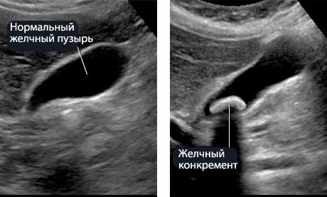
- Gallbladder: abnormalities in the appearance of the bladder and bile ducts, cholelithiasis and its complications are determined, inflammatory pathologies(acute and chronic cholecystitis), polyps, benign tumor lesions, malignant lesions.
- Pancreas: developmental abnormalities are detected, inflammatory diseases: acute and chronic pancreatitis and its complications (pseudocysts, abscesses), non-neoplastic lesions (cysts, fatty infiltration); benign and malignant tumor lesions, age-related changes.
- Spleen: ultrasound this body most appropriate for suspected malformations, as well as for damage to the spleen, which are most often observed with physical damage to the abdominal organs. Moreover, an increase in the spleen with its inflammatory lesions and with concomitant liver pathologies is established, cysts, heart attacks, abscesses, neoplasms, changes in systemic diseases blood.
- Vessels: analysis of the placement of the main and intraorgan vessels, their sizes, the state of the lumen, the detection of blood clots.
Indications
An abdominal ultrasound is done to a child or adult if the patient suspects chronic or acute pathologies inflammatory nature: liver cirrhosis, hepatitis, pancreatitis, cholecystitis, malignant or benign tumors... This type of diagnostic research must be carried out every time even the slightest suspicion of a disease in the body arises.
Due to a timely preventive examination, which is usually carried out annually, there is a high probability of detecting and preventing the disease if it is at an early stage.
An abdominal ultrasound is necessary if the patient has symptoms such as:
- heaviness in the stomach and a feeling of fullness after eating;
- heaviness in the right hypochondrium;
- pain in the abdomen (especially in the upper section);
- sharp pain in the lower abdomen;
- bitterness in the mouth;
- active gas formation.
How is the survey carried out?
An abdominal ultrasound is done in horizontal position patient on his back. In some cases, in order to obtain a better image, the doctor asks the patient to turn on the right or left side, take a deep breath, and hold his breath. Some patients with individual characteristics(for example, with a high position of the spleen) it is necessary to examine while sitting or even standing. In the process of carrying out this type of research, the specialist diagnoses, specifies and tracks the state of the abdominal organs in dynamics, determines the diseases and pathology of the spleen, changes in its density and increase in size, the presence of damage. The specialist should determine the factor of the occurrence of vague pain, accompanied by cramps in the abdominal cavity. He must exclude the presence of formations, calcifications, hemangiomas, hepatosis, cysts. Based on the results of the ultrasound, the doctor writes out a study protocol with a conclusion.
What shows
By means of ultrasound examination, it is possible to exclude or confirm real hypertension, pancreatic pathology, for example, oncological processes and pancreatitis. Ultrasound helps to monitor the biopsy, confirm ascites and paracentesis. This method diagnostics is simply necessary before performing abdominal surgical interventions... Nowadays, this diagnosis helps to identify inflammatory changes in the body, additional education, bulky neoplasms and chronic diseases.
Thanks to ultrasound, it is possible to detect in a timely manner wrong development and the formation of internal organs, because most of the defects occur in a person even during the period intrauterine development... Often, together with an ultrasound of the abdominal cavity in the female part of the population, an examination of the uterine cavity is carried out at the same time. As a rule, this diagnostic method is used when it is necessary to detect pregnancy or to establish the presence of polyps in the uterus, ovarian cysts and other formations. Due to this, you will not need to carry out additional numerous and expensive studies, pass a number of numerous tests, and immediately you will be able to start surgical and effective way treatment.
The cost of an abdominal ultrasound will be available to everyone. Many patients wonder where is the best place to perform given view survey. Who to trust: doctors from state clinics or private medical centers? In fact, it makes absolutely no difference. It all depends on whether a particular room has sufficiently innovative and high-quality medical equipment, as well as on the experience of a team of professionals who will directly carry out the ultrasound procedure itself.
During the examination of the peritoneal organs, no unpleasant sensations should not occur. After applying the gel, patients experience light feeling cold, which then passes. As the sensor moves over the body, different sensations... There may be pain when the sensor touches the hypochondrium. The emergence pain of an acute nature is a sign that the patient urgently needs an examination by a surgeon and the ultrasound procedure itself has nothing to do with it. The duration of the study is about twenty minutes. It is worth taking the time to prepare for the procedure itself in order to perform it correctly. This is, first of all, diet, smoking cessation, timely bowel cleansing, during ultrasound of the kidneys - timely filling of the bladder.
Ultrasound examination (ultrasound) - informative, non-invasive, practically safe research internal human organs.
The main obstacle to an ultrasound scan is the presence of air. therefore the main task preparation for an ultrasound scan is to remove all excess air from the intestines. Preparation for ultrasound is especially important for obese people, since fat is the second most important obstacle in the way of ultrasound.
Training:
Diet:
2–3 days do not consume black bread, milk, carbonated water and drinks, vegetables, fruits, juices, confectionery, alcohol.
In the absence of contraindications, you can also take any enterosorbent (polysorb, polyphepan, "white coal", enterosgel) in a standard dosage, it is also desirable to make a cleansing enema 1.5-2 hours before the study.
The study is carried out strictly on an empty stomach (at least 6, and better - 12 hours after eating). For example, the pancreas in a living person is located behind the stomach, and when the stomach is full, it is practically invisible on ultrasound.
Ultrasound examination of the abdominal organs.
With the help of ultrasound, parenchymal organs can be examined, as well as hollow organs filled with fluid. In the abdominal cavity, these include liver, gallbladder, pancreas and spleen, bile ducts. Kidney anatomically located in the retroperitoneal space, but usually they are examined together with the above-named organs of the abdominal cavity.
The intestines and stomach are hollow organs in which air is almost always present, so it is extremely difficult to examine them. And although very good preparation the patient to an ultrasound scan allows partially to examine the walls of the stomach and colon, these techniques are extremely difficult, time-consuming and painful for patients (the colon is first completely emptied using siphon enemas, and then filled with liquid). Therefore, a simpler and more informative method is used to study the intestines - colonoscopy.
Ultrasound is performed with the patient lying on his back. Sometimes a doctor to receive the best picture asks the patient to turn on the right or left side, take a deep breath, hold the breath. Some patients with individual characteristics (for example, with a high position of the spleen) must be examined while sitting or even standing.
In the process of ultrasound, the dimensions liver, her position, shape, ability to transmit ultrasonic waves, structure, condition of blood vessels and bile ducts, presence of foreign inclusions(for example, stones), shape, condition of the walls, size of the gallbladder, its position, the state of bile, the presence of foreign inclusions, the structure, shape, position, the ability to transmit ultrasonic waves, the state of the pancreatic duct, are studied the state of the biliary tract (with measurement of their lumen), portal, inferior vena cava and splenic veins. The same scheme is used to evaluate pancreas, spleen, kidney. At the end of the study, the general state top floor abdominal cavity.
Based on the results of the ultrasound, the doctor writes a study protocol with a conclusion.
Important note. We have all seen photographs of internal organs obtained with the help of ultrasound machine- echograms. They are not the subject of study, they are not commented on. and serve only as an additional, optional supplement to the ultrasound examination protocol.
| № | Name | Price |
|---|
| 706 | Liver ultrasound | 1200 | |
| 707 | Ultrasound of the gallbladder | 1000 |
Modern ultrasound examination is performed using quality equipment, therefore is irreplaceable assistant when examining the state of health. A specialist with its help can thoroughly study the functioning of the liver, bladder, kidneys with spleen and other organs.
Finding out what exactly includes an abdominal ultrasound, it must be borne in mind that it makes it possible not only to assess the size, shape, location and structure various bodies, but also in time to identify many diseases, abnormalities, not only congenital, but also acquired. Thanks to this, the patient receives effective treatment and recovers.
The study is notable for its painlessness, absolute safety, high efficiency, and it takes no more than half an hour. With its help, diseases are diagnosed not only in adults, but also in small patients, and the normality of development is checked.
It is advisable that people undergo this comprehensive ultrasound examination at least once every couple of years. Preventive examination using ultrasound examines the liver, the state of the gallbladder, spleen, kidneys with adrenal glands, retroperitoneal space and pancreas, whether there is free fluid. It is to this study that the doctor instantly sends people complaining of various pains in the abdomen, any incomprehensible and unpleasant symptoms.
People with inflammation, stones and sand, cysts in various organs and disorders of urine outflow are looked at for ultrasound. An abdominal ultrasound is the first procedure after any trauma or injury, even if at first the person does not complain of pain. Indications can be not only painful sensations but also bitterness in the mouth, unpleasant severity in the stomach, too active formation of gas, nausea. Patients are sent there with hints of oncological diseases, various infections, inflammation, and functional problems.
Despite the ease of passing the examination for the patient, ultrasound of the abdominal cavity still requires some preparatory procedure, because the correctness and effectiveness of the data obtained depends on it. When figuring out what the research includes, you must not forget about the preparatory measures.
They differ from what kind of organ will be subject to inspection.
- For examination of the liver with the gallbladder, as well as the pancreas with the spleen, nothing should be taken by mouth twelve hours before the appointed date. Make your last meal light and small. Ideally, it should include a slice of dietary fish or chicken fillets garnished with vegetables.
- Kidney examination prescribes a liter pure water an hour before the procedure. A full bladder is critical to the doctor's outcome. In addition, it is impossible to eat food the day before that causes an active separation of gases, since they also distort the resulting image.
- Examination of the aorta only requires refusal to eat twelve hours before the ultrasound.
- Regardless of the diseased organs, general rules prescribe at least three days before the ultrasound to be limited to low-fat cereals, dietary meats or fish, lean on low-fat cheese, eggs. You need to remove everything that contributes to the release of gases, that is, fruits, pastries, sweets, milk, all drinks with gases.
- Under no circumstances should you smoke! Nicotine will cause stomach cramps and the ultrasound results will be incorrect.
If you need to examine little child, you will have to give up one feeding right before the ultrasound and do not give drink. Babies under three years old will have to give up food four hours before the procedure, and after three years they will not eat for at least six hours.
How is the procedure carried out?
Whatever includes in different cases Ultrasound of the abdominal cavity, it is always based on the properties of high frequency ultrasound. A special apparatus sends out an impulse, part of which is reflected and returned back from various tissues. Thanks to the achievements modern science the specialist receives a three-dimensional moving picture in the current time mode. 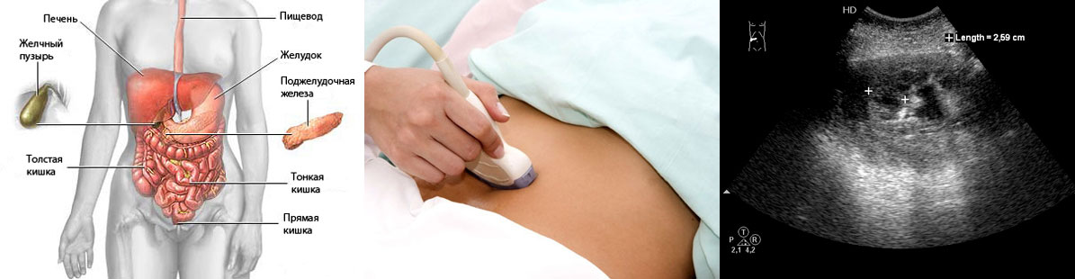
To undergo the procedure, a person just needs to undress and lie down as the doctor says. In this case, there will be no pain, no discomfort, no harmful effects on the body. The patient's skin is smeared with a special gel, thanks to which the sensor glides better and the air pockets between it and the skin are removed. If necessary, the doctor tells the person to turn or hold their breath. Depending on the complexity of each particular case, the entire process can take up to sixty minutes.
What organs are checked for such an ultrasound scan?
Thanks to the ability to use ultrasound, modern doctors can see the state of various organs without penetrating the body itself. Most often, an area survey abdominal space on ultrasound involves the study of examination of the following organs:
- Liver.
- Kidneys.
- Pancreas.
- Spleen.
- Genitourinary system.
- The gallbladder.
- Vessels.
- Is there liquid.
Usually, the reason for referring to such an ultrasound is the complaints of patients about the severity after eating, general weakness, malaise, pain in the abdomen, and a feeling of bitterness in the mouth. If it comes to a pregnant lady, then she is sent for an ultrasound scan if there is any suspicion of any wrong work of any internal organs. The same happens if the doctor suspects cancer, development diabetes mellitus or pancreatitis.
Ultrasound can easily detect cysts, various neoplasms, hepatitis, polyps and sand stones, both in the kidneys and in gallbladder, mononucleosis, cholecystitis, inflammatory processes and the consequences of any abdominal trauma. The specialist will immediately discern even the smallest changes in the shape or size of various organs. This is especially true in cases where the organs are displaced due to the proliferation of adjacent tissues, formed cysts or tumors.
The same happens with the appearance of aneurysms, enlargements of the aorta, ducts, and thickening of the walls in the gallbladder. After passing the examination, the doctor is able to schedule the most effective treatment, as well as monitor the patient's recovery process.
If the ultrasound shows fluid accumulation
The appearance of fluid in the abdominal cavity is called ascites, and it is diagnosed with the help of ultrasound. Usually, the cause of this phenomenon is inflammatory processes, as well as circulatory disorders.
Symptoms of this malaise include:
- Increased intra-abdominal pressure.
- An increase in the size of the abdomen, its weight.
- Fluid causes shortness of breath, belching, heartburn.
- Swelling in the legs. When it comes to men, scrotal edema may develop.
- Bursting pains are also caused by fluid.
- Bulging of the navel.
- The fluid causes white stretch marks in women.
- If the cause of everything is a violation of pressure in the hepatic vessels, then veins clearly appear on the abdomen.
- If the vessels located under the liver are to blame for everything, the patient will suffer from vomiting, nausea, and jaundice will appear.
- When ascites is associated with tuberculosis, the person suffers from weakness, rapid heartbeat, constant fatigue and headaches.
All these symptoms appear when about half a liter of fluid accumulates in the abdominal cavity. Such free fluid in the abdominal cavity is clearly visible on ultrasound. This pathology leads to disruption of the functioning of all organs. digestive system, even lungs. However, most often it is problems with any organs that cause the development of ascites.
Usually, fluid accumulates due to cirrhosis of the liver, but there may be other reasons: cancer, heart failure, various gynecological problems, pancreatitis, kidney disease, and tuberculosis. If a pregnant woman has any diseases or unhealthy habits, the baby may suffer from ascites, and therefore fluid will be visible on an ultrasound of the abdominal cavity.
What else are experts checking? The presence of congenital problems with gastrointestinal tract, edema, because they can also cause the accumulation of fluid.
Survey results
Having received all the necessary data, the doctor examines them on the monitor, proceeds to write the conclusion. Within a few minutes, the patient receives in his hands all the papers containing general information, all deviations from the norm, displacement of various organs, inflammation or damage. 
If necessary, a person will go for new examinations: this happens if tumors or cysts, fluid accumulation are found in the abdominal organs. If stones are found in the gallbladder or kidneys, the patient will also be sent to further treatment, most often surgical.
Conclusion
Ultrasound examination, especially if it concerns the abdominal cavity, allows examining all organs located in a given part of the body, giving a comprehensive and extremely informative picture of the state of each organ, its size, shape.
Due to its high efficiency, safety and painlessness, ultrasound is highly popular in the treatment of many diseases, as well as monitoring the condition of patients after surgery, when planning surgical interventions... It must be remembered that the reliability of the data obtained is affected by the correct preparation of the patient for the procedure.
In addition, the data will be inaccurate if the person is severely obese. The only condition under which it is impossible to carry out this survey, Is open wounds or a bandage over the area to be examined.