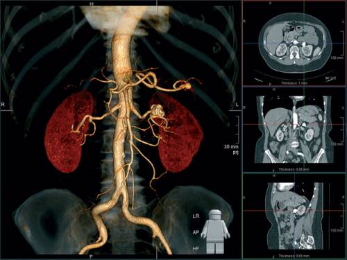
Computer tomography of the abdominal cavity and retroperitoneal space. Computer tomography of retroperitoneal space
Kt. abdominal cavity And the retroperitoneal space is the most informative study in surgical and therapeutic medical practice. To date, this study allows you to visualize an absolutely any point of the test zones and identify the formation of various densities that differ from each other by 0.5%. It is no coincidence that the computer tomography of the abdominal cavity and the retroperitoneal space is the most reliable method of oncological search, and in general early diagnosis pathogical changes in this anatomical area.
Application area:
This diagnostic procedure is indispensable in the study of the gastrointestinal tract. The CT abdominal cavity well visualizes bulk education, cholangitis, appendicitis, the accumulation of fluid in the abdominal cavity, necrosis, inflammatory processes, helps to identify the malignant process and its stage, etc. MSCT of the retroperitoneal space allows you to diagnose the abdominal vessels, investigate the activities of the kidneys, urinary system, adrenal glands, retroperitoneal space, lymphoid tissue, carry out a virtual colonoscopy of the large intestine and reveal. Including the accumulation of exudate, fresh blood, lesion lymphatic nodes with various malignant tumors Blood, Brill-Simmesis disease, Hodgkin's disease, abscess, etc.
Varieties Services:
CT abdominal organs can be carried out without contrast (native research). But most often the study of the abdominal organs is carried out using a contrast agent (with oral contrast) and a targeted study of the organ. And for research vascular system MSCT with intravenous contrasts is used. In our clinic you can pass next species Studies of the MSCT abdominal cavity and retroperitoneal space:kidney; liver; hepato-pancreatododenal zone;pancreas;spleen; gallbladder; intestines; retroperitoneal space; ureters; bladder; adrenal glands; gastrointestinal
Preparations for CT abdominal cavity and retroperitoneal space:
CT scan This anatomical area is always carried out with the preparation, namely 2-3 days before the study is necessary special dieteliminating products capable increased gas formation (No apples, beans, cabbage, carbonated drinks, etc.) The study is conducted on an empty stomach. Specify all nuances of the conducted research when writing to the procedure or your attending physician.
CT of retroperitoneal space is a method of studying tissues using X-ray rays, in which the doctor gets a layered image necessary organs in a given plane. High accuracy and informative of this research method allows large probability Put a correct diagnosis and make up the correct scheme Treatment.
Computer tomography of retroperitoneal space - one of best ways Diagnostics
In the retroperitoneal space concentrated a large number of Organ systems performing vital functions to maintain human life. These include:
- kidney;
- adrenal glands;
- ureterals;
- regional lymphatic ducts and nodes;
- pancreas;
- downward and horizontal part of the duodenum intestinal department;
- ascending colon.
Indications for research:
- post-traumatic study when suspected damage internal organs, for example, after stupid injuries of the abdomen obtained at an accident;
- assessment of the state of the internal organs before surgery;
- suspicion of the oncological process, determining the degree of illustrative;
- stones in the kidneys and a ureter;
- cystic education in organs;
- purulent diseases - phlegmon, abscess - clarification of localization and degree of prevalence;
- anomalies of the development of the brancheshaft bodies;
- insolvency of other research methods (for example, non-informative urography).
Contraindications
If there are contraindications, it is worth a preference to other research methods
CT of retroperitoneal space is considered safe method Studies, but still has its own contraindications. They are separated into absolute and relative (the use of the method is possible in the case when the benefit from it exceeds the harm).
Absolute
- Pregnancy - X-rays have a teratogenic property, therefore, they can significantly affect the development of the fetus. The use of this method is impossible on any gestation.
- Overweight - tomograph withstands patients with weight up to 120 kg, the device will not withstand a larger load.
- Mental deviations in behavior - manipulation can cause a patient in an inadequate reaction.
- Diseases accompanied by hyperkinosis (pathological mobility). For the accuracy of the study, the patient needs to be fully fixed during the procedure, otherwise it does not make any sense and it will not work clear pictures.
- Allergies to the contrast (in the event that it is used) - the contrasting agent based on iodine is quite allergenic.
Relative
- Patient age up to 14 years.
- A sharp inflammatory process in the kidneys and a ureter, chronic renal failure - in the study of kidney with contrasting, a contrast agent is used, additional load falls from the body to the inflamed organ.
- Cardiovascular diseases, diabetes, some neurological diseases.
CT with contrasting
A computed tomography with a contrast agent is used to study some systems and organs. The contrast is a substance based on iodine, which is introduced intravenously, orally or rectally (depending on the object under study), and allows you to clearly see the structure of the body. Excreted from the body natural way During the day.
Important! Contrast stands out with breast milkTherefore, it is recommended for one or two days to interrupt breastfeeding.
Preparation for the study of bodies
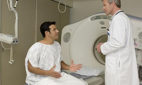
The doctor must prepare the patient for the upcoming procedure and tell it in detail about it.
CT of retroperitoneal space is carried out on an empty stomach, so by the time the gastrointestinal tract should be as frequently released from food. Patients suffering from constipation, on the eve of the cleansing enema or to take a light laxative. It is not necessary to use products that cause increased gas formation and slowing intestinal peristalsis (bean, peas, soda). It is preferable to study in the morning, but depending on the time of tomography, it is recommended:
- light dinner (the study is scheduled for morning);
- liquid lightweight breakfast (study at lunchtime);
- do not lunch if the study will be held in the evening.
Tip! In the study of the organs of the urinary system, the fresh will most likely need biochemical analysis blood. Take care of it.
Methodology
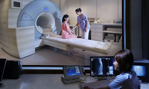
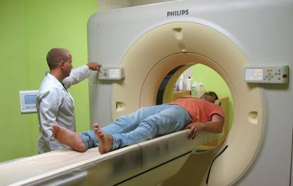
During the examination of the organs, the patient is fixedly on the couch, around which a special scanner is moving. The procedure takes about 15 minutes, with contrast - half an hour, and all this time must be installed motionless, so it is recommended to cook comfortable clotheswhich will not cause any discomfort, or ask the medical staff a disposable set of clothes.
Be sure to remove all metal products and remove the electronic devices, the device can harm them. The doctor will all the time be behind the glass and follow the process if it became necessary to ask a question - this can be done with two-way communications. Conclusion The radiologist will give up for 1-2 hours.
Advantages of tomography of retroperitoneal space before other methods
- Non-invasiveness. To assess the state of the organs, surgical cuts, laparoscopy, the method is painless and extremely accurate is not required.
- In the same picture shows bone structures, and vessels, and soft fabrics organs. This allows you to do with one research procedure.
- Study at the desired depth and in the desired plane. Ultrasound and the usual X-ray picture have no such opportunity.
- Fast result. Saves vital important time for emergency situations (for example, bleeding from internal organs).
- Less sensitive to the mobility of the studied, unlike MRI.
- Allows you to diagnose diseases on early stagesthat is especially important if the oncological process is suspected. In this case, the doctor can start treatment in a timely manner to prevent the development of the process.
- Convenient for the patient, from which active actions are not required and do not endure painful manipulations.
Useful recommendations! Carefully collect all your results of the tests, before studying the doctor may need any of them. Perform all regulations, in no case neglect the diet before the procedure, this will avoid the direction to a re-image due to the non-informativeness of the result. In the event that the noise of the device is confused during the manipulation process, you can use headphones. Relax, close your eyes, imagine yourself in another place, do not panic, it will help you keep stillness. With strong excitement, the eve of a light soothing agent can be taken.
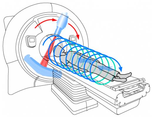
The MSCT abdominal cavity is one of the types of computer-type tomography. The study is carried out using X-ray rays. At the same time, the MSCT apparatus applies detectors that are located two-dimensional, but the computer tomograph has a linear type sensors.
MSCT is a multispiral computed tomography that shows the "section" of each organ during the survey. Most often, such a study is necessary for those who have detected diseases associated with gastrointestinal tract. Thanks to him, it is better to consider the stomach and intestines.
The MSCT apparatus almost displays radiation radiation. At the same time, pictures are obtained high Qualitywhat allows you to consider the most indemplifications Details. Due to the pictures with the MSCT, you can see very thin cuts of the abdominal organs with a thickness of less than 1 mm. Due to the three-dimensional presentation of the stomach, the attending specialist can accurately diagnose the disease.
Tomography of the abdominal cavity
Tomography of the abdominal cavity with contrasting quite quickly finds various pathologies, due to which the patient will not lose time during the survey. For this reason, multispiral computed tomography is prescribed for severe patients and for stratifying claustrophobia.
The one who is inside the MSCT apparatus will be able to see what is happening next to it. This happens because the body is not fully placed in the device. Also hit this examination The patient's peritoneum organs will not detain his breath for a long time. This is a plus in comparison with a number of other studies.
Due to the tomography of the type of MSCT, it is possible to obtain sufficiently high-quality snapshots of the abdominal organs in a thin cut. It does not compare with images resulting from classical tomography. The device for this tomography makes it possible to make the scan of the internal organs of a person in a few minutes, especially when administered inside the contrast agent.
As for the safety issues of the procedure, the following should be noted here: despite the use of X-rays, negative influence It does not have health. Due to the fact that tomographs of the latter models emit the minimum radiation, the MSCT with contrast does not harm the body significantly. This is true, even if the procedure is carried out several times for a short time.
Doctors often resort to this study, because it allows you to find a lot of diseases. Additional advantage is the lack of the need for special recommendations to patients regarding training. But for more convincing results, MSKT must be adhered to some rules.
How is the MSKT of the abnormal organs?
Preparations for the MSCT is quite simple and lies in the failure of the evening meals. 8 hours before the procedure also prohibited to consume food. Cleaning the digestive organs from gases is better occurs if within a few days to tomography, limit itself from dairy, bakery products, kvass, cabbage, apples and carbonated drinks.
In addition, MSCT abdominal organs A person requires preparation of documentation, namely the results of previous surveys plus directions from the attending specialist.
The remaining moments are more related to psychology, because the patient who fell to the examination should be in a fixed lying position for some time.
Multispiral computed tomography of abdominal organs will not bring painful sensations. If the study is carried out for the patient with claustrophobia, the latter takes sleeping pills.
Advantages of MSCT
The use of MSCT allows a radiologist to diagnose for most reasons that cause abdominal pain or injury pain. High degree accuracy helps to get rid of the need for new procedures invasive diagnostics. If the pain is a consequence of infections or inflammation, the MSCT will reduce the risk heavy complications Due to speed, accuracy and simplicity. Competently pronounced diagnosis helps to set the most effective treatment tactics.
MSCT is quite painless, non-invasive, is informative way diagnostics. Compared to a conventional x-ray, this tomography allows you to get an image in a more detailed form.
MSCT has some important advantages Compared from MRI:
- The sensitivity of the MSCT is less than the MRI.
- Metal in body or implants do not interfere with the ICST.
- Due to images in the present time mode, it is reduced to a minimum number of invasive procedures under visual control.
- The need for such complex procedures as laparoscopy and biopsy of the surgical species is eliminated.
- There is no residual radiation after the MSCT.
Thus, advantages over MRI are obvious, and this will help to make patients the right choice.
If pregnancy, tomography is not recommended. This is possible only if serious grounds. It is quite rare, but some risk associated with allergies for contrast agents can arise, which contains iodine. The contrast procedure for nursing mothers requires that child feeding is not carried out within 1-2 days after the introduction of a contrast agent into the body.
MSCT may in some points to give up ultrasound: it concerns the detection of stones in bile bubble and other studies. Also, if necessary, diagnose the diseases of the MSCT of the abdominal cavity organs and the retroperitoneal space is inferior to MRI. Due to the fact that a significant part of the MSCT devices are kept maximum mass 120 kg, patients with great weight will not be able to count on it.
What shows CT abdominal cavity and retroperitoneal space
Computer tomography allows you to effectively diagnose the presence of the following pathologies and diseases in the patient:
MSCT allows not only to detect them, but also to establish an accurate diagnosis that will allow in the future to assign proper treatment.
MSCT abdominal cavity in the early stages of cancer
Progress in terms of diagnostic methods for the type of radiological studies and methods of their application helps to diagnose oncology in the first stages. It depends on this, whether a person has a chance to survive and cure from this disease. The MSCT is necessary to determine how much metastases spread with oncological lesions of the endometrium, esophagus, bladder and colorectal cancer.
Due to the fact that computed tomography has ample opportunities, you can make a complete and detailed analysis research areas, get all necessary information For each organ, muscles and bones. With the help of tomography, it is possible to identify liquid, solid, cystic pathological elements and formations, tumors with different density.
In a number of countries, today, computed tomography is deservedly considered the most clear and professional methodWith which it is possible to detect oncological education in the exposed area of \u200b\u200bthe body. MSCT also helps to diagnose other states, including:
- cholecystitis;
- pathology associated with the occurrence of stones in the bustling bubble;
- the occurrence of small stones and polyps in bile ducts;
- deterioration of the indicators of bile and so on.
Contrasting allows you to fully improve the clarity of visualization. On average, the study of the abdominal cavity takes about 20 minutes. In certain cases, an option is possible with an increase in the time of the procedure until half an hour at the same cost.
Thus, the CSD is quite efficient. It allows you to start to deal with a number of serious diseases by detecting them at the most initial stages. Doctors strongly recommend this procedure, especially since its conduct is possible when severe condition patient.
Additionally
The above advantages are not the only for MSCT. Additionally, this procedure allows you to accurately determine the degree and foci degenerative changes Spine and contributes to the identification of hernia in the lumbar department.
In addition, the MSCT contributes:
- Determining the degree of arthrosis of the joints.
- Opportunities for accurate diagnosis of any injuries and fractures.
- Determine the degree of circulatory disorder, evaluating the degree of damage to blood arteries in the presence of embolism.
And it is ne full list Diagnostic procedures conducted using MSCT. Basically, the feasibility of the MSCT determines only the attending physician, depending on the specific problems in the body. This procedure Today it is common and allows as soon as possible Conduct diagnostics, which is important for effective and timely treatment.
\u003e CT (computed tomography) of retroperitoneal space
This information cannot be used during self-treatment!
Be sure to consult with a specialist!
Retropher space is an anatomical area located along back wall Abdominal cavity. From above, it extends to the diaphragm, and below - to a small pelvis. In this space there are adrenal glands, kidneys, ureters, part duodenal gut, pancreas, part of the colon, bottom hollow vein, part of aorta, trunks and plexus vegetative nervous system, The lymph nodes. The space between the organs is filled with fatty tissue.
Computed tomography of this area identifies the pathology of all organs located in it. For better visualization of renal vessels and neoplasms, intravenous administration of a contrast agent is used.
When is the KT of the retroperitoneal space appointed?
Most often, the CT of the retroperitoneal space is prescribed by suspected the combined pathology of the organs located in this field, or their abnormal structure. It is shown in closed lens or belly injuries, suspected the presence of tumors and metastases, lesions of lymphatic nodes, urolithiasis, kidney polycystic and inflammatory processes In them, kidney omit. The survey allows us to properly plan the move and the volume of the upcoming operation and evaluate the effectiveness of the treatment.
Who gets the direction and where to get the KT of the retroperitoneal space?
Direct the inspection of urologists, traumatologists, surgeons, gastroenterologists, oncologists. You can go through the KT of the retroperitoneal space in medical institutionsequipped with a computed tomograph. This can be both diagnostic and therapeutic state or commercial institutions.
Who can not do CT of transfroaching space?
The examination is carried out using the effects on the body of the X-ray rays, the radiation dose is quite low in comparison with the usual X-ray study. The procedure is contraindicated on any gestation period. It can be held for children, but only if there are reasonable testimony and it is impossible to replace it with other methods of surveys.
CT S. contrast amplification It is contraindicated in allergies to a contrast agent. Contrast can also cause complications in patients with renal failure, liver failure and diabetes. In these cases, the issue of the possibility of conducting CT is solved by a radial diagnostician individually for each patient.
Preparation for CT of the retroperitoneal space and the methodology for its conduct
If the scanitoneal space is to be surveyed, 2-3 days before the procedure, it is necessary not to use products that contribute to the formation of gases in the intestine and slowing its peristaltics (carbonated drinks, beans, peas, etc.). For chronic constipation It is recommended on the eve of the study to adopt laxatives or to make an enema. CT of retroperitoneal space is better to spend in the morning, as the patient must come hungry.
During scanning, the patient lies motionless on a special table in a tomograph tunnel. The ring of the apparatus will rotate around the body of the surveyed, it should not be metal objects and electronic devices. The medical staff at this time is in the neighboring office and observes what is happening through the glass. To communicate a patient with a doctor, a double-sided connection is provided.
Deciphering CT results occurs immediately after the procedure. Rade diagnostics describes the condition of all organs in the retroperitoneal space. If necessary, research can be carried out repeatedly.