Clinical anatomy of the nose. Nose: Hoc is the beginning of the airway. At the same time he serves
The nasal cavity (Fig. 1, 2) - the initial section of the respiratory tract, lies in the facial skull, upward from the oral cavity and is separated from it by the hard palate. The upper part of the muzzle that defines the nasal cavity dorsally is called the nose.
The skeleton of the nasal cavity is formed by the bones of the skull and cartilage. Outside, it is covered with muscles and skin, inside it is lined with a mucous membrane. The nasal cavity is communicated with: external environment- inlets, or nostrils, with the cavity of the pharynx; - outlets, or choanas. The median cartilaginous nasal septum divides the nasal cavity into two halves - right and left. In each half protrude two turbinates - the upper and lower - and the labyrinth of the ethmoid bone.
From the anterior section of the upper and lower edges of the cartilaginous nasal septum, lateral cartilage extends to the right and left, forming the lateral walls of the nasal cavity. Pterygoid cartilages are connected to the initial end of the nasal septum, which serve as the skeleton of the nostrils. Each pterygoid cartilage consists of a cartilaginous plate and a curved arch. The shape of the nostrils in animals different types different: from crescent to circular-oval. The nostrils are bounded by the inner and outer wings of the nose, formed by folds of skin based on cartilage. The wings of the nose are controlled by the muscles that dilate the nostrils. In the lower corner of each nostril is the opening of the nasolacrimal canal.
Rice. 1. Cartilage of the nose of cattle: 1 - the end of the nasal bones; 2 - incisor bone; 3 - upper jaw; 4 - lateral cartilage of the nose; 5 - plate of pterygoid cartilage; 6 - its process (bow); 7 - the end of the nasal septum; B - nasolabial speculum with glands.
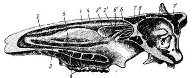
Rice. 2. The nasal cavity of cattle; 1 - upper nasal passage; 2, 2-1, 2-2-middle nasal passage; 3 - lower nasal passage; 4 - upper nasal concha; 6 - inferior turbinate, 6-1 - labyrinth; 7, 7-1-frontal sinus; 8 - sinus of the sphenoid bone; 9 - palatine sinus.
In horses, most of the lateral wall of the nasal cavity is devoid of a cartilaginous basis and forms a soft nose, consisting of a connective tissue plate, skin and mucous membrane. In the upper part of the soft nose, there is an invagination of the skin of a conical shape, the blind apex of which is turned posteriorly. This invagination is called a nasal diverticulum or nasal drum. Its length is 5-7 cm. The entrance to the diverticulum is located in the upper part of the nostril.
Skin covering Between the nostrils it is supplied with glands and forms: in large ruminants - a nasolabial speculum, in small ruminants and dogs - a nasal speculum, in omnivores - a proboscis. The mirror and proboscis are always wet and, due to the evaporation of moisture from them, cooled.
The mucous membrane of the walls of the nasal cavity, concha and the labyrinth of the ethmoid bone forms folds, thereby creating a huge surface for contact with the inhaled air.
The nasal cavity is divided into three areas: the vestibule, olfactory and respiratory. The vestibule area is located at the entrance to the nasal cavity and is lined with flat, multilayered Epithelium. The olfactory area is called the postero-superior part of the nasal cavity. The mucous membrane of her yellowish, it contains olfactory cells, which serve as the organ of smell. The respiratory region occupies most of the nasal cavity - from the vestibule to the choanas. It is lined with prismatic ciliated epithelium with goblet cells and contains mucus-secreting glands. The ciliated hairs are directed towards the choanas. The secretion of the mucous glands moisturizes the mucous membrane of the nose, and, consequently, the inhaled air.
In the submucosal layer of the nasal cavity, there are many large arterial and venous vessels that form the venous plexus. Therefore, the inhaled air is not only humidified here, but also warmed up.
The nasal concha (upper and lower) divide the nasal cavity into four nasal passages: 1) the upper, or olfactory, passage is narrow, lies between the nasal bones and the upper concha; 2) middle stroke lies between the shells and is both olfactory and respiratory; it communicates with the sinuses, or sinuses, of the bones of the skull; 3) the lower passage is wide, lies between the inferior concha and the bottom of the nasal cavity (or hard palate); it leads through the choanas into the pharyngeal cavity, therefore it is called respiratory; 4) all these passages are connected in the general nasal passage, located between the turbinates and the median cartilaginous nasal septum.
The paranasal sinuses, or sinuses, which communicate with the nasal cavity through the middle nasal passage, are filled with air. Their mucous membrane is covered with prismatic ciliated epithelium.
In horses, the following sinuses (or sinuses) are distinguished: the frontal, maxillary, palatine, sphenoid bone, the cavity of the shells and the curls of the olfactory labyrinth of the ethmoid bone.
In cattle, the frontal sinus is extensive and extends into the cornea, the parietal bone, and the scales of the occipital bone. The maxillary sinus is large and communicates with the palatine sinus and with the sinus of the lacrimal bone.
In pigs, the maxillary sinus is located in the maxillary and lacrimal bones, and in older animals it extends into the palatine and zygomatic bone... The sphenoid sinus is extensive, extending into the scales of the temporal bone.
Dogs do not have a maxillary sinus; it is replaced by an expansion of the nasal cavity between the ethmoid and maxillary bones.
The nose is the initial section of the upper respiratory tract, the peripheral section of the olfactory sensory system, in speech function - part of the extension tube of the vocal apparatus. The nose consists of the outer nose and the nasal cavity with its paranasal sinuses. The outer nose covers the nasal cavity, formed by the bone-cartilaginous skeleton, muscles, covered with skin. The muscles provide the expansion and contraction of the nostrils. Thanks to the cartilage, the nostrils are open and separated from each other. The nasal cavity is divided into two halves by the nasal cartilaginous septum. Upper rear part the septum is formed by a perpendicular plate of the ethmoid bone, the lower posterior part is formed by a vomer, fixed on the maxillary and palatine bones. The anteroposterior part of the septum is formed by elastic cartilage. Atmospheric air enters the nasal cavity through the nostrils, from the nasal cavity into the nasopharynx through the openings of the choanae. Each half of the nasal cavity has 4 walls: upper, lower, inner and outer. Top wall or the roof is formed mainly by a perforated plate of the ethmoid bone, it forms part of the base of the skull, is pierced by numerous holes through which fibers of the olfactory nerves pass into the cranial cavity. Through these holes, an infection can easily penetrate into the cranial cavity; a purulent process in the nasal cavity is especially dangerous. The lower wall or floor of the nasal cavity is at the same time the upper wall of the oral cavity, formed by the hard palate. The inner wall of the nasal cavity is formed by the nasal septum and is common to both halves. The outer (lateral) wall is the most complex, formed by several bones of the skull. It has 3 projections in the form of curved plates - the turbinates. The upper (smaller) and middle (longer) shells are formed by outgrowths of the ethmoid bone, the lower shell is an independent bone. There are 3 nasal passages between the shells:
- lower - between the bottom and the lower shell;
- middle - between the lower and middle shells;
- the upper one is between the middle and upper shells.
The slit space between the nasal septum and the nasal passages is called the common nasal passage. The turbinates increase the overall surface of the nasal cavity. The nasolacrimal canal opens into the lower nasal passage, through which excess tear fluid flows out of the eye cavity. From the inside, the nasal cavity is lined with a mucous membrane, which is covered with ciliated epithelium, only in the initial part of the nose, in anticipation, is lined with squamous epithelium, contains hair, sebaceous and sweat glands... Under the layer of ciliated epithelium are mucus-secreting glands. Suspended dust particles settle on the hairs of the vestibule, mucus, and the movements of the cilia of the epithelium together with these particles are removed from the walls of the nasal cavity, providing purification and moistening of the inhaled air. Thanks to the lysozyme contained in the mucus and having bactericidal properties, the inhaled air is rendered harmless. The mucous membrane of the nasal cavity is richly supplied with blood vessels, therefore, the inhaled air, passing through the narrow spaces of the nasal cavity, is warmed up. The temperature and humidity of the atmospheric air we breathe can fluctuate very widely depending on the season of the year. However, in any case, the temperature of the air entering from the nasal cavity into the nasopharynx is 28-300 C. Normal breathing is possible only with free patency of the nasal passages. Any obstacle to the passage of air in the nasal cavity (hypertrophy of the turbinates, polyps, swelling of the mucous membrane during inflammation, etc.) violates nasal breathing and it is carried out through the mouth. In this case, the protective function of the nasal mucosa is disrupted, which leads to frequent inflammation of the airways.
Olfactory receptors are located in the mucous membrane of the upper nasal passage, this part of the nasal cavity is called the olfactory region, the middle and lower nasal passages are called the respiratory tract. In the mucous membrane of the nasal concha, especially in the lower one, there is the so-called cavernous tissue formed by the dilated venous plexuses. At different influences(chemical, temperature, emotional, as well as under the influence medicines) there is a swelling of this tissue due to reflex expansion of blood vessels and filling them with blood, which causes a stuffiness in the nose. In the mucous membrane of the middle part of the nasal septum, approximately 1 cm posterior to the entrance to the nose, there is an area with a superficially located network of blood vessels — a bleeding zone that is the source of nosebleeds. In speech function, the nasal cavity plays the role of a resonator for sounds generated in the larynx. With the correct pronunciation of sounds, nasal resonance is involved only in pronouncing the sounds m and n and their soft variants.
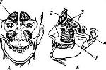
Hoc is the beginning of the airways. At the same time, it serves as an organ of smell, and also participates in the formation of the so-called extension tube of the vocal apparatus. The nose consists of the outer nose and the nasal cavity with its paranasal sinuses.The external nose consists of a bone-cartilaginous skeleton and soft parts. The upper narrow end of the nose, starting from the forehead, is called the root of the nose; from top to bottom and in front of it stretches the back of the nose, ending with the tip of the nose. The lateral moving parts of the nose are called the wings of the nose, and their free edges form the outer nasal openings, or nostrils. The skeleton of the external nose includes the frontal processes of the maxillary bones, nasal bones and cartilage of the nose (Fig. 42). The soft parts are formed by the muscles and skin. The purpose of the muscles is mainly to widen and narrow the nostrils (Fig. 43, 44).
1 - nasal bone; 2 - lateral cartilage of the nose; 3 - large wing cartilage; 4 - wing of the nose; 5 - small wing cartilage; 6 - frontal process upper jaw
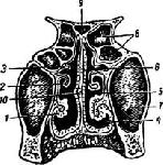
/ - lower shell; 2 - middle shell; 3 - upper shell; 4 - lower nasal passage; 5 - middle course; 6 - upper nasal passage; 7 - maxillary sinus; 8 - trellised cells; 9 - the main sinus; ten - nasal septum
The nasal cavity consists of two halves, separated from each other by a nasal septum. The back-upper part of the septum is bony, and the antero-lower part is cartilaginous.
Each of the two halves of the nasal cavity has four walls: upper, lower, inner and outer.
The upper wall, or roof, of the nasal cavity is mainly formed by the sieve plate of the ethmoid bone. This plate is pierced with numerous holes. On its upper surface, facing the cranial cavity, lies the bulb of the olfactory nerve. Thin branches extend from the bulb downward - the so-called olfactory filaments (fibers of the olfactory nerve), which penetrate into the nasal cavity through the openings of the sieve plate.
The lower wall, or bottom, of the nasal cavity is also the upper wall of the oral cavity (hard palate). The bottom of the cavity is formed by two intergrown midline The palatine plates of the maxillary bones and is complemented behind by the horizontal plates of the palatine bones.
The inner, or median, wall of the nasal cavity - common to both halves - is formed by the nasal septum.
The outer, or lateral, wall of the nasal cavity is the most complex in structure. It has three horizontally located bony protrusions resembling a half of a bivalve shell in shape. These are the turbinates - lower, middle and upper. The largest of them - the lower one - is an independent bone, while the middle and upper are the processes of the ethmoid bone. There are three nasal passages under the nasal conchas: between the inferior concha and the bottom of the nasal cavity - the lower nasal passage, between the middle and lower concha - the middle one, between the upper and middle concha - the upper nasal passage. The slit space between the nasal septum and the surfaces facing it of all three turbinates is called the common nasal passage.
In front, the nasal cavity is covered by the external nose and only in its lower part opens outward through the entrance to the nose - the nostrils. The nasal cavity does not have a posterior wall and communicates from the back with the pharyngeal cavity through large oval holes - choanas (one hole in each half of the nose). The entire nasal cavity is lined with mucous membranes. In the part of the mucous membrane that covers upper part the nasal septum, the superior and partly the middle turbinate, branches of the olfactory nerve, ending in olfactory cells. This part of the nasal cavity is called the olfactory region. The rest of the nasal cavity is called the respiratory region.
The mucous membrane of the respiratory region is lined with ciliated epithelium. There are many mucus-secreting glands under the epithelium.
The so-called cavernous tissue, consisting of dilated venous plexuses, is embedded in the mucous membrane of the nasal concha, especially the lower one. The walls of these plexuses contain a large number of smooth muscle fibers. When exposed to various irritants (temperature, chemical), as well as mental factors the cavernous tissue is capable of swelling rapidly due to the reflex expansion of the venous plexus and filling them with blood. This swelling sometimes causes a sudden stuffiness in the nose.
In the mucous membrane of the middle part of the nasal septum, approximately 1 cm posterior to the entrance to the nose, there is an area with a superficially located network of blood vessels. This area is called the bleeding zone of the nasal septum and is the most frequent source of nosebleeds.
The nasal cavity has a number of paranasal sinuses. They are cavities filled with air and are located in the bones involved in the formation of the walls of the nasal cavity. These sinuses communicate with the nasal cavity through openings located in the upper and middle nasal passages.
All paranasal sinuses are paired (Fig. 45). The frontal bones contain the frontal sinuses; in the upper jaw - maxillary, or maxillary, sinuses; in the main bone - sphenoid and in the ethmoid bone - ethmoid cells. The walls of the paranasal sinuses are lined with a thin mucous membrane, which is a continuation of the nasal mucosa.
Rice. 45.
(paranasal) sinuses (A - front. B - side):
1 - maxillary sinus; 2 - frontal sinus; 3 - trellised cells; 4 - the main sinus
In a newborn, the paranasal sinuses are in their infancy, and the frontal sinuses are absent. Lattice cells develop faster than others. The maxillary sinuses reach full development only by the end of the eruption of permanent teeth, and the frontal sinuses begin to form only at the age of 4-6 years and finish development by 20-25 years.
Sensitive innervation of the nose and paranasal sinuses is obtained from the 1st and 2nd branches of the trigeminal nerve (V pair). The motor nerves to the muscles of the wings of the nose and to the so-called "muscle of the proud" (the muscle wrinkling the skin of the forehead above the nose) are branches of the facial nerve (VII pair).
on the topic: "Embryology and anatomy of the nose"
Nose- the organ of smell; is also the initial part of the respiratory tract.
Embryology. From the end of the third embryonic week at human embryo under the endbrain and anterior to the eye vesicles, the olfactory fields are laid on both sides in the form of a thickening of the integumentary epithelium. By growing and deepening the epithelium, these thickenings are transformed into olfactory pits. At the beginning of development, the opening of the oral cavity is a wide pentagonal hole surrounded by five protrusions: an upper unpaired (frontal process), two paired upper (maxillary processes), two lower (mandibular processes). At the same time, one so-called differentiate on the frontal process. the median frontal process and two lateral nasal processes. Further, the fossa plunges into the depths in the form of an olfactory sac, bounded, in addition to the nasal processes, by the adjoining maxillary process of the first visceral fold from the side. The latter also covers the olfactory sac from below; the role of the septum between it and the median nasal process is played by the thin and small pharyngeal membrane lying in the continuation of the nasal groove, which is part of the septum between the olfactory sac and the primary oral cavity... This membrane subsequently breaks through by deepening olfactory fossa, which with their posterior ends open into the primary oral cavity, forming small openings (primary choanae). The maxillary processes grow significantly and gradually grow together with the lateral nasal processes (giving rise to the subsequently disappearing nasolacrimal groove) and with the median frontal process (forming together with the latter top edge mouth opening).
Subsequently, on the inner side of both maxillary processes, protrusions begin to form - palatal ridges growing towards each other and gradually dividing the primary oral cavity into the upper section, or the nasal cavity, where the primary choans open, and the lower section - the oral cavity itself. During the second month of embryonic life, the formation of the face and N. occurs. The latter appears first in the form of a transverse, so-called nasal ridge, which forms on the median frontal process and represents the anlage of the tip of the nose; the openings of the olfactory fossa turn into external nasal openings, and the lateral nasal processes give the wings of N. At the same time, on the inner side of the median frontal process, a septum perpendicular to the palatine processes, the so-called nasal septum, appears, growing gradually inward and dividing the nasal cavity formed above the palatine processes into two halves ... The edges of the palatine processes gradually grow together with the lower edge of the nasal septum and with each other, which leads to the formation of the palate. The posterior edges of the palatine processes form small outgrowths, from which the uvula is subsequently formed; the two openings remaining above them and leading into the nasal cavities are secondary choans. The importance of for the formation of the nasal cavity, they have shell-like formations that appear on its outer wall. Here you can distinguish between them formed by the lower shell and the lattice labyrinth. From the epithelium of the olfactory field, and later from the epithelium of the fossa of the sac, by means of special differentiation, the epithelium of the nasal mucosa with its olfactory and supporting cells occurs. The folds that appear on the lateral surfaces of the nasal cavities subsequently form the nasal concha, of which there are four in a newborn.
Anatomy. The nose is divided into an external nose and a nasal cavity with paranasal sinuses. The shape of the nose varies considerably, not only due to the structural features of the facial skeleton, but also depending on the age (during growth) in the same subject. For the purpose of anthropological study, Martin divided all forms into 15 groups. Each group is characterized by 5 main features: the shape of the back (straight, convex, concave), its length (short, long, medium), the location of the root (deep, high, medium), the shape of the tip (obtuse, acute, medium) and the direction of the base ( up, down, horizontally).
The outer nose has the shape of an irregular triangular pyramid, the base of which coincides with the skeleton of the face, the top is the tip of the nose, and one of the faces, which is obliquely anteriorly and downwardly, is the nasal bridge. The upper narrow end of the bridge of the nose at the forehead is called the root; above it there is a slightly deepened platform between the brow ridges - the glabella. The lateral surfaces of the nose are convex downward, delimited by a pronounced groove, mobile and constitute the wings of the nose; between their lower free edges, a movable part of the nasal septum is formed. The outer nose consists of bony, cartilaginous (hyaline cartilage) and soft parts. The bone framework in the upper part is formed by the nasal part of the frontal bone and the nasal bones. From below and from the side, the frontal process of the upper jaw adjoins the nasal bones on each side. The nasal bones fill the space between the frontal processes of the upper jaws and participate in the formation of the nasal dorsum; their upper edge is connected to the frontal bone, the outer one - with the frontal process of the upper jaw, the inner one - with the bone of the same name on the other side, the lower free edge forms the upper border of the pear-shaped opening.
The cartilaginous framework of the nose is a continuation bone skeleton and is tightly welded around the pear-shaped opening to the latter. The lateral wall of the external nose on each side is formed by a cartilaginous plate of an irregular triangular shape. The upper edge of this cartilage extends somewhat backward, under the nasal bones and the frontal process of the upper jaw, attaching to them through dense connective tissue. The inner edge at the nasal dorsum connects to the anterior edge of the cartilage of the nasal septum. The antero-inferior angle of the triangular cartilage reaches the large cartilage of the wing of the nose. The large cartilage of the wing of the nose is also paired, especially thin and has varied form... It consists of two plates, of which one wider, called the outer leg, forms the wing of the nose, and the other, the inner one, is located with the same leg of the other side and is part of the movable nasal septum, loosely connecting with it and with the cartilage septum of the nose. The small cartilages of the wing of the nose are small irregular shape pieces of cartilage, in various quantities located in the posterior part of each of the wings of the nose.
Small cartilage plates in the amount of one or two, located between the triangular cartilage and the large cartilage of the wing of the nose, are called accessory cartilage. The part of the nasal wing in the posterior inferior part does not contain cartilage and is formed only by duplication of the skin. The cartilaginous section of the external nose also includes the quadrangular cartilage of the nasal septum, which is an irregularly quadrangular cartilaginous plate, which is lower part the nasal septum and is inserted between the perpendicular plate of the ethmoid bone, and the posterior inferior edge lies in the groove of the vomer and the anterior part of the nasal ridge of the upper jaw. The anterior-inferior edge of the cartilage forms in front of the upper border of the movable nasal septum; posteriorly it descends somewhat lower and attaches to the anterior nasal spine. Under the name of Jacobson's vomer-nasal cartilage, a small cartilaginous strip is described that directly adjoins on both sides of the cartilage of the nasal septum immediately below the anterior nasal spine.
The muscles of the external nose in humans are rudimentary and have almost no practical value. From muscle bundles of greater or lesser importance. This is a superficial muscle that lifts the wing of the nose and the upper lip; it starts from the frontal process of the upper jaw and attaches to the posterior edge of the wing of the nose, partly passes into the skin upper lip... The other muscle of the nose consists of two bundles: the transverse one, which narrows the nasal openings, and the pterygoid, which pulls the wings of the nose downwards. The muscle that precipitates the nasal septum pulls down the nasal septum. The skin of the nose is very thin and is connected to the underlying parts at the top by means of loose, fat-poor connective tissue, and on the wings of the nose it is closely connected by means of elastic connective tissue to the underlying muscle cover. The skin of the nose has numerous sebaceous glands, which, especially in the posterior part of the nasal wings at the tip of the nose, are exceptionally large; their holes are visible with a simple eye... In the skin of the nose, in addition to the hair follicles and thin hair, there are also sweat glands. In the area of the nostrils, the skin is wrapped inside the nose and in the area of the fold inner surface wing, called the threshold of the nose, gradually passes into the mucous membrane of the nasal cavity. In the initial part, directly at the entrance to the nose, the nasal cavity is also lined with skin, which bends inward and is supplied with hairs and sebaceous glands. Hair located here; they can be of considerable length. This is followed by an intermediate belt, which further passes into the actual mucous membrane.
The outer nose is very richly vascularized. The arterial network of the external nose, located mostly under the skin, has numerous anastomoses with arterial system nasal cavity. The veins of the external nose, branching almost analogously to the arteries, but not everywhere accompanying the latter, flow into several branches.
Lymph, nasal vessels pour out into large limf, face vessels, which in turn are directed to limf, nodes of the submandibular region. The abundance of blood and lymph vessels in the external nose contributes to fast healing wounds that have arisen in this area, engraftment of skin flaps after plastic surgery and the resolution of local inflammatory processes. Along with this, the presence of a rich circulatory and lymphatic systems under certain circumstances facilitates the spread of an invading infection through the blood and lymphatic pathways.
The nasal cavity (inner nose) is located between the anterior third of the base of the skull, the eye sockets and the oral cavity. In front, it opens with nostrils located on the lower surface of the outer nose and bordered only by skin, which have a very varied shape and are located somewhat obliquely. Behind the nasal cavity communicates with the upper part of the pharynx (nasopharynx) through two adjacent oval posterior nasal openings, called choans. The median septum usually deviates partially to one side or the other (the entire cavity is divided into two more or less identical halves. The upper and posterior part of the septum is bony, its anterior part is formed by quadrangular cartilage, in front and below it is adjoined by a membranous septum. Each half the nose has four walls forming it: inner, outer, upper and lower.
The inner wall is the nasal septum, the bony part of which in the posterior superior section is formed by the perpendicular plate of the ethmoid bone, and in the posterior inferior section - an independent bone of the nasal septum - by the vomer. In the lower part of the partition, closer to the front, on the border of the opener, there is the opening of the opener organ; in humans, it is almost undeveloped, is several millimeters long, while in animals it has the form of an elongated sac lined with olfactory epithelium. Posteriorly and below the vomer organ, at the bottom of the nasal cavity, there is often a small opening leading into the tubule, called the incisal duct; this latter usually ends blindly, although it can open with a very thin unpaired opening on the incisor papilla of the hard palate. This canal is a rudiment of the Stenon canal, which is well developed in many mammals.
The outer, or lateral, wall of the nasal cavity seems to be the most complex. The composition of its bony skeleton includes, then the medial surface of the body of the upper jaw with the frontal process, then the lacrimal bone is adjacent to the posterior, followed by the cavity system of the ethmoid bone and, finally, most of the posterior half of the outer wall is formed by the perpendicular part palatine bone and the inner plate of the pterygoid process of the main bone. On the bony part of the outer wall are the bases of three turbinates: lower, middle and upper. The free space between the nasal septum and the turbinates on the one hand and between the fornix and the nasal floor on the other is the so-called common nasal passage. In addition to him, separate nasal passages are distinguished under each of the nasal concha; between the inferior concha and the bottom of the nasal cavity is the inferior nasal passage, between the middle concha and the lateral wall of the nose - the middle nasal passage and above the middle concha - the upper nasal passage. The most posterior part of the nasal cavity behind the posterior ends of the middle and inferior concha, which is directly adjacent to the choanas, is called the nasopharyngeal passage. The inferior turbinate is an independent bone that attaches to the crest of the upper jaw and palatine bone. Average and top sink represent parts of the lattice maze. V highest point of the lower nasal passage, under the arch of the inferior concha in the anterior third of the canal (in an adult, approximately 14 mm from the anterior end of the concha) is the opening of the nasolacrimal canal. The width of the lower nasal passage depends on the size of the concha and on the position of the nasal septum. Above the inferior shell is the middle one, which extends neither anteriorly nor posteriorly as far as the inferior one; its free anterior vertical edge converges at right angles to the lower horizontal edge. Often, one of the cells of the ethmoid labyrinth develops in the bone itself, which makes up the anterior part of the middle shell, and the latter, significantly increasing in size. In the middle nasal passage, almost all paranasal sinuses (maxillary, frontal and anterior cells of the ethmoid labyrinth) open. Due to this anatomical relationship to the paranasal sinuses, the middle nasal passage clinically represents the most important part of the lateral wall of H. and downward slightly convex anteriorly slit 2-3 mm wide, which, according to its shape, is called a semilunar slit; sometimes it is very pronounced. This gap was first described by N.I. Pirogov under the name "oblique half-channel". This course in front and behind is limited by the hook-shaped process of the ethmoid bone, and from above - by one of the cells of the ethmoid labyrinth. The semilunar slit in the rear part expands in a funnel-like manner, forming a special depression - a kind of funnel. At the bottom of this funnel, near the posterior end, there is the inlet of the maxillary sinus. If you trace the lunar slit with a probe anteriorly and upward, then they mostly fall through it into the frontal sinus, less often into the opening of some other cell of the ethmoid labyrinth. If the end of the probe is directed along the bottom of the lunar slit, then it falls into the opening of the maxillary sinus. On the front and back walls of the lunar slit or near it, usually several anterior cells of the trellised labyrinth open. By means of tender bony processes, going posteriorly and downward, the large opening is divided into several smaller ones; both lower ones are covered with a membrane. Bone holes are called anterior and posterior fountains. In the posterior fountain, in almost 10% of cases, there is a second opening communicating with the jaw cavity. The superior concha, the smallest of the nasal concha, is a weakly pronounced bony protrusion of the ethmoid labyrinth in the region above the middle concha. Under the upper concha is the upper nasal passage, in the area of which the posterior cells of the ethmoid labyrinth open. Its anterior end is usually common with that of the middle shell. The opening of the main sinus opens above the posterior end of the superior concha. In newborns, the superior concha, or rather, the posterior end of the superior concha, appears to be divided by a longitudinal groove, and a sort of separate turbinate is formed, which in such cases is called the fourth turbinate.
The lower wall of the nose (nasal floor) is formed mainly by the palatine process of the upper jaw and behind the horizontal plate of the palatine bone. The floor of the nasal cavity is slightly concave in both the frontal and sagittal planes. The upper wall of the nasal cavity, or fornix, is formed by a horizontally located perforated plate of the ethmoid bone, through the holes of which the olfactory nerves pass from the cranial cavity into the nasal cavity. Behind the nasal cavity is connected to the nasopharyngeal space through the choanas. The latter are limited medially by the vomer, laterally by the main bone, from the bottom-horizontal plate of the palatine bone. Joanes from the lateral side are separated by means of the nasal part of the pharynx, into which they pass.
In the mucous membrane of the nasal cavity, with the exception of a small space of the vestibule of the nose, two areas are distinguished - the respiratory and the olfactory. The vestibule of the nose is first lined with skin, which is folded into the nose. The olfactory region is limited by the surface of the superior concha, part of the middle concha and the corresponding part of the nasal septum. The rest of the nasal cavity belongs to the respiratory region.
The mucous membrane of the respiratory region has a multilayered cylindrical ciliated epithelium, the hairs of which move towards the choanas. The mucous membrane is closely adhered to the periosteum and perichondrium and in different parts of the nasal cavity differs only in thickness, which in the lower turbinates reaches 4 mm. Under the epithelium there are branching alveolar-tubular glands of a mixed nature. In the mucous membrane, next to the cylindrical cells, there are also special goblet cells, the process of mucus formation in which is especially intense in inflammation. A feature of the mucous membrane of the respiratory region is the presence in it of numerous venous vessels and venous plexuses, and in some areas the mucous membrane takes on the appearance and character of cavernous tissue. This cavernous tissue is especially developed on the medial surface and on the edge of the inferior shell, on the edge of the middle shell and at the posterior end of the middle shell. It consists of a deeper, coarser, and superficial, shallower venous network. The walls of the vessels are characterized by an abundant content of muscles and elastic fibers. Due to the extreme lability of the cavernous tissue, the nasal mucosa under the influence of physical, chemical and psychogenic factors easily swells and just as easily contracts. The filling and emptying of blood vessels occurs mainly under the influence of nerve impulses emanating from the main palatine node.
In the anterior part of the nasal septum, and sometimes at the bottom of the nasal cavity at the base of the septum, there is a superficially located network of arterial vessels, in the walls of which there are few muscle and elastic fibers. As a result, these vessels are easily traumatized, which leads to the appearance of nosebleeds. This circumstance is of great clinical importance, since up to 95% of all nosebleeds occur from this area of the mucous membrane, so that in fairness it can be called a bleeding zone of the nasal septum (V.S. Preobrazhensky). Passing on to all paranasal sinuses, the mucous membrane lines their walls in the form of a thin (up to 0.02 mm) cover relatively poor in glands. The color of the mucous membrane is reddish, with varying degrees of intensity.
Clinical anatomy of the nose
The nose is the initial part of the upper respiratory tract and is divided into three sections: - The external nose. - The nasal cavity. - Paranasal sinuses. Outer nose The outer nose is a bone-cartilaginous pyramid covered with skin. The following elements of the external nose are distinguished: root, back, slopes, wings and tip. Its walls are formed by the following tissues: bone, cartilage and skin. 1. The bone part of the skeleton consists of the following elements: paired nasal bones; frontal processes of the upper jaw; the nasal process of the frontal bone. 2. The cartilages of the external nose are paired: triangular; winged; additional. 3. The skin covering the nose has the following characteristics: sebaceous glands, mainly in the lower third of the external nose; a large number of hairs on the eve of the nose, performing protective function; an abundance of blood vessels that anastomose to each other. The blood supply to the external nose is carried out as follows: arterial blood comes from the system of the external and internal carotid arteries; venous outflow occurs through the facial vein into the orbital vein, then into the cavernous sinus located in the cranial cavity and further into the internal jugular vein. This structure of the venous system is of great clinical importance, since it can contribute to the development of orbital and intracranial complications. Lymphatic drainage from the tissues of the external nose is carried out mainly into the submandibular lymph nodes. Innervation is provided by branches of the facial nerve, the first and second branches of the trigeminal nerve. Nasal cavity The nasal cavity is the space between the anterior cranial fossa and the oral cavity. The nasal cavity is divided by a septum into the right and left halves and has anterior openings - nostrils and posterior - choans leading to the nasopharynx. Each half of the nose has four walls. The medial wall, or septum of the nose, is formed by: quadrangular cartilage in the anterior section; perpendicular plate of the ethmoid bone in the upper section; opener in the lower-rear section. The upper wall consists of a perforated plate of the ethmoid bone, through which branches of the olfactory nerve and blood vessels pass. The lower wall, or the bottom of the nasal cavity, is formed by: the alveolar ridge of the upper jaw; palatine process of the upper jaw; a horizontal plate of the palatine bone. The lateral wall, which has the greatest clinical significance, is the most complex in structure. It is formed by the following bones: nasal, lacrimal, ethmoid, basal and palatine. On the inner surface of the lateral wall, there are three bony protrusions - the turbinates. The superior and middle turbinates are processes of the ethmoid bone, and the inferior one is an independent bone. The corresponding nasal passages are located under the shells - the upper, middle and lower. The space between the nasal septum and the edges of the turbinates forms a common nasal passage. In children early age the inferior turbinate fits snugly to the bottom of the nasal cavity, which leads to a complete shutdown of nasal breathing even with minor inflammation of the mucous membrane. Anatomical formations located in the nasal passages are of great clinical importance: the outlet of the nasolacrimal canal opens into the lower nasal passage, a delay in its opening leads to a violation of the outflow of tears, cystic expansion of the canal and narrowing of the nasal passages in newborns; in the middle nasal passage, the maxillary sinus opens, in the anteroposterior section - the canal of the frontal sinus, in the middle part of the passage - the anterior and middle cells of the ethmoid bone; the sphenoid sinus and the posterior cells of the ethmoid labyrinth open into the upper nasal passage. The nasal cavity can be divided into three areas: vestibule, respiratory, and olfactory. The vestibule is limited by the wings of the nose, its edge is lined with a strip of skin 4-5 mm, equipped with a large number of hairs that perform a protective function, but also create conditions for the occurrence of boils and sycosis. The respiratory region occupies the space from the bottom of the nasal cavity to the lower edge of the middle turbinate and is lined with mucous membrane with a cylindrical ciliated epithelium. It contains a large number of goblet cells that secrete mucus, and branched alveolar glands that produce serous secretions. The movement of the cilia of the ciliated epithelium is directed towards the choanas. Under the mucous membrane of the nasal concha there is a tissue consisting of a plexus of vessels and resembling cavernous tissue. The latter promotes instant swelling of the mucous membrane and narrowing of the nasal passages under the influence of physical, chemical and psychogenic stimuli. The olfactory region is located in the upper-posterior part of the nasal cavity, its border is the lower edge of the middle turbinate. This zone is lined with an olfactory epithelium containing olfactory spindle-shaped cells, supporting cells and glands that produce a special secretion to dissolve organic matter. Blood supply to the nasal cavity: branches of the external carotid artery provide the lower posterior parts; branches of the internal carotid artery supply the upper anterior parts of the nasal cavity; the venous vessels accompany the arteries. Through the venous plexus, there is a connection with the veins of the skull, orbit, pharynx, which creates the possibility of the spread of infection and the development of complications. In the anterior third of the nasal septum, there is a section of the superficial capillary network, called the bleeding zone, or Kisselbach's zone. Lymphatic drainage is carried out into the submandibular and deep cervical lymph nodes, in addition, it has a connection with the cranial cavity along the olfactory pathways. The innervation is divided into the following types: sensitive, which is provided by the first and second branches of the trigeminal nerve olfactory, represented by the olfactory epithelium, the olfactory bulb and the central part of the olfactory analyzer; secretory, which is provided by the fibers of the sympathetic and parasympathetic nervous system... And now it is more clear, detailed and clever.
The basis of the cartilaginous part of the external nose is lateral cartilage, the upper edge of which is bordered by the nasal bone of the same side and partially with the frontal process of the upper jaw. The upper edges of the lateral cartilage constitute a continuation of the nasal dorsum, adjoining in this section to the cartilaginous part of the upper sections of the nasal septum. The lower edge of the lateral cartilage is bordered by the large wing cartilage, which is also paired. The large wing cartilage has a medial and lateral pedicle. Connecting in the middle, the medial pedicles form the tip of the nose, and the lower portions of the lateral pedicles are the edge of the nasal openings (nostrils). Between the lateral and greater cartilages of the wing of the nose in the thickness of the connective tissue, sesamoid cartilages can be located, different shapes and magnitudes. The wing of the nose, in addition to the large cartilage, includes connective tissue formations, from which the posterior-inferior parts of the nasal openings are formed. The internal sections of the nostrils are formed by the movable part of the nasal septum. The outer nose is covered with the same skin as the face. The outer nose has muscles that are designed to squeeze the nasal openings and pull the wings of the nose down. The blood supply to the external nose is provided by the ophthalmic artery (a. Ophtalmic), dorsal nasal (a. Dorsalis nasi) and facial (a. Facialis) arteries. Venous outflow is carried out through the facial, angular and partially ocular veins, which in some cases contributes to the spread of infection with inflammatory diseases external nose to the sinuses of the dura mater. Lymphatic drainage from the external nose occurs in the submandibular and upper parotid lymph nodes. The motor innervation of the external nose is provided by the facial nerve, the sensory one by the trigeminal (I and II branches). The anatomy of the nasal cavity is more complex. The nasal cavity is located between the anterior cranial fossa (above), the orbits (laterally) and the oral cavity (below). In front, the nasal cavity communicates with the external environment through the nostrils, and behind, with the help of the choanas, with the area of the nasopharynx. There are four walls of the nasal cavity: lateral (lateral), internal (medial), upper and lower. The most complex structure is the side wall of the nose, formed by several bones and bearing the turbinates. Of the bony formations, it is composed of the nasal bones, the upper jaw, the lacrimal bone, the ethmoid bone, the inferior turbinate, the vertical plate of the palatine bone and the pterygoid process of the sphenoid bone. The side wall has three longitudinal projections formed by shells. The largest is the inferior turbinate, it is an independent bone, the middle and superior concha are outgrowths of the ethmoid bone. The lower wall of the nasal cavity (the bottom of the nasal cavity) is actually a hard palate, it is formed by the palatine process of the upper jaw (in the anterior regions) and the horizontal plate of the palatine bone. At the front end of the fundus of the nose there is a canal that serves for the passage of the nasopalatine nerve (n. Nasopalatinus) from the nasal cavity to the oral cavity. The horizontal plate of the palatine bone limits the lower choanal regions. The inner (medial) wall of the nasal cavity is the nasal septum (Fig. 2). In the lower and posterior sections, it is represented by bone formations (the nasal crest of the palatine process of the upper jaw, the perpendicular plate of the ethmoid bone and an independent bone - the vomer). In the anterior sections, these bone formations are adjoined by a quadrangular cartilage of the nasal septum (cartilage septi nasi), the upper edge of which forms the anterior section of the nasal dorsum. The posterior edge of the opener limits the choanas medially. In the antero-inferior part, the cartilage of the nasal septum adjoins the medial processes of the greater cartilage of the wing of the nose, which, together with the cutaneous part of the nasal septum, make up its movable part
The upper wall of the nasal cavity (roof) in the anterior regions is formed by the nasal bones, the frontal processes of the upper jaw and the partially perpendicular plate of the ethmoid bone. In the middle sections, the upper wall is formed by the ethmoid (perforated) plate (lamina cribrosa) of the ethmoid bone, in the back - the sphenoid bone (the anterior wall of the sphenoid sinus). The sphenoid bone forms the superior wall of the choanae. The ethmoid plate is penetrated by a large number (25-30) holes through which branches of the anterior ethmoid nerve and the vein accompanying the anterior ethmoid artery and connecting the nasal cavity with the anterior cranial fossa go. The space between the nasal septum and the turbinates is called the common nasal passage. In the lateral parts of the nasal cavity, respectively, there are three nasal conchas. The lower nasal passage (meatus nasi inferior) is bounded from above by the inferior turbinate, from below by the bottom of the nasal cavity. In the anterior third of the lower nasal passage, at a distance of 10 mm from the anterior end of the concha, there is an opening of the nasolacrimal canal. The lateral wall of the lower nasal passage in the lower parts is thick (has a spongy structure), closer to the place of attachment of the lower nasal concha it becomes significantly thinner, and therefore the puncture of the maxillary sinus is performed precisely in this area: 2 cm away from the anterior end of the lower one.
The middle nasal passage (meatus nasi medius) is located between the lower and middle turbinates. Its lateral wall is represented not only by bone tissue, but also by a duplication of the mucous membrane, which is called "fountains" (fontanelles). If you partially remove the middle turbinate, then a lunar cleft (hiatus semilunaris) will open, in the antero-inferior regions bounded by a bone plate (hook-shaped process), in the posterior-superior ones by a bony vesicle (bulla etmoidalis). In the anterior sections of the lunar fissure, the mouth of the frontal sinus opens, in the middle sections - the anterior and middle cells of the ethmoid sinuses, and in the posterior sections there is a depression formed by a duplication of the mucous membrane and called a funnel (infundibulum), which ends with an opening leading to the maxillary sinus. The upper nasal passage (meatus nasi superior) is located between the upper and middle turbinates. The posterior cells of the ethmoid bone open into it. The sphenoid sinus opens into a sphenoid-ethmoidal depression (recessus spheno-ethmoidalis). The nasal cavity is lined with a mucous membrane that covers all the bony sections of the walls, and therefore the contours of the bony section are preserved. The exception is the vestibule of the nasal cavity, which is covered with skin and has hairs (vibrissae). In this area, the epithelium remains stratified, flat, as in the area of the external nose. The mucous membrane of the nasal cavity is covered with multi-row cylindrical ciliated epithelium. Depending on the structural features of the mucous membrane of the nasal cavity, the respiratory and olfactory sections are distinguished. The respiratory section occupies the area from the bottom of the nasal cavity to the middle of the middle turbinate. Above this border, the ciliated columnar epithelium is replaced by a specific olfactory epithelium. The respiratory part of the nasal cavity is characterized by a large thickness of the mucous membrane. Its subepithelial section contains numerous alveolar-tubular glands, which, by the nature of the secretion, are divided into mucous, serous and mixed. The respiratory part of the mucous membrane is characterized by the presence of cavernous plexuses in its thickness - varicose-dilated venous sheaths that have a muscular wall, due to which they can contract in volume. The cavernous plexus (cavernous bodies) regulate the temperature of the air passing through the nasal cavity. Cavernous tissue is contained in the thickness of the mucous membrane of the inferior turbinates, located along the lower edge of the middle turbinate, in the posterior parts of the middle and superior turbinates. In the olfactory department, in addition to the specific olfactory epithelium, there are supporting cells, which are cylindrical, but lack cilia. The glands in this part of the nasal cavity secrete more liquid secret than the glands in the respiratory tract. The blood supply to the nasal cavity is carried out from the system of the external (a. Carotis externa) and internal (a. Carotis interim) carotid arteries. From the first artery originates the main palatine artery (a. Sphenopalatina); passing through the main palatal opening (foramen sphenopalatinum) into the nasal cavity, it gives off two branches - the posterior nasal lateral and septal arteries (aa. From the internal carotid artery, the ophthalmic artery originates, from which branches of the anterior and posterior ethmoid arteries (aa. Ethmoidales anterior et posterior) depart. The anterior ethmoidal arteries pass into the nose through the ethmoid plate, the posterior ones through the posterior ethmoid opening (foramen ethmoidale post.). They provide nutrition to the ethmoid labyrinth area and the anterior nasal cavity. The outflow of blood is carried out through the anterior facial and ocular veins. Features of the outflow of blood often determine the development of orbital and intracranial rhinogenic complications. In the nasal cavity, especially pronounced venous plexuses are found in the anterior sections of the nasal septum (locus Kilsselbachii). Lymphatic vessels form two networks - superficial and deep. The olfactory and respiratory regions, despite their relative independence, have anastomoses. Lymphatic drainage occurs in the same lymph nodes: from the anterior regions of the nose to the submandibular, from the posterior to the deep cervical. Sensitive innervation of the nasal cavity is provided by the first and second branches of the trigeminal nerve. The anterior part of the nasal cavity is innervated by the first branch of the trigeminal nerve (anterior ethmoidalis anterior-branch of the nasociliary nerve - n. Nasociliaris). The nasociliary nerve from the nasal cavity penetrates through the nasal opening (foramen nasociliaris) into the cranial cavity, and from there through the ethmoid plate into the nasal cavity, where it branches in the region of the nasal septum and the anterior sections of the lateral nasal wall. The external nasal branch (ramus nasalis ext.) Between the nasal bone and the lateral cartilage extends to the dorsum of the nose, innervating the skin of the external nose. The posterior parts of the nasal cavity are innervated by the second branch of the trigeminal nerve, which penetrates into the nasal cavity through the posterior ethmoid opening and branches in the mucous membrane of the posterior cells of the ethmoid bone and the sinus of the sphenoid bone. From the second branch of the trigeminal nerve, the nodal branches and the infraorbital nerve depart. The nodal branches are part of the pterygopalatine node, but most of them pass directly into the nasal cavity and innervate the posterior-superior part of the lateral wall of the nasal cavity in the region of the middle and superior nasal conchas, posterior cells of the ethmoid bone and sinus of the sphenoid bone in the form of rr. nasales. A large branch, the nasopalatine nerve (n. Nasopalatinus), runs along the nasal septum in the direction from behind in front. In the anterior parts of the nose, it penetrates through the incisal canal into the mucous membrane of the hard palate, where it anastomoses with the nasal branches of the alveolar and palatine nerves. Secretory and vascular innervation is carried out from the upper cervical sympathetic node, the postganglionic fibers of which penetrate into the nasal cavity as part of the second branch of the trigeminal nerve; parasympathetic innervation is carried out through the pterygopalatine ganglion (gang.pterigopalatinum) due to the nerve of the pterygoid canal. The latter is formed by the sympathetic nerve extending from the superior cervical sympathetic node, and the parasympathetic nerve originating from the geniculate node of the facial nerve. Specific olfactory innervation is carried out by the olfactory nerve (n. Olfactorius). Sensory bipolar cells of the olfactory nerve (neuron I) are located in the olfactory region of the nasal cavity. The olfactory filaments (filae olfactoriae) extending from these cells penetrate into the cranial cavity through the ethmoid plate, where, when connected, they form an olfactory bulb (bulbus olfactorius), enclosed in the vagina, formed by the dura mater. The fleshy fibers of the sensitive cells of the olfactory bulb form the olfactory tract (tractus olfactorius - II neuron). Further, the olfactory pathways go to the olfactory triangle and end in the cortical centers (gyrus hippocampi, gyrus dentatus, sulcus olfactorius).
Clinical Anatomy of the Paranasal Sinuses
The paranasal sinuses are air cavities that are located around the nasal cavity and communicate with it through the outflow openings or ducts. There are four pairs of sinuses: maxillary, frontal, ethmoid labyrinth, and wedge-shaped (main). The clinic distinguishes between the anterior sinuses (maxillary, frontal and anterior and middle ethmoid) and posterior (posterior ethmoid cells and sphenoid). Such a subdivision is convenient from the point of view of diagnosis, since the anterior sinuses open into the middle nasal passage, and the posterior ones - into the upper nasal passage. The maxillary sinus, (aka the maxillary sinus), located in the body of the maxillary bone, is an irregular pyramid with a size of 15 to 20 cm3. The front or front wall of the sinus has a depression called a canine fossa. A sinus opening is usually done in this area. The medial wall is the lateral wall of the nasal cavity and contains a natural excretory opening in the region of the middle nasal passage. It is located almost under the roof of the sinus, which makes it difficult for the contents to drain and contributes to the development of stagnant inflammatory processes. The upper wall of the sinus is at the same time the lower wall of the orbit. It is quite thin, often has bone openings, which contributes to the development of intraorbital complications. The lower wall is formed by the alveolar ridge of the upper jaw and usually occupies the space from the second premolar to the second molar. The low position of the sinus floor contributes to the close position of the roots of the teeth to the sinus cavity. In some cases, the tops of the roots of the teeth will stand in the lumen of the sinus and are only covered with a mucous membrane, which can contribute to the development of odontogenic infection of the sinus, the ingress of filling material into the sinus cavity, or the formation of a persistent perforation during tooth extraction. The posterior wall of the sinus is thick, bordered by the cells of the ethmoid labyrinth and the sphenoid sinus. The frontal sinus is located in the thickness of the frontal bone and has four walls: the lower orbital - the thinnest, the anterior - the thickest up to 5-8 mm, the posterior, separating the sinus from the anterior cranial fossa, and the inner one - the septum. The frontal sinus communicates with the nasal cavity through a thin tortuous canal that opens into the anterior part of the middle nasal passage. The size of the sinus ranges from 3 to 5 cm3, and in 10-15% of cases it may be absent. The ethmoid labyrinth is located between the orbit and the nasal cavity and consists of 5-20 air cells, each of which has its own outlet openings into the nasal cavity. There are three groups of cells: anterior and middle, opening in the middle nasal passage, and posterior, opening in the upper nasal passage. The sphenoid, or main, sinus is located in the body of the sphenoid bone, divided by a septum into two halves, which have an independent exit into the region of the upper nasal passage. The cavernous sinus, the carotid artery, the intersection of the optic nerves, and the pituitary gland are located near the sphenoid sinus. Therefore inflammatory process the sphenoid sinus is a serious hazard. The blood supply to the paranasal sinuses occurs due to the branches of the external and internal carotid artery. The veins of the maxillary sinus form numerous anastomoses with the veins of the orbit, nose, sinuses of the dura mater. The lymphatic vessels are closely related to the vessels of the nasal cavity, the vessels of the teeth, the retropharyngeal and deep cervical lymph nodes. Innervation is carried out by the first and second branches of the trigeminal nerve. Features of the structure of the paranasal sinuses in childhood Newborns have only two sinuses: the maxillary sinus and the ethmoid labyrinth. The maxillary sinus is a fold of mucous membrane about 1 cm long in inner corner the orbit, laterally, under the lower wall of the orbit, there are two rows of primordia of primary and permanent teeth. By the end of the first year of life, the sinus acquires rounded shape... By the age of 6-7 years, the teeth gradually take their position, and the sinus becomes multifaceted. In early childhood, the canine is closest to the sinus; at 6 years old, there are two premolars and a molar. By the age of 12, the volume of the sinus increases and the topography approaches that of an adult. The cells of the ethmoid labyrinth in newborns are in their infancy and fully develop by the age of 14-16. Frontal and sphenoid sinuses in newborns are absent and begin to form from 3-4 years of age. Frontal sinuses develop from the anterior cells of the ethmoid labyrinth and by 6 summer age have a volume of about 1 cm3. Sphenoid sinuses are formed from cells of the ethmoid labyrinth located in the body of the sphenoid bone. The final development of the sinuses ends by the age of 25-30.