Computer tomography CT. What is this - Computed Tomography: Survey Types and Procedure
Tomography is generally called a layer-by-country study of any object. CT scanused in medicine is a layered study of human tissues and organs based on X-ray pictures made under different angles and processed on the computer. Under what problems it is recommended to make computer tomography and how it is done - read in the article.
Computer tomography method
Abbreviations CT (CATE), RTC means a study based on X-ray radiation. The methodology of the study, invented in 1972, was awarded the Nobel Prize, since it became a real breakthrough in the diagnosis. The tomograph device assumes that the scanner directs X-rays on the studied area with a certain frequency. They are absorbed by the tissues of the body, and the degree of this absorption is different; Not only computed tomography is based on this principle of operation, but any radiation diagnosis.
The duration of the scanning of one layer is 3 seconds, and each subsequent snapshot is performed already under a different angle. Data obtained with such X-ray exposure is transmitted to the computer. He conducts their analysis and issues ready-made exposures of the body that has been studied. With the help of CT surveys, you can get a clear three-dimensional image of the organ with the definition of its localization, the relationship with the surrounding tissues and pathologies.
Also distinguished two species based on the principles of computed tomography:
- Angiography on tomograph - a method for studying the state of veins, vessels and capillaries. Based on the data obtained after scanning, the model is built blood system in three-dimensional format. This allows you to estimate the vessels and the nature of blood flow.
- Perfusion, which is usually applied for. With the help of the injected contrast, a visual clear picture of the organ of blood supply is created, and the doctor is able to determine the pathology zones.
Advantages and disadvantages
If you compare CT and X-ray, the advantages of the first procedure are obvious:
- Resolving ability is 20 times higher than that of ordinary X-ray.
- The ability to get a three-dimensional image of the zone under study.
- No imposition of tissues and organs in the picture.
However, it is impossible to deny that this method of diagnosis has drawbacks. They are not connected with how the study is made, first of all, with the fact that X-ray, and tomography suggest a certain dose of radiation. On average, it is 2-11 mW (depending on the type of apparatus, the use of contrast and other factors). Taking into account the maximum allowable dose of radiation for a person, this examination is recommended no more than 3 times a year, and strictly by appointing a doctor.
What does computed tomography show?

X-ray computed tomography is used in most sectors of medicine. It is able to identify the pathology of the following organs and systems:
- Organs abdominal cavity. With the help of the survey, you can see the increase in lymph nodes, neoplasms, inflammatory processes, evaluate their size and localization.
- Liver: reveals bleeding, neoplasm, allows you to determine the cause of jaundice. Defines cysts, inflammation, all varieties of liver dystrophy. MRI liver -
- Rib cage. CT equipment determines cancer, tuberculosis, in some cases - inflammation of the lungs (list of centers where you can make CT lungs). With the help of the examination, you can identify heart disease, evaluate the state of vessels and tissues chest, determine the stenosis of the esophagus and violations in.
- Brain. Evaluation of tissue density and thereby identifying both neoplasms and aneurysms, strokes and other diseases.
- Kidney I. urinary Ways. Detection of stones and cysts, congenital anomalies, hydronephrosis.
- Spine. Allows you to evaluate the state of all spinal departments, detect injuries, hernias, cracks, fractures, foci of infection.
- Limbs. Diagnosis of bone diseases, muscles, both upper and lower extremities.
- Intestines. the main task Research is to identify neoplasms, and polyps. It is often recommended to conduct this examination by everyone who has reached 50 years of age twice a year as the prevention of cancer. .
- . Tomography is carried out both for women and for men. It allows you to install inflammatory diseases In this area, chronic processes, determine the causes of bleeding and secretions.
Indications and contraindications
Why is the procedure appointed? Indications to CT are as follows:
- Screening test with chronic pains, fainting, with suspected malignant neoplasms.
- If necessary, carry out emergency diagnosis in injuries, cramps, bleeding and other states.
- Planned diagnostics when the direction on CT is issued after other studies to confirm the diagnosis.
- If you do diagnostic manipulations when the scanner for computed tomography is used as a tool for clarifying the localization of the organ or pathology (for example, during biopsy).
Since the study uses X-ray radiation, tomography has contraindications. These include:

Tomography has contraindications.
- Pregnancy.
- Sugar diabetes.
- Renal failure.
- Allergic reactions to iodine, as well as hyperthyroidism, if recommended to pass a computer tomography with contrast.
With breastfeeding, you can do CT, but after the procedure must pass at least a day before the woman can be breastfeed again.
Preparation
As a rule, special preparation for the procedure is not required. Exceptions are cases when the CT abdominal cavity is prescribed. In this case, there are CT on an empty stomach, so it is necessary to refrain from meals not only before the study, but also, preferably, on the eve of the evening. In addition, you need to warn your doctor about the following factors:
- Receiving a patient suffering diabetesMetmorphine. The introduction of contrast in this case can lead to the development of acidic acidic acid. It is necessary to refrain from his reception a day before the procedure and during the day after it.
- The presence of a pacemaker. X-ray computed tomography is not prohibited for such patients, but the dose of contrast should be adjusted.
- If the body has traces of barium and bismuth. Barium acts as a contrast of a substance during the intestinal examination, so if you have to do CT repeatedly after a short period of time, it is necessary to inform you about this. Bismuth is contained in some preparations appointed during gastritis and stomach ulcers.
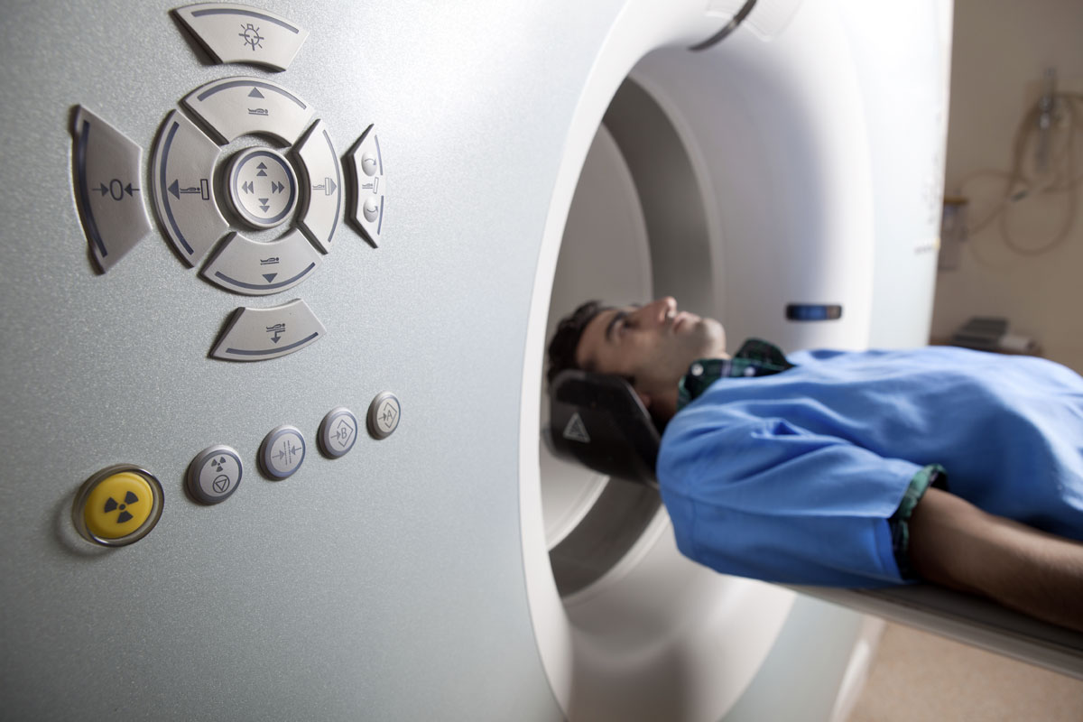
CT procedure
How is the procedure of computed tomography? The equipment consists of an apparatus itself with a table and a tunnel and a computer connected to it, where the results of tomography of the CT will be processed. The table will slide through the ring rotating around the studied area. While the CT scanner is working, it is impossible to move, so for the convenience of the patient part of its body can be fastened with belts.
Quick description CT looks like this:
- It is necessary to remove all metal decorations.
- The patient falls on the table that will move through the tunnel.
- The required time must be lying motionless, you can communicate with your doctor through a negotiation device.
- Some information, the doctor may report immediately after the completion of the procedure, but the images themselves with decoding are transmitted to the attending physician after 1-2 days.
How much will the scanning continue and what time it takes to it - depends on the tasks that the study should solve, as well as what kinds of CT are practiced in the clinic. Medium duration Procedures - from 15 to 30 minutes.

Some clinics today offer the CT service of the whole organism - as a preventive and diagnostic measure.
What is computed tomography with contrast and how do this study do? The algorithm is the same, except for the introduction of a contrasting agent. It can be carried out in two ways:
- Osually: the patient drinks a solution based on barium.
- Intravenously - manually or through a special injector (bolus method). Modern equipment Most often implies a bolus administration at which it is possible to adjust the supply time of the substance and the speed of its receipt.
Decoding KT.
How is the transcripts of photographs of computed tomography, which is necessary to inform the patient? These questions are the competence of an exclusively radar doctor. However, we list some signs of pathologies so that it is clear what is the result of a computer tomography.
- Changes in contours and sizes internal organs.
- Detection of foreign bodies.
- The growth of the tissue, the heterogeneity of its structure.
- Changes in tissue density.
- The presence of pathological fluid.
- Increase lymph nodes.
- The obstruction of blood vessels.
The survey results are not always unambiguously reliable. In some cases, the indicator is incorrect due to the fault of the patient itself - for example, if it moves when scanning, although it is prohibited. It also happens that the doctor himself makes an opinion on the basis of the data obtained. Reducing the likelihood of such errors - the appeal to the proven clinic, chosen not least prices, and for decent reviews and the availability of serious experts in the state.
Some clinics today offer the CT service of the whole organism - as a preventive and diagnostic measure. It is rather expensive, and these expenses are unlikely to be appropriate and justified. Tomography suggests a radiation load, so the body will subjected to it without the presence of objective readings - is not the most correct solution.
One of the modern and informative methods of diagnosing internal organs is. Thanks to tomograph, you can get an image of a high-resolution organ a short time. This type of survey is often used to diagnose various diseases.
Computer tomography is, in which it is possible to determine the state of the internal organs of a person, not penetrating inside.A procedure is performed using a tomograph - an instrument that emit X-rays that affect the patient's body at different angles. Then these rays fall on top-sensitive sensors and transmit received information as pictures.
In the future, these images are processed and three-dimensional pictures are obtained, which allows the doctor to explore the necessary organ of the patient more thoroughly.
The main mainly CT:
- X-rays used in tomography do not have adverse Reactions. After examining the traces of radiation in the body, the patient does not remain.
- Computed tomography is prescribed to diagnose changes in organs and tissues, which cannot be detected when performing other research methods.
- This method allows you to study any area of \u200b\u200bthe body, including soft fabricswhich are not amenable to radiography.
- Computer tomography helps to identify oncological diseases, pathology of cardio-vascular system, infectious diseases et al. Research allows in case of injury to identify injuries, lungs, vessels, and other organs.
- CT is used to conduct or implement therapeutic and diagnostic procedures. Also such a survey allows you to check the results operational treatment. With the help of computed tomography, you can determine the development stage and further treatment plan.
The survey is appointed only towards the doctor. Many diseases can be identified using affordable and simple methods diagnostics.
Types of examination
CT - area of \u200b\u200bexamination
There are several varieties of computed tomography:
- CT brain. The procedure allows you to determine possible pathology in cerebral shells or vessels. It will help accurate to determine the focus of inflammation.An examination is assigned during skull injuries, blood circulation disorders, meningitis, encephalitis, etc. In the resulting images, the structure of its shell, bones of the skull, blood vessels are clearly seen.
- CT. The study makes it possible to estimate the condition of all organs of the gastrointestinal tract and the retroperitoneal space. Tomography can show pathological changes, focus of inflammation, borders, degree of distribution.A study is appointed to confirm the presence or absence of foreign bodies, congenital anomalies, kidney stones ,. You can also identify the lesion of lymph nodes, atherosclerosis, blood disease, and other liver pathologies, digestive disorders, etc.
- CT lungs. The survey is assigned to confirm or exception: pulmonary tuberculosis, lung cirrhosis, pneumonia, diffuse pathologies, etc. You can also determine the state pulmonary artery, trachea, vessels and veins hollow.With the help of computed tomography, you can reveal a tumor at an early stage, trace the presence and number of metastases. With tuberculosis, you can define local and occurring in the pulmonary fabric.
- . The study is carried out in order to detect pathology this organ. This type of diagnosis allows you to determine the condition of the kidneys. Examination is conducted to identify congenital defects, pathological accumulation of fluid around the kidneys, urolithiasis, Polycystrosis, etc.The procedure is performed after removing the kidneys to monitor the state of the kidney bed. When performing the biopsy of this organ, using tomography, you can track the correctness of the fence of the tissue.
- CT chest. It helps to differentiate the foci of tuberculosis from the neoplasm, emphy of the lung from the abscess. You can also detect pleural effusion, pulmonary embolism, infectious diseases, mediastinal pathology, etc. The doctor prescribes tomography at chest injuries, occurrence pain sensations In the chest area, when preparing for operation.
- CT. The study is carried out to diagnose the spinal channel stenosis, osteochondrosis, intervertebral hernia, during injuries, abscesses and others.
- In rare cases, assigned. Such a survey is shown in the case of severe nasal injury. CT CT may be assigned to a plastic surgery to eliminate the nose defect.
In the early detection of pathology, using computed tomography, it is possible to begin treatment and prevent possible negative.
Preparation

Special this procedure does not require. Before research, you need to remove all metal objects: studs, rings, earrings, glasses, dentures. They can cause interference during the study. It is better to leave these items at home.
A few hours before the survey can not be eaten. This is especially important if you need the introduction of a contrast agent. It is necessary to tell the doctor about the preparations used or the presence of allergies.If an allergic reaction is observed in the introduction of a contrast agent, then the doctor will prescribe a medicine to eliminate signs of allergies.
Before performing CT, you need to take care of gastrointestinal was released from food.
The procedure is performed. A couple of days before the study, it is necessary to abandon the products that cause the bloating: sauer cabbage, apples, legumes, dairy products, carbonated drinks, alcohol. If possible, avoid solid and difficult food. On the eve of the study, it is necessary to make a cleansing enema.
When examining the kidneys, organs, the abdominal cavity, it is necessary to increase the volume of the fluid consumed. From the evening and until the moment of study, it is necessary to drink about 4 liters of net non-carbonated water, in which the urille of 76% or triombrus is 60% (2 ampoules).In the case of pregnancy, it is necessary to inform the doctor.
Examination procedure
Computer tomography is performed as follows: the doctor lays the patient on a special table and fastens with special belts. This is necessary to maintain proper position During the survey. The procedure is performed in the lying position on the back or on the side.
If you need to use a contrast material, it is administered intravenously. It is possible to introduce through the mouth or in the rectum. It depends on the type of CT studies. There is a feeling of heat, a metal taste in the mouth will appear at the site of the introduction of the substance. These signs disappear in a few minutes. Some may have a call for urination, which also passes.
The contrast agent is used for a better and informative study of a certain portion of the body.
When dizziness occurs, breathing difficulties must be said to the doctor. Specialists will provide medical care.The survey is performed when the table slowly starts moving through the scanner. When the apparatus is working, you can hear a small buzz.During research, the doctor may ask to hold his breath. It is strictly prohibited to move, as this will lead to defects on the tomogram. As a result, the image will be blurred.
More information about CT can be found from the video.
While staying in the scanner to monitor body position, special lighting can apply.The patient is located in the room one. The doctor talks to the patient during the entire study. If a person suffers from claustrophobic, then computed tomography will be a complex test for him.
The procedure is painless and lasts for 30 minutes. After the examination, the obtained data of the radiologist will analyze, and the results of the survey departure. At the end of the procedure to remove a contrast agent. a large number of liquids.
Contraindications

Computer tomography is not performed in the following cases:
- Pregnancy
- Excessive body weight
- Heavy Stage
- Renal failure
- Reception of adrenoblocators
It has been proven that even minimal X-ray radiation is negatively affected by the formation of the fetus. Therefore, apply this method Diagnosis during is strictly prohibited.If the study was conducted after giving birth during breastfeeding, it is possible to resume feeding only 2 days after tomography. And irradiation, and the contrast substance negatively affects the composition of milk.
In renal failure, tomography is prohibited, since this pathology slows down the purification of a contrast substance from the body.
If a person is in severe physical condition And it cannot control its actions, the procedure is also not conducted. The doctor appoints other methods of examination.Procedure for myelomic disease is prohibited, since the contrast agent can exacerbate the course of the disease and lead to.Patients with body weight above 120 kg are contraindicated.
Children under 14 this examination It is carried out only in rare cases if other research methods have not allowed to establish an accurate diagnosis. Contrast is not used in severe liver pathologies and some diseases of the cardiovascular system.
Side reactions after survey
 When performing tomography, the patient is subjected to a certain radial load. Even the minimum dose of X-rays increases the risk of oncological diseases. A major danger bears the implementation of several such procedures. The CT for children is appointed strictly according to the testimony. younger age, as well as women of childbearing age.
If it is performed with the introduction of a contrast agent, then the patient may be observed allergic reactions. This substance consists of iodide compounds.
The effect of iodide drugs can lead to a violation of the work of the kidneys.
To a greater or lesser extent, all contrast substances are toxic for kidney cells, which violates their functioning.
Noticed a mistake? Highlight it and click Ctrl + Enter.To let us know.
Computer tomography refers to one of the most modern and exact methods diagnostics. Studies are carried out without disrupting mucous membranes and skin Pokrov. CT is the most informative way if there are deviations in the development various organs and diseases. The high accuracy of modern tomographs allows you to put a correct diagnosis without surgical operation and appoint the most effective treatment schemes.
What is the difference between CT and MRI?
It is better to study the density and condition of the tissue allows X-ray radiation, which is based on computed tomography. The basis of MRI is based on radio frequency radiation in aggregate with action. magnetic field. As a result of research on a magnetic resonance tomograph, you can get detailed visualization of the organ. MRI is assigned to study soft tissues and helps to determine the diagnosis of head lesions, spinal cord and cartilage. Choose CT, if there is a need to explore bone structures, with diseases of the pelvis, chest, the base of the skull.
Diagnostics is needed on a computed tomograph in brain injury, damage to the bones of the skull, acute intracranial hematomas. The indication of the CT may be the assumption of the aneurysm of various localization. MRI will make it possible to determine with greater accuracy overall picture in the study of the pituitary, brain tissue inflammation, sclerosis scarm and stroke. In cases of determining the stage of oncological diseases, an MRI study with contrasts is prescribed.

Not a passing feeling of discomfort and pain in the intestinal, stomach, pancreas, liver, kidneys - reason to urgently undergo a study. CT can be excellent alternative surgical methods of diagnosing internal organs, and a diagnosis can be diagnosed in as soon as possible. Doctors in many cases prefer to assign CT to diagnose the pathologies of the spine, lungs, hearts. Timely measures will allow to avoid serious complications related to the pathology of the vessels, namely their bundles when various diseases. It is for this that computer tomographs are used in studies of the state of the pulmonary artery and aorta.
Multispiral computed tomography - an indispensable way of detailed study of previously detected pathologies. An indicative of the NOS CT will be indicative after detecting the curvature of the partition or the sinusitis during the radiographic study of the sinuses. Identification of benign and malignant formations on the most early stages Perhaps only thanks to the performance of computed tomography. With the help of CT, it is possible to carry out a detailed study of the maxillofacial region, so the method is well in demand and dentistry.
The cost of computed tomography varies from 3 to 20 thousand rubles. The high cost relative to X-ray is justified by the greater informativeness of similar studies, the presence of unique studies using contrast and testing vessels. The accuracy of CT is comparable to data obtained by conducting diagnostic operations.

And whether the daily background radiation is harmful present in environment? 2-10 MSV - radiation load during CT. In urban conditions, a similar dose receives every 3-5 years of life, the radiation background of the Earth, the flights and many other factors are taken into account, which cannot be avoided for an hour. The impact of CT on the body is compensated by the possibility of detailed diagnosis of the body, without which health control is not possible.
Contraindication for CT may be pregnancy. Doctors try to choose alternative methods Research to avoid risk to health fetus. Children are more sensitive to radiation, so it is prescribed it only if it is impossible to diagnose in a different way.
An exception can be a CT study of pregnant in the threat, a possible or existing disease, the life of a mother and a child. In similar situations, CT nursing women, with research with the introduction of contrast, it will be necessary to take a break in feeding for 24 hours. CT with contrast is really necessary, with allergies to iodine-containing substances, the safer contrast without iodine is selected.
How is the procedure?
Computer tomography is carried out on an empty stomach, so in 3-4 hours it is necessary to refrain from meals, especially before topography with contrast. The diagnosis of one zone takes about 20 minutes. Discomfort is possible in the presence of claustrophobia, but in comparison with the diagnosis on the MRI apparatus closed typeHe is minimal. During the study, it is important to maintain immobility in order to get the most clear pictures.
The procedure for administering the contrast is painless, for a minute after administration, the patient feels warm, spilling through the body. After computed tomography, you can get behind the wheel, go to work or do homemade deals, i.e. come back to the usual image Life.
Computer tomography - the second in popularity after the MRI method, allowing in detail to consider the smallest details of the state of the organs in the human body and identify the most tiny foci of damage in them. Magnetic resonant tomography of the CT is only inferior to the fact that is not 100% safe (irradiation with which it is carried out this species diagnostics, although minimally, but still takes place). BUT chief flaw MRI in comparison with the CT is that a computer study covers everything without exception of the human body zone, and the magnetic resonance "blind" in relation to thick cruise bones and the structures of the ribs. And, of course, the differences in two techniques are noticeable.
The scope of the study of CT covers the entire body of a person, includes a brain, all spoken departments, organs in the chest cavity, the contents of the abdominal cavity together with the retroperitoneal space or separately, the organs of the small pelvis. In addition, computed tomography makes it possible to examine the whole body entirely and its individual organs.
Whole body
The study of the whole organism is appointed by specialists rarely - for planning treatment strategy, to assess the results of the conducted operational interventions, To control the state of transplanted organs, to monitor the reaction of oncology to chemotherapy. In addition, CT of the whole body is carried out for:
- diagnostics of processes of pathology developing secretive;
- detection and estimates of the prevalence of various tumor formations, the degree of coverage of other bodies / systems;
- identifying metastases;
- estimates of the state of the vascular system;
- clarifications of the presence of complex diseases of the chest organs, abdominal cavity, a small pelvis.
Since the comprehensive diagnosis is prescribed quite rarely, and the duration and coverage of the procedure is large, such a scan is not cheap. Within Moscow, the price of a CT of the whole body ranges from 20.000-25.000 to 35.000-40.000 rubles, on the periphery costs may be lower, but only 1,000-2,000 rubles are equipped with all medical centers for such diagnostics.

Brain
The unique opportunity to study the brain is informatively and without external intervention, with the identification of pathologies in structures, tissues, brain vessels, in the bones of the skull provides modern medicine Computer tomography head. Standard testimony for CT brain is:
- often and suddenly manifested obscure pain;
- confusion of consciousness;
- dizziness;
- cramps.
This procedure is assigned with the objectives of identifying:
- stroke;
- neoplasms (including cystic) and metastases, their localization, distribution, penetration into neighboring tissues;
- consequences of head injuries;
- anomalies in the size of the ventricles of the brain;
- diverse pathologies;
- anomalies in the structures of the brain;
- inflammatory processes in the brain itself and in his shells;
- narrowing and spasms of vessels;
- defeats of the bones of the skull.
The brain scanning procedure is one of the most common in medical institutions, so the cost of CT of this direction ranges from 3,200-4.100 rubles, and the price can be reduced due to discounts for the service at night and weekends, for pensioners and children. A separate price for a study with the introduction of contrast - the cost increases to 5.900-8.500 rubles.

Spine
CT spine visualizes a detailed image of intervertebral discs, vertebrae with processes, bundles, blood vessels, spinal cord with nerves passing in it. Timely diagnosis Problems with this department human body It is very important because the spine is an element on which the mobility and normal human activity depends. Most often, such research is carried out to clarify the nature. owl syndrome In the back area, to detect the hernia of intervertebral disks, as well as for:
- pre- and postoperative examination;
- identifying all types of tumors (including metastatic) spinal column;
- estimates of the vertebral bone fabric for the diagnosis of vertebral fractures;
- control over the conduct of some diagnostic procedures;
- identifying the narrowing of the spinal channel, diseases of infectious-inflammatory, degenerative origin.
The cost of the spinal survey method by computer tomography in Russia varies from 2.100-2.500 for each department in the Republic of Bashkortostan to 3.200-4.000 in the Central Federal District. For the service with the introduction of a contrast agent that makes a study still more clearly, will have to pay another 3.000-5.000 rubles more.
Rib cage
Computed tomography of the chest organs is used to diagnose diseases, pathologies, abnormalities of light, esophagus, vascular and lymphatic system, soft tissues, mediastinum organs. A study is assigned to confirm the results of conventional X-ray and fluorography, to establish nature with identifying the following problems:

An examination of the chest is often carried out using a contrast agent, which is why its cost is sufficiently high. The price of additional services for the introduction of contrast ranges from 1.800-3.500 (depends on the dose and the amount of ampoules at the calculation of the patient's weight), thus, the final cost is 8.000-10.000 rubles in Moscow. The minimum was recorded on the territory of the Southern Federal District - 4.000-4.300 rubles with contrast.
Abdominal cavity and retroperitoneal space
The examination of the abdominal cavity and the retroperitoneal space can be carried out individually and as a comprehensive study. CT scanning of the organs of this part of the human body gives the most detailed and accurate results, allowing you to identify minimal pathologies in a state of organs and tissues. In the trouser tomography inspects such organs as the liver, with bubble, pancreas, spleen and intestines. In the retroperitoneal region, kidneys, urinary pathways, adrenal glands are scanned. Limph nodes of local localization, blood vessels that permeate the organs of this region are also inspected.

An exemplary list of diseases, the detected CT in the peritoneum and in the space behind it, includes:
- tumors of cystic, primary, secondary genesis;
- abscesses;
- anomalies of the development of organs;
- circulatory disorder due to atherosclerosis or aortic aneurysms;
- pathological processes;
- inflammatory processes;
- mechanical jaundice;
- foreign bodies, volume formations (stones);
- injuries with consequences;
- obstruction of ureters;
- renal colic.
The price of the CT-examination of the abdominal and retroperitoneal organs depends on whether a comprehensive study of both zones is carried out, or the peritonean and the retroperitoneal space is scanned separately. In the first case, the cost of average in Russia averages from 3.100-3.500 to 5.000 rubles, in the second - 2.100-2,800 rubles.
Low pelvis organs
The bodies of a small pelvis include bladder, straight intestine, appendages and uterus (women), seed bubbles and prostate gland (men). CT of this body area identifies diseases such as:
- injury damage;
- inflammatory diseases;
- malignant / benign neoplasms, metastases;
- anomalous development of organs;
- pain of obscure nature;
- cyers of appendages.
The cost of CT organs of a small pelvis is in the capital 7.500-8.000 rubles (10,000 rubles for a contrast procedure), in Ufa, the average price is fixed at 4.000-4.600 rubles, in St. Petersburg, the scanning can be passed in 4.500-5.200 rubles. Many centers offer discounts on the passage of CT people pension age, Children, patients who agree on the passage of the procedure at night.
CT for everything
Computer tomography allows you to scan both large groups of organs and separate parts Body. You can pass the eye examination, sinuses, teeth, joints or limbs. There are also special complex CT-programs: Computer Urography for the study of kidneys, ureters and bladder (cost in Moscow 3.800-4.100), CT-angiography of vessels (price depending on body departments varies from 6.800 to 11.000 rubles), CT-coronorography for Consideration of coronary arteries (cost - 8.000-8.500 rubles).
Obviously, computed tomography helps to collect information on the health status of the whole body - from head to feet, from bones to cavities internal organs. It is worth this pleasure cheaper than MRI, issues results better ultrasound, therefore is worthy alternative The main methods of diagnosis.
The method of computed tomography is the most modern and informative method medical examination. CT is practiced relatively recently - since 1988, and during this time made it possible to significantly improve the diagnosis of diseases. There was no need for surveys requiring introduction to the body of additional devices, and other inconveniences for the patient. Based on the CT, another method of a layered study of the organism was developed in the future. So, computed tomography, what is it?
Computed tomography is the study of human internal organs using X-ray radiation.
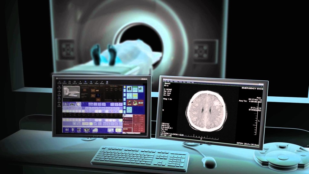
On the patient's body using a radius tube of the CT-tomograph, under different angles with low doses of X-rays, the result of which is registered by special super-sensitive detectors, obtaining many layered images of the body under study.
Next, the computer using complex software produces processing and analysis of the obtained CT-shots, creating a three-dimensional image of a patient body that allows the doctor to study it in various angles. This is the main advantage of CT to conventional radiography.

Computer technologies allow you to carry out a detailed study of all tissues, coordinating the process.
With this method, almost any area of \u200b\u200bthe body can be studied, including soft tissues that are not amenable to conventional radiography. It became possible to perform measurements, adjust the work of the scanner, directing it to a particular area.
When diagnosing back diseases with a computer are recreated volumetric models Spine and muscle tissues. This allows you to explore both bone tissue and intervertebral discs and adjacent areas.
Varieties of computed tomography
All types of CT are based on the same radiation impact method. They differ mainly technical features devices, as well as applications.
- Spiral KT. - The earliest, but at the same time the most popular and accurate type. He got its name due to the fact that the annular part of the tomograph, in the walls of which the radiation source is located relative to the horizontally moving table, on which the patient is located. Thus, the movement of the radiation source scanning the desired area is reminded by the movement of the spiral. This reduces the study time and increase the anatomical coating zone.
- Multispiral CT - Improved variety of first type. It is distinguished by bunched radiation, which increases the range of the area being viewed. Sometimes tomographs can have several radial tubes. Changes contribute to the accelerated passage of the procedure, and also reduce the number harmful effects In case of inspection.
Check out the video about the possibilities of multispiral computed tomography.
- Cone-ray CT - A narrower variety, focused on the study of bones and tissues of the head, is used including in dentistry. The device has smaller size, only the patient's head falls under the ring. Localization helps to make sharper, large and surround pictures, and discover the disease even at an early stage.
- Emisy CT. - The most rare type used mainly in oncology, cardiology and other areas, where to recognize the hearth disease is not always simple. The essence of the principle in conducting a patient of radionuclides, which "highlight" organs. Equipment for such a procedure is far from each clinic, and it is applied only in specialized centers diagnostics.
Opportunities KT.
The method is great for primary diagnostics and disease detection. At the same time, the CT can also be used to confirm the diagnosis established when using other clinical methods.
The range of bodies that can be studied with CT, extremely wide. This includes the abdominal cavity, the region of the chest, gOOD SYSTEM, liver, pancreas and other parts and body organs. Thanks to CT, it became possible to study the diseases of the brain.
In some cases, patients are performed computed tomography with a contrast - a special substance that is used to improve the clarity of the structures of the body under study.
The drug is introduced into the vein and accumulates in tissues, improving their visualization in the pictures. Especially well, it penetrates into richly blood supply and tissues, because of which it is often used when identifying pathological foci with a strengthened blood flow: plots of inflammation, malignant neoplasms. A contrast agent without consequences is completely excreted from the body for one and a half days.
CT is extremely effective in the diagnosis of diseases of the spine.
Thanks to the data received by the computer, it is possible not only to consider each individual vertebra, to establish the bone density, but also determine the state of intervertebral discs, joints, recognize the localization of inflammation of soft tissues and the degree of compression of nerve roots.

Computed tomography, contraindications
There are no categorical contraindications to CT. Radiation, which affects a person during the survey, is so insignificant that it is not worried about what. The process will not harm the body even with repeated CT.
In some centers to CT, children under 14 are not allowed. In addition, if the introduction of contrasting substances is planned, you should make sure that you do not have allergies. For this, tests are carried out or anti-allergic drugs are used.
Methodology of procedure
If a decision is made to apply a contrast agent, the composition is administered to the patient before CT (usually intravenously, or by ordinary adoption).
Before starting the study, it is necessary to remove clothing and decorations from yourself, you can usually leave underwear or a special bathrobe.

The patient falls on the mobile table, which will move into the beginning of the procedure inside the scanning rings. During the examination, it is desirable to preserve immobility. The table will perform minor horizontal movements, the ring - rotate around the patient.
The procedure is absolutely painless. In case the patient has any inconvenience, it can always consult a technique sitting in the next room. On average, the procedure takes from 15 to 30 minutes.
How to prepare for research
Usually, special training Before conducting CT does not require, with the exception of the following cases:
- CT using contrast preparations is available on an empty stomach;
- in studies in the area of \u200b\u200bthe small pelvis, the bladder must be filled in moderate;
- when examining the abdominal cavity, on the evening before, it is necessary to empty the intestine using the laxative or with the help of the enema.
Also necessary for several days before the procedure try not to use products capable of causing meteorism.
Warn attending your doctor in case you:
- have chronic diseases;
- recently passed radiography using barium (this substance may interfere with the clarity of the obtained images);
- we suffer from claustrophobia (finding inside tomograph in this case may be unpleasant for you).
It is necessary to have certificates with themselves regarding the course of your illness, including: the direction, an extract from the history of the disease, snapshots or results obtained during other methods of survey.
At the end of the procedure, the patient receives pictures on hand, in some cases a CD disk with three-dimensional images can be attached to them. The doctor who issued the direction decides on further treatment Depending on the results obtained.
If you have passed the survey on your own initiative, about further action You can consult with the specialists of the Diagnostic Center.
In the clinics of St. Petersburg, the cost of the CT procedure of one site approximately begins from 2600 rubles and depends on which body is examined, and whether the contrast agent is used.
In Moscow, it will cost somewhat more expensive: the minimum cost will be 3,700 rubles.