The head of the fetus is less than a week. At what stage of pregnancy should the baby's head take the correct position? Norm and deviations from it
During pregnancy female body is undergoing tremendous changes. It is worth noting that for each of the fairer sex, they can occur in different ways. In the same way, subsequent pregnancies can be very different from previous ones.
While waiting for the baby, a woman is faced with various diagnoses and unknown terms. So, some ladies who are in an interesting position are told that the fetal head is low. It is about this feature that will be discussed in this article.
The fetal head is low: what does this mean?
When an expectant mother hears such a statement from a gynecologist, she often panics. This is absolutely impossible to do. The low position of the fetal head is not a pathology. A similar condition will not harm your unborn child in any way. However, if you find such a feature, it is worth following certain recommendations.
Diagnostics
Is it possible to independently determine the low position of the fetal head? Answer to this question negative. Doctors say that a woman may have suspicions about the presence of this feature. However, it is possible to say with confidence that the fetal head is located low only after examination. There are two ways to determine this condition.
Manual inspection
During the routine, the doctor can determine the position of the child. When an experienced obstetrician-gynecologist determines how far the child's body is from the entrance to the cervical canal.
Ultrasound diagnostics
At this survey you can also find out that the fetal head is low. In this case, the doctor must necessarily determine the state of the cervix and describe its position in the ultrasound protocol.

What if the baby's head is low?
When this feature is found, a pregnant woman is most often not prescribed any treatment, but the doctor always gives recommendations and advice. They can be different depending on the gestational age. Let's figure out how to behave with a low position of the child?
Limiting physical activity
In case of premature prolapse of the fetus in women, it is always recommended to limit any load. Any sports training and lead a relaxed lifestyle. It is strictly forbidden to lift heavy objects and walk in heels.
Refusal of sexual intercourse
The low head of the unborn child already presses on the entrance to reproductive organ, provokes the expansion of the cervix.
Psychological peace
When the baby's head is low, the woman is advised to take sedatives. These include the drug "Valerian" and the drug "Motherwort". These medicines are completely safe for the unborn child.
These drugs should only be taken on the advice of a specialist. Never self-medicate. Otherwise, you can only aggravate the current situation.
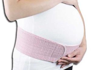
Using a bandage
It is always recommended to use a bandage when the child is placed low. This device can be purchased at any pharmacy chain or a medical supply store. The bandage will gently support big belly and will not let the baby be born prematurely.
It should be noted that the bandage can only be used in the second half of pregnancy. It is always necessary to put on the device in horizontal position... Only in this case will the efficiency be maximized.
Treatment for premature head drooping
Correction is carried out only when the premature opening or shortening of the cervix has begun. That is why women with this diagnosis should always be under the supervision of a doctor.
Treatment consists in putting a device called the Pessary on the cervix. He will hold in its original position until the very birth. This is what will help to avoid premature birth of a child.
Normal pregnancy
Normally, the fetal head descends into the small pelvis after 36 or even.In this case, the woman is not given any recommendations and is allowed to lead familiar image life. It is worth noting that with this outcome of events, the child is full-term and ready for birth.

Conclusion
If you have found low location the head of the child, it is worth carefully remembering all the recommendations of a specialist. Subject to the rules, you will be able to endure and give birth to your baby on time.
Ultrasound for pregnant women is a screening examination method. The medical term "ultrasound screening" is an examination of absolutely all pregnant women in deadlines to identify intrauterine defects fetal development.
The screening study is carried out three times during pregnancy:
- I screening - at 11-14 weeks;
- II screening - at 18-22 weeks;
- III screening - at 32-34 weeks.
Ultrasound of the fetal head at 1 screening
The expectant mother at the end of the first trimester is prescribed in order to exclude such gross malformations of the fetal head as pathology of the brain, skull bones and facial skeleton in utero.
The doctor evaluates the following structures of the fetus:
- the contours of the bones of the cranial vault for their integrity;
- structures of the brain, which normally look like a "butterfly";
- measures the length of the nasal bone of the fetus (at 11 weeks indicate its presence or absence, and at 12-14 weeks - the norm is from 2 to 4 mm);
- biparietal size (BPD) of the head - measured between the most prominent points of the parietal bones of the fetus. The average normative value of BPD in the period of 11-14 weeks is from 17 to 27 mm. The doctor will look at these indicators in a special table.
If everything is in order with your fetus, the doctor will write down the following in the ultrasound protocol:
- bones of the cranial vault - the integrity is preserved;
- BPR -21 mm;
- the choroid plexuses are symmetrical, in the shape of a "butterfly";
- the length of the nasal bone is 3 mm.
What pathology of the head occurs during the first ultrasound screening?
Particular attention is paid to assessing the length of the fetal nasal bone. This is an informative criterion. early diagnosis Down syndrome.
Examination of the bones of the skull already at the end of the first trimester makes it possible to identify such severe developmental abnormalities as:
- acrania;
- exencephaly;
- anencephaly;
- cranial hernia.
Anencephaly- the most common defect of the central nervous system, in which the brain tissue and skull bones are completely absent.
Exencephaly- the bones of the skull are also missing, but there is a fragment of brain tissue.
Acrania- a developmental defect in which the fetal brain is not surrounded by the bones of the skull.
It's important to know! With these three vices, the death of the child occurs. Therefore, if they are detected at any stage of pregnancy, it is proposed to terminate it by medical indications... V further woman consultation of a geneticist is required.
Cranial hernia- This is a protrusion of the meninges and brain tissue through a defect in the bones of the skull. In this case, a neurosurgeon's consultation is required to find out whether it is possible to correct this defect with an operation after the birth of the child.
Decoding of ultrasound of the fetal head at 2 screening
During this time, close attention is paid to the brain and facial skeleton. Identification of the pathology of fetal development allows you to warn future parents about possible consequences and get information about the long-term forecast.
Important indicators on examination are biparietal size (BPD), frontal-occipital (LZR) and fetal head circumference. All these important measurements are carried out in strictly cross-section at the level of certain anatomical structures.
The doctor evaluates the shape of the fetal head by the cephalic index (BPD / LHR ratio). A variant of the norm is:
- dolichocephalic form (oval or oblong);
- brachycephalic form (when the skull is rounded).
Important! If the fetus has a lemon-shaped or strawberry-shaped head, this is bad. It is necessary to exclude genetic diseases and concomitant malformations.
A decrease in these indicators ( small head of the fetus) is an unfavorable sign in which microcephaly must be excluded (a disease characterized by a decrease in brain mass and mental retardation). But not always a small head circumference speaks of pathology. So, for example, if all other sizes (tummy circumference, thigh length) are also less than normal, this will indicate intrauterine fetal growth retardation, and not a malformation.
With an increase in BPD and head circumference ( big head fetus) can talk about dropsy of the brain, about the presence of a cerebral hernia. If, during fetometry (fetal measurement), all other indicators are also higher than normal, then an increase in BPD indicates a large size of the fetus.
By the time of the second screening, all the anatomical structures of the brain had already been formed and they are well visualized. Measurement of the lateral ventricles of the brain is of great importance. Normally, their dimensions should not exceed 10 mm (on average - 6 mm).
Note! If the lateral ventricles of the fetal brain on ultrasound are expanded from 10 to 15 mm, but the size of the head is not increased, this condition is called ventriculomegaly.
Chromosomal abnormalities can lead to enlargement of the lateral ventricles and ventriculomegaly, infectious diseases mothers during pregnancy, intrauterine hypoxia fetus.
Ventriculomegaly can be:
- symmetric (when the lateral ventricles of both cerebral hemispheres are expanded);
- asymmetric (expansion of one of the ventricles or its horn, for example, left-sided ventriculomegaly);
- can exist in isolation from developmental defects;
- or be combined with other vices.
With light and medium careful dynamic observation of the size of the ventricles of the brain is necessary. In severe cases, this pathology can turn into dropsy of the fetal brain (or hydrocephalus). The earlier and faster the transition from ventriculomegaly to hydrocephalus occurs, the worse the prognosis.
It is very difficult to answer the question of parents, how pronounced with such a deviation will be neurological manifestations in their future baby and what will be his psychomotor development... And if there is a question of terminating pregnancy after the detection of this pathology, the recommendations of the doctors should be followed.
Hydrocephalus - another pathology of the brain, which is detected by ultrasound. This is a condition when there is an increase in the size of the ventricles of the brain more than 15 mm due to the accumulation of fluid (cerebrospinal fluid) in their cavities with a simultaneous increase intracranial pressure and leading to compression or atrophy of the brain. As a rule, this pathology is characterized by an increase in the size of the fetal head.
It should be said that the most unfavorable prognosis will be when ventriculomegaly / hydrocephalus is combined with other malformations, chromosomal abnormalities, as well as with isolated hydrocephalus.
At the second screening special meaning assigned to the assessment of the anatomy of the cerebellum (it consists of two hemispheres, which are interconnected, the so-called cerebellar vermis). The cerebellum - translated means "small brain", is responsible for the coordination of movements.
Hypoplasia (underdevelopment) of the cerebellar worm can lead to disastrous consequences:
- the ability to maintain balance is lost;
- lack of muscle coherence;
- loss of smoothness in movements;
- problems with gait appear (it becomes staggering, like a drunken one);
- trembling appears in the limbs and head of the child, delayed speech.
Measurement of the interhemispheric size of the cerebellum is very important for detecting this pathology.
Making a "cut" through the cerebellum, the doctor estimates the size of the cerebellum, determines the cerebellar worm. Normally, the interhemispheric cerebellar size (MRM) in the 2nd trimester is equal to the gestational age.
Fetal cerebellum size by week of pregnancy: table
|
Pregnancy period, weeks |
|||
Subject to careful study:
- reflection of ultrasound - a signal from the median interhemispheric fissure (M-echo);
- the cavity of the transparent septum;
- visual hillocks;
- the shape of the horns of the lateral ventricles;
- corpus callosum.
On the second screening, abnormalities of such a structure of the brain as the corpus callosum may be detected. It is a plexus of nerve fibers that connect the right and left hemispheres.
If the corpus callosum is not clearly visualized on the median section of the brain, then one can think about dysplasia, hypoplasia or agenesis of the corpus callosum. The cause of this deviation can be hereditary, infectious factors and chromosomal diseases.
The doctor compares all the obtained digital indicators with the average statistical norms indicated in special tables.

Examination of the facial skeleton in the II trimester
The fetal face is another important subject of study during ultrasound screening.
When examining the face of the fetus and the nasolabial triangle on an ultrasound scan, you can see the lips, nose, eye sockets and even the pupils. With certain skills, the doctor will see lip movements, including protruding the tongue, chewing, and opening the mouth.
It is possible to diagnose defects such as cleft lip and hard palate:
- Cleft on both sides upper lip popularly called "hare lip".
- The splitting of the tissues of the hard and soft palate, in which there is a communication between the mouth and nasal cavity, is called the "cleft palate".
It is not difficult to imagine the confusion of the expectant mother when she is informed about such tricks of nature. Of course, the pathology is complex and unpleasant. But modern medicine able to carry out surgical correction and help such babies.
Why do you need an ultrasound of the head at the 3rd screening?
The purpose of the third screening is to confirm or deny the identified deviations and malformations suspected during the second screening.
V mandatory an examination of all the same structures of the brain and facial skeleton is carried out.
The purpose Ultrasound screening the fetal head is a careful study of the structures of the brain and the structure of the face in order to identify abnormalities. If the diagnosed malformation is incompatible with life, then obstetricians-gynecologists recommend interrupting such a pregnancy. If the prognosis is favorable, then the parents will be able to get advice from specialists in the surgical correction of the defect and promptly begin treatment after the birth of the baby.
Oksana Ivanchenko, obstetrician-gynecologist, specially for the site
From the 14th week of pregnancy, during an ultrasound examination of the fetus, it is possible to assess the biparietal size (BPD). This fetometric indicator is quite informative, since it not only reflects the degree of development of the embryo, but also allows one to conclude about the presence or absence of pregnancy pathology. BPR is currently required element second and third screening studies. Correct interpretation of its values is extremely important for an adequate interpretation of the ultrasound results.
Definition and research method
The biparietal size is the distance between the most prominent points of the parietal bones of the fetal head. This ratio represents the width of the baby's head. As a rule, an ultrasound examination is performed using a transabdominal method. Only in the case of unsatisfactory visualization, a transvaginal method is used, which has a higher resolution.
The main principle of the study of BPD is to conduct an ultrasound cut through the fetal head in a horizontal plane. After obtaining a clear picture, the distance between the outer surface of the upper contour and inner surface the lower contour of the received echographic picture. Thus, the value of the biparietal size is obtained, with an accuracy of 1 mm. The nuances of ultrasound are important only for diagnosticians.
Normal indicators of bipolar disorder

In addition to the average value, for each gestational age, the table contains variants of the norm of bipolar disorder. Currently, among neonatologists and obstetricians-gynecologists, it is considered pathological sign deviation of the biparietal size from the gestational age by 2 weeks or more.
BPR value
Evaluation of this fetometric indicator on ultrasound allows us to draw conclusions about the following nuances pregnancy:
- delivery process - the study of bipolar disorder and the anatomical dimensions of the pelvis is widely used in obstetric practice... The degree of their correspondence to each other determines the tactics of future childbirth. With the proportionality of these structures, delivery is performed per vias naturalis (through the natural birth canal). If there is a significant discrepancy (for example, with congenital narrow pelvis), it is recommended to perform the operation "cesarean section";
- gestational age - in the absence of pregnancy pathology, the biparietal size can be used for indicative definition term. The need for this is extremely rare. As a rule, this is true for those mothers who long time avoided consulting doctors or have no idea about the date of conception;
- the presence of pathology of pregnancy and abnormal intrauterine development.
It is most important to determine the presence of deviations from normal flow pregnancy or fetal development. To make an adequate interpretation of the results, you need to know under what conditions an increase or decrease in this parameter occurs.

Reducing BPD
The deviation of the biparietal size to the smaller side indicates that the fetus has a head that is too small for it gestational age... Most probable cause this state - intrauterine retention development (IUGR), which can occur under the influence of the following factors:
- chronic intoxication of a woman before or during pregnancy (smoking, alcohol consumption, substance abuse, drug addiction);
- an unbalanced diet with a deficiency of vitamins, polyunsaturated fatty acids, protein and other nutrients;
- multiple pregnancy;
- the mother has arterial hypertension which has not been normalized before pregnancy, or endocrine diseases;
- development of infection during pregnancy.
There are two forms of pathology. A small head in the fetus can be observed only with a symmetrical form of developmental delay. In addition to a decrease in BPD, a decrease in all other fetometric parameters will also be observed. The prognosis is unfavorable, as there is a high risk of having a child with multiple congenital malformations development.
To assess the severity of intrauterine growth retardation, obstetricians-gynecologists distinguish three degrees of IUGR by ultrasound:
The tactics of managing a pregnant woman depends on the degree of delay, which is determined individually by the supervising physician.
Increased BPD
A large head in the fetus, which is determined by ultrasound as an increase in bipolar disorder, most often indicates dropsy of the brain (hydrocephalus). There are two types of this pathology, each of which requires additional ultrasound structures of central nervous system child.
The first is external hydrocephalus, when cerebrospinal fluid (CSF) fills a significant space between the membranes of the brain. In this case, it is necessary to examine the subdural space (under the outer meninges) of the fetal head by ultrasound.
Internal hydrocephalus is the second type of pathology, more unfavorable. With it, there is an accumulation of cerebrospinal fluid in the cavities of the brain, which is detected during the study of the ventricles on ultrasound. As a result of compression of the surrounding tissues, atrophy of the cortical structures and the brain stem occurs, which can be the cause of intrauterine fetal death in a pronounced process.
A large fetal head may be a variant of individual variation. Therefore, it is important to evaluate BPD in relation to fetometric parameters of the trunk (diameter of the chest and abdomen) and extremities. With an increase in only the biparietal size and a large volume of cerebrospinal fluid is detected, the diagnosis of hydrocephalus can be confidently made.
Prevention of bipolar disorder
The best prevention of any developmental abnormality is the correct pre-gravid preparation of both parents. This is a set of measures for the maximum possible recovery of the organisms of men and women before pregnancy. Neonatologists recommend starting preparations for pregnancy at least 6 months before the planned conception. Adequate pre-gravid preparation, prescribed by a qualified doctor, allows 98% of cases to avoid the development of pregnancy pathology. Have healthy people there are no children with birth defects.
Evaluation of the biparietal size during ultrasound of expectant mothers has great importance... It reliably determines some violations of fetal development and allows you to choose the tactics of further pregnancy management. It is important to evaluate bipolar disorder in combination with other fetometric indicators. This approach excludes the setting misdiagnosis if an increase in biparietal size is a manifestation of the child's individual variability.
Pregnancy is special for all women life stage... At this time, the expectant mother experiences new sensations and, on the other hand, learns her essence. At the same time with positive emotions and fantasies about the future baby, the young mother has to go through many consultations and take a lot of tests. Such visits to the clinic sometimes make you nervous. But tests are needed to control normal growth and the development of the baby in the woman's tummy.
When an ultrasound is needed
When the expectant mother comes to see her doctor, she is explained the need and timing of observations under an ultrasound machine. There are two types of screenings and selective studies. Screening is a compulsory examination of all pregnant women using ultrasound in certain terms... Usually, the planned expectant mother takes place at a period of 10 to 12 weeks, from 22 to 24 weeks, at 32 and 37-38 obstetric week pregnancy. When carrying out this type of examination, the size of the fetus and their compliance with the norms, the state of the uterus and placenta are measured. Selective studies are prescribed by the attending physician if there is a suspicion of a complication of pregnancy. In the case of determining the pathology of pregnancy, such examinations can be carried out an unlimited number of times.
Fetometry - what is it and why

One of important procedures fetometry is considered. When carrying out, the doctor analyzes the size of the fetus and their compliance with the norm. The procedure is ultrasound examination, the data of which the specialist verifies with the tables of norms. The check helps to detect defects and deviations in the development of the baby in time. When carrying out fetometry, the fetal head circumference is determined by weeks - the norm is an important indicator. For weeks, the doctor fixes the ultrasound values and draws conclusions about the baby's health. When the doctor states smaller sizes fetus than established for this period, then they talk about a slowdown in fetal growth. If, over the course of pregnancy, a lag of a couple of weeks appears, then doctors talk about intrauterine growth retardation. This delay can be caused by bad habits mothers, internal infections, chromosomal abnormalities or placental insufficiency.
How the circumference of the growing head of a normally developing fetus changes

Fetal head circumference by week - important indicator in mommy's womb. As you know, the baby's head in the mother's belly grows unevenly. At the beginning of development, its size significantly exceeds the size of the body. And by the end of pregnancy, the size of the fetus becomes uniform and proportional. If you trace how the fetal head circumference changes by week, you will notice that the greatest increase occurs in the second trimester. It is from the 15th to the 26th week of pregnancy that the head circumference of the crumbs increases by an average of 12-13 mm. This increase occurs every week. With a further increase in gestation, the growth of the head circumference slows down. By the end of the third trimester - about a month before the birth of the baby - this figure becomes more than 12-15 mm.
How to measure the circumference of a crumb's head

In order to measure the fetal head circumference by week, regular diagnostics are used using the device ultrasound examination... The study is carried out by a specialist in several projections to obtain the most correct and accurate result... In addition to diagnosing the head circumference, the doctor diagnoses such fetometric parameters as biparietal (BPD) and sagittal size, length of the femur bone, abdominal circumference, frontal-occipital (LZR) size, and others.
The table of norms used for diagnosis helps the specialist to determine the development of the fetus and potential deviations. If the doctor discovers significant deviations from the norm, then the woman is offered to terminate the pregnancy.
Formula for calculation
The fetal head circumference by week is determined according to the same principle as the biparietal size: it is measured by a method such as computer planimetry, or by the formula. The biparietal and LZR are preliminarily determined. The formula has the following form: OG = 1/2 * (LZR + BPR) * 3.1416. This indicator is rarely used to calculate the weight of the fetus and does not depend on the shape of its head.
The value of the indicator of the circumference of the child's head

What can such an indicator as the circumference of the fetus's head tell the doctor by week? The table of norms for this indicator has boundaries. If they are exceeded, this indicates the presence of developmental disorders. In this case main task the doctor becomes earlier identification of deviations and their correction. For example, an increase in head circumference may indicate a condition such as hydrocephalus. The disease manifests itself in the accumulation of fluid in the cavities. This process leads to an increase in pressure inside the skull and, as a result, to a decrease in the volume of the brain. Immediately after birth, the baby is punctured. With the help of the procedure, the accumulated fluid is removed and the condition of the child is alleviated.
The value of indicators for childbirth

In most cases, exceeding the parameters is attributed to individual characteristics crumbs. For example, if the parents are large, then it is assumed that the child will also be large. As already mentioned, the table shows the fetal head circumference by week of pregnancy. An increase in the indicator towards the end of pregnancy can lead to problems in generic process... For example, to a ruptured perineum. In this case, an episiotomy is done, that is, a small incision to facilitate labor.
The importance of the indicator
So, the definition of indicators of head circumference on different dates pregnancy and comparison of other indicators helps the doctor in time to identify pathologies in the development of the fetus, and development, as well as possible difficulties. A woman should not independently interpret or try to decipher the results of ultrasound diagnostics and draw conclusions about the health of the baby. The doctor takes into account multiple factors and observations, and only then makes an objective conclusion. It is important to remember that the development of each baby is individual and may not occur according to the tabular values.
32 weeks - why it matters

An important stage in ultrasound diagnostics is the 32nd week of pregnancy. Around this period, the fetus takes correct position for childbirth - head down. The fetal head circumference (32 weeks gestation) is approximately 283-325 mm. This gestational age is quite significant. Little baby in mom's tummy it is almost formed and even has eyelashes and eyebrows.
Fetal head circumference: table
As already mentioned, the first important ultrasound diagnostics the expectant mother spends 10-12 weeks interesting situation... The table shows the data starting from The calculation takes place from the day last menstruation... Tabular data for the 10th, 50th and 95th percentiles are presented. Most often, doctors are guided by the 50th percentile, but fluctuations from 10 to 95 are considered the norm. The percentile is the percentage that is below a certain amount percent in the sample. That is, the 50th percentile indicates that 50% of these values are below this level.
Weeks of pregnancy | Percentiles |
||
Of course, for every woman a fortune little miracle more important than anything else. While the baby is still in the tummy, the only way it remains to be seen by ultrasound. The importance of studying indicators of head circumference, height, weight and others is due to the need for constant monitoring of fetal development. Such monitoring not only helps experienced specialist manage pregnancy correctly, but also soothes expectant mother who wants to quickly hold her baby in her arms.