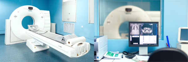
Preparing for the abdominal CT scan. Multispiral computed tomography (msct) of the kidneys
X-ray diagnostics is a research method that helps to identify many pathologies. A modern type of such research is called MSCT. abdominal cavity... This type of diagnostics refers to a type of computed tomography performed using the latest generation devices, which allow making thinner "slices".
MSCT has another name - doctors call it spiral computed tomography, or multilayer X-ray tomography... Unlike a standard CT unit, a spiral tomograph is equipped with several rows of emitters and detectors that pick up the signal. Such design features allow:
- reduce the duration of the diagnostic procedure;
- get a clearer picture;
- reduce radiation exposure to the body;
- to get an image of a whole organ in one rotation (additional "rolling" is not needed);
- to fix the processes taking place in the organs;
- reduce the number of defects in images as a result of natural organ movements.
Such tomographs differ from the MRI unit in the type of radiation and the technologies used:
- when examining an MRI, a magnetic field is used, while CT and MSCT involve the use of X-rays;
- magnetic resonance imaging better shows the structure and structure of soft tissues and organs, and CT is used when it is necessary to examine in detail the bone structures and tissues with high density;
- the MRI procedure is not always suitable for examining the intestines and other hollow organs, while CT or MSCT examination using contrast solutions reveals the slightest changes in the intestinal mucous membranes and internal cavities organs.
Before sending a patient to study the abdominal cavity with contrast, the doctor weighs all the points - indications and contraindications, the patient's current condition, indications of previous examinations - and then chooses which is better: standard or contrast MRI, CT scan or more advanced multilayer tomography with contrast.
The use of a multispiral tomograph in the diagnosis of abdominal and retroperitoneal pathologies makes it possible to create flat images of organs and structures located in the study area, as well as to simulate a 3D image of the abdominal organs (ABP).
What MSCT OBP shows
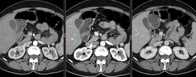
With the help of MSCT of the abdominal cavity and retroperitoneal space, the doctor examines the organs and structures in this area. Unlike a computer scan, this diagnostic method is used when it is necessary to examine the hollow organs. In the presence of symptoms that indicate violations in them, the use of a contrast solution is indicated (most often it is the drug Trazograf with triiodide or a barium solution).
Examination by the MSCT method with contrasting establishes the position (including mutual), condition, structure of organs located in the abdomen. Diagnostics reveals:
- dysfunction of the OBP;
- pathological changes (inflammation, necrotic processes and infectious foci);
- trauma;
- tumor processes, including at stage 1.
It is important to understand what MSCT shows abnormalities in the form of a black and white image that can only be read by a radiologist.
When MSCT is prescribed
Prescribe MSCT of the abdominal and retroperitoneal organs in the case when other diagnostic methods cannot give clear unambiguous results. Direct indications for diagnostics on a spiral tomograph are:
- jaundice, especially asymptomatic;
- sharp weight loss for no apparent reason;
- chronic digestive disorders;
- chronic forms of dysfunction of the intestines, stomach and other organs;
- acute anterior injury abdominal wall and penetrating wounds of the abdominal cavity.
Also, patients who are preparing for an operation on the abdominal organs or undergoing a complex course of therapy that require monitoring of even minimal changes are referred to MSCT.
Contraindications to MSCT
Radiologists say that such a study as MSCT is much safer than conventional computed and magnetic resonance imaging. The only significant limitation to the procedure is pregnancy. Lactating women, if necessary, are recommended to temporarily interrupt diagnostics. breast-feeding.
The list of relative contraindications for MSCT includes:
- intolerance to the drug Trazograf or its analogues, as well as barium;
- children's age (up to 14 years old MSCT is carried out in case of urgent need);
- renal failure, in which barium or the contrast agent Trazograph is excreted too slowly from the body;
- severe form of diabetes mellitus (in this disease, the Trazograph contrast solution cannot be used);
- the presence of metal elements in the patient's abdomen, which can distort the result.
MSCT is also not performed on obese patients weighing more than 130 kg, since the installation is not able to accommodate large patients.
Important! In the presence of relative and strict contraindications, MSCT is replaced by MRI, X-ray or ultrasound examination.
Preparing for the procedure
![]()
8 hours before the diagnosis on a spiral tomograph, the patient is advised to refuse food. Also, preparation for a study using contrast agents requires a short-term diet. A day before the start of the procedure, it is advisable to exclude from the menu products that cause increased gassing:
- carbonated drinks and alcohol;
- legumes;
- baked goods and bread;
- apples;
- milk products;
- cabbage.
It is important to remember that it is better to ask the doctor in advance about the use of contrast and the method of administration of drugs, since it is possible to prepare correctly for MSCT only if all recommendations are followed.
How is the procedure
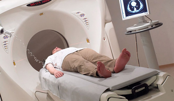
The examination procedure does not cause discomfort and pain. The patient puts on disposable clothing and is placed on the tomograph's couch. If it is necessary to check the liver and other internal organs (not hollow), a contrast agent (Trazograph or its analogs) is injected into a vein at the elbow bend. Do it as a bolus (using a special pump) or in the classic way(with an ordinary syringe). If it is planned to examine the intestines with the help of contrast, the patient is given a barium solution to drink.
Important! Immediately after the contrast enters the body, the patient may experience nausea, dizziness, or headache... About the appearance unpleasant symptoms you need to inform the radiologist.
Next, the scan begins, during which the doctor will be in the adjacent room. This procedure lasts about half an hour. At the end of it, the doctor proceeds to decipher the data, and the patient can change clothes and wait for the result at home.
Diagnostic results
The use of X-rays in diagnostic medicine has significantly increased the level of early diagnosis serious illnesses. Today, one of the most accurate methods based on the effects of ionizing radiation is MSCT - multislice computed tomography.
Multispiral computed tomography
How is MSCT different from conventional CT?
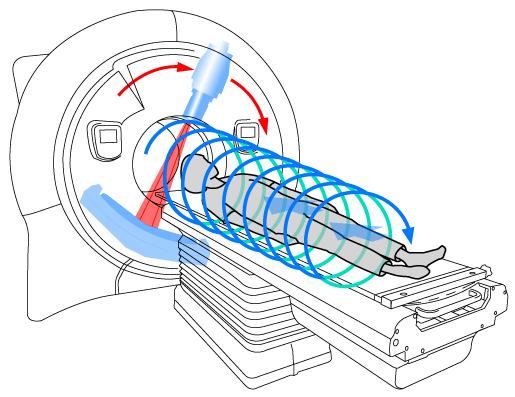
Spiral computed tomography physical fundamentals is no different from CT - the method is also based on X-rays. The main difference between MSCT and conventional computed tomography is that scanning is carried out along a spiral trajectory - the device rotates around the couch with the patient's body, while the couch itself moves into the depth of the tomograph. One turn of the spiral corresponds to a snapshot of one zone in different planes. With the help of MSCT, it became possible to obtain accurate three-dimensional models of the examined organs of the abdominal cavity, while the specialist has the ability to adjust the size of the cut - the most modern tomograph scans with slices less than 1 mm - this is much less than with simple computed tomography.
Advantages of MSCT in the examination of the abdominal organs:
- No interference caused by fluid movements inside the body - due to the fact that scanning occurs from different planes, the image is not blurred during natural physiological processes.
- Slices less than 1 mm thick improve the understanding of the structure of the examined organ.
- Due to the spiral trajectory of movement, the study area increases, while the radiation load on the body decreases.
- For multispiral computed tomography, less contrast agent is required, thereby reducing the burden on the patient's kidneys.
- With the help of MSCT, it is possible to make three-dimensional three-dimensional models of the organs under study.
- Simple preparation for the examination.

MSCT of the abdominal cavity
Indications for the purpose of the study
As a rule, multispiral computed tomography of the abdominal organs is prescribed after other examination methods - most often after ultrasound or radiography. In clinical practice, MSCT is prescribed to clarify the diagnosis or a more detailed study of the detected pathologies. Computed tomography is ideal for planning surgical treatment and observation of postoperative patients.
The organs of the abdominal cavity to be scanned on a tomograph include:
- Gastrointestinal tract - stomach, small intestine and large intestine.
- Pancreas, biliary tract, liver, spleen, gallbladder.
- Kidneys, ureters, adrenal glands.
- Lymph nodes and vessels of the abdominal cavity.
- The abdominal aorta and other large vessels that lie in this area.
- Fiber of the abdominal cavity.
The main indication for MSCT examination of the abdominal organs is abdominal pain of unknown origin, differential diagnosis of diseases of the abdominal organs, as well as examination for oncological diseases. Spiral computed tomography shows benign and malignant tumors, inflammatory diseases of the abdominal organs, ascites (accumulation of fluid in the abdomen), as well as purulent diseases - abscesses. In addition, MSCT helps to identify stones in the kidneys and ureters, obstructive jaundice, aortic aneurysm, and various traumatic lesions.
Contraindications to the examination
The radiation level during multispiral computed tomography is significantly lower than with CT, so there are fewer contraindications to this method. All contraindications to the examination are relative, which means the possibility of scanning in emergency situations... These include:
- The patient's age is up to 14 years.
Childhood is a relative contraindication to MSCT
- Pregnancy and breastfeeding - During pregnancy it is highly undesirable to allow exposure to X-rays.
- Excess permissible weight patient's body - patients weighing more than 120 kg are not allowed for examination.
- The presence of metallic elements in the body can distort the picture.
- In the case of MSCT with contrast, it is undesirable to conduct an examination in patients with kidney disease. Allergy to contrast is also a contraindication for examination.
What is involved in preparing for a survey?
To improve the image quality and increase the information content of the method, it is recommended to prepare gastrointestinal tract... To do this, you must follow a diet - remove gas-forming products from your diet. These include: carbonated drinks, bread, fruits and vegetables, dairy products. Scanning is best done on an empty stomach, so it is recommended that you refrain from eating eight hours before the scheduled examination. If the abdomen is bloated, it is recommended to take a carminative drug. Preparation for the study is quite simple and does not require significant efforts from the patient.
How is the research going?
The multislice computed tomography procedure does not cause any discomfort or discomfort in the patient. painful sensations... Before starting the scan, the patient should remove all jewelry and metal objects; in the presence of removable dentures, it is also recommended to remove them from the oral cavity. The patient is placed on a couch and fixed in the desired position, most often on the back. After that, the table drives in and scanning begins: the couch gradually moves in a horizontal plane, and the main part of the apparatus will move in a spiral around the couch with the patient. The study does not take much time - in most cases, 10-15 minutes is enough. Medical staff at the time of the examination, he is removed to the next room, if necessary, you can contact him using two-way communication built into the tomograph.
X-ray technicians prepare the patient for the study
MSCT with bolus contrast
The use of a radiopaque substance during the study increases the contrast of tissues, which allows differential diagnosis of the identified pathologies. A substance based on iodine is used as a contrast - it is he who is opaque to X-rays. The contrast agent is injected intravenously just before the start of the study using an automatic injector - the speed of administration is controlled by the program built into the tomograph. Contrast helps diagnose:
In the case when multispiral computed tomography is impossible due to different reasons, you can conduct research using other methods:
- Magnetic resonance imaging of the abdominal organs allows you to obtain a more accurate image of soft tissues, while there is no harmful effect of ionizing radiation. However, the cost of this procedure is much higher. The method is allowed during pregnancy.
Man undergoing magnetic resonance imaging
- Conventional computed tomography is a cheaper method, but the clarity of the images suffers. It is highly undesirable to have a CT scan during pregnancy.
- Ultrasonography - safe method express diagnostics, but the information content of this study is much lower.
Multispiral is a study in which X-rays pass through human tissue. This results in an image in different shades gray, depending on tissue density or existing pathology. Its huge advantage is the ability to view organs shown almost as accurately as in the anatomical atlas.
Such a study is called multispiral because the X-ray emitter moves along a circular line around the patient's body, at the same time moves in the direction of the longitudinal axis. X-ray tubes are located around the perimeter of the chamber, which resembles a tunnel, due to which the layers of the body are examined with different sides and at different angles.
If you draw the trajectory of the emitter for the entire duration of the procedure, then a spiral emerges. Each of the turns of the spiral corresponds to a snapshot of one zone of the body from different angles. Snapshots are called slice. One slice can be from 0.5 to 11 mm thick.
Computed tomography can distinguish between the components of the abdominal cavity, such as the stomach, small and large intestine, and parenchymal organs such as the liver, spleen, kidneys, adrenal glands and pancreas. The tomogram also shows the biliary tract, and in special cases you can do cholangiography that more accurately reflects them.
With the ability to view multiple areas of the body in as soon as possible, computed tomography of the digestive system has become a frequently performed research. It is especially useful for diagnosing emergencies in hospital wards. emergency care: trauma to the abdomen or acute pain in the abdomen, as it is well tolerated by patients, and at the same time very effective.
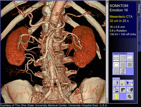
Studies can be carried out both with the introduction of a contrast agent (angiography), and without it. Angiography is helpful in identifying malformations vascular diseases(birth defects), aneurysm and angioma, renal artery stenosis. It happens that it is performed before the embolization procedure - the targeted closure of the vessel lumen for treatment.
2 Indications for
- Cancer of the intestine, stomach, pancreas, biliary tract, liver (for diagnosis and assessment of the degree of development).
- Inflammatory bowel disease (Crohn's disease, ulcerative colitis).
- Acute conditions such as obstruction, perforation, appendicitis, trauma.
- Spleen injury.
- Cholelithiasis.
- Acute and chronic pancreatitis (for differentiation and assessment of complications).
- Kidney inflammation.
- Calcification of the kidneys.
- Nephrolithiasis (sensitivity is almost 100%).
- Diseases of the renal vessels.
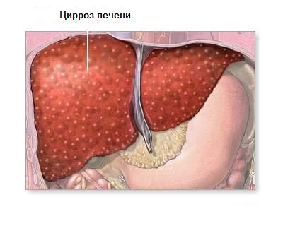
- Cirrhosis of the liver.
- Budd-Chiari syndrome (i.e., thrombosis of the veins of the liver or inferior vena cava).
3 Preparatory activities
The study is safe and painless for the patient, but some preparation is required before MSCT. Before the study, the doctor will take an anamnesis (sometimes you need to fill out a prepared questionnaire), you must report an allergy or an early reaction to the injection of a contrast medium. It is also necessary to report pregnancy, lactation, claustrophobia, and a tendency to bleeding. It is worth delivering the results of other imaging methods for research.
Before CT scan, the patient must remove all metal things. Remove the mobile phone.
The patient should come for an examination of the MSCT of the digestive system on an empty stomach and after consumption a large number liquid (2-3 l). It is important to have an up-to-date creatinine test result with you.

Before scanning the small intestine, the patient is given an intravenous contrast agent such as diluted barite. It is a liquid substance that absorbs X-radiation to a greater or lesser extent than the surrounding tissue.
When preparing for the procedure, the following rules must be observed:
- In order for the results of the diagnosis of the large intestine to be reliable, 2 days before the study, it is necessary to follow a diet (exclude milk, bread, cheese, fruits and vegetables) and use laxatives.
- The day before the CT scan, a liquid diet plus a laxative is used, and on the day of the study, the patient is given an enema. A relaxant (eg, hyoscine) can also be administered to the patient.
- In preparation for the examination of the stomach, the patient drinks 750 ml of water on an empty stomach immediately before the procedure in order to obtain the correct contrast between the stomach wall and the surrounding structures.
- Before examining the pancreas, drink 500-1500 ml of water about half an hour before performing a computed tomography to fill the intestines.
In some cases, people who are unable to lie still during the study time need to be given sedatives, or even used general anesthesia... In the case of examination of children, they are administered depressant, or applies general anesthesia... Before the study, you need to inform the child about the course of the MSCT procedure. He should know that computed tomography is painless method that does not last long.

All questions regarding preparation for a tomographic examination must be discussed with a doctor in advance!
4 How is the diagnosis carried out?
The study of multislice computed tomography is carried out in a special laboratory where the tomograph is located. The patient lies on his back on a table that automatically moves. The investigated area of the body is installed in the area of action of X-ray lamps located around the circumference.
X-rays passing through various fabrics the patient's body is weakened.
The amount of attenuation depends on the type of fabric. Images are obtained in the transverse plane, but appropriate reconstruction is also possible.
During computed tomography, a doctor or technician is in an adjacent room and instructs the patient through a microphone about when he should lie still or hold his breath to improve the quality of photographs, he monitors the progress of the study. In this case, the patient should report any alarming symptom, which occurs in him during the study: nausea, dizziness, shortness of breath.

5 Possible complications
Complications after MSCT can be associated, first of all, with the use of a contrast agent, but this is very rare. Contraindications are:
- pregnancy;
- renal failure;
- previously identified allergies to contrast agents.
Before performing MSCT, each time you should inform your doctor about chronic diseases, such as diabetes(especially which is treated with pills). Computed tomography is a test that can be repeated several times and can be performed on children as well.
Doing research in women in the second half should be avoided. menstrual cycle, since they have the possibility of fertilization.
Fortunately for us, the time when the majority of diseases were tried (and often unsuccessfully) to reveal only thanks to the patient's complaints and a short external examination are in the past.
Modern medicine has a huge variety of ultra-precise and ultra-modern technological devices and techniques that make it possible to significantly expand diagnostic and healing possibilities our time. One of them can be safely called Multispiral Computed Tomography.
Multispiral computed tomography or MSCT, one of the most accurate and highly informative examination methods internal organs human body... Scanning with ultra-thin slices allows you to identify pathological changes in size from a few millimeters. Such a study makes it possible to determine diseases at the most early stages, which means - to start prevention or treatment as early as possible.
MSCT is a layered X-ray examination. For simplicity, it can be compared to slicing bread into thin slices. The more “slices”, the higher the resolution of the apparatus, and hence the possibility of visualizing small foci of pathology.
After scanning, the result is processed by a powerful computer, which displays all the obtained sections on the screen, and also, connecting the sections, forms a three-dimensional image. In addition, MSCT with contrast allows you to "highlight" necessary organs or vessels by injecting a special contrast agent. This greatly simplifies the diagnosis of many diseases, especially tumors.
Multispiral Computed Tomography is universal method for the diagnosis of a huge number of diseases of various organs and systems.
Survey musculoskeletal system effectively visualizes the smallest changes and damage to the bone structure. The method is very effective for detecting destructive processes in the spine, bones and joints, displaying the smallest fractures invisible by other methods, including intra-articular ones. In addition, the tissues surrounding the bone are visualized - muscles, ligaments, articular capsule, which makes it possible to assess the extent of their damage during trauma or inflammation.
MSCT 3D reconstruction
A great advantage of the method is the ability to assess the state of bone tissue in people after osteosynthesis, which is a contraindication to MRI. MSCT allows you to assess the correctness of the performed surgical reduction and, if necessary, to correct the treatment.
In addition, due to its high resolution, the method is indispensable for detecting inflammatory and tumor diseases of the abdominal cavity, retroperitoneal space, brain, and thyroid gland.
The speed of the examination allows scanning for the presence of traumatic injuries of internal organs and the musculoskeletal system, the brain and peri-cerebral spaces.
Thanks to 3D modeling, MSCT is an indispensable method in maxillofacial surgery, especially when it comes to reconstructive operations.
MS computed tomography of the abdominal cavity: general characteristics
I would like to pay special attention to multispiral computed tomography of the abdominal organs. Thanks to the multispiral slice system, such an examination allows visualizing the desired organ in any projection at any level. This allows you to identify tumor processes and metastases in the abdominal organs in size from two millimeters.
Scheme of operation of a multispiral computed tomograph
MSCT is indicated in the following cases:
- If you suspect abscesses, cysts, cirrhotic lesions of the liver.
- If there are signs of obstructive jaundice.
- Hepato- and splenomegaly of unknown etiology.
- With symptoms of hepatocerebral dystrophy.
- Acute and chronic pancreatitis.
- Spleen hemorrhages and infarctions.
- Traumatic injuries of the abdominal organs.
MS computed tomography with contrast
In the case of serious fears or suspicions, MSCT using contrast is often prescribed. This method is more complicated and expensive, it requires additional preparation of the patient, but it can significantly enhance the visualization of even this exact method like Multispiral Computed Tomography.
When examining the organs of the abdominal cavity, the introduction of contrast allows you to "highlight" the areas of interest to the doctor, which is especially effective for diagnosing neoplasms several millimeters in size. In addition, contrasting allows you to determine the normal or pathological course of blood vessels (including tumor), to reveal an increase in vascularization in inflammatory diseases OBP or foci of ischemia, for example, with heart attacks of internal organs.
Preparation for MSCT of the abdominal cavity
For Multispiral Computed Tomography, as for most medical examinations, required special training... When carrying out a computed tomography, the examined person must remove all metal objects and jewelry - rings, hairpins, hairpins, dentures, which can interfere with scanning.
The examination is carried out on an empty stomach, the last meal should be six to eight hours before the scan. This point is especially important for patients who are planning to administer contrast, since dyspeptic reactions such as nausea and vomiting are possible on its introduction with a full stomach and intestines.
Bowel preparation is carried out in two days. Foods that can cause increased gas formation in the intestines, such as cabbage, legumes, milk and dairy products, black bread, carbonated drinks, are excluded from the diet.
Before the examination, it is necessary to inform the doctor about the presence allergic reactions and concomitant diseases to prevent the consequences of the examination. In the case of pregnancy, it is very important to indicate its timing, since MSCT is contraindicated in the third trimester due to the high radiation exposure for the unborn baby.
Detected pathologies
As already mentioned, MSCT is most often used to detect a wide variety of OBP diseases:
- Abscesses of the abdominal organs are visualized as thick-walled cavities with uneven walls, fluid contents and sometimes a small amount of gas.
- Simple cysts have thin walls, homogeneous contents without the presence of gas.
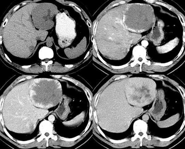
- Hemangioma on CT looks like a round formation with clear contours and low density. With the introduction of contrast, the hemangioma fills from the periphery to the center, which is its clear hallmark from other tumors.
- With cirrhosis of the liver, a decrease in its size, tissue compaction, and the appearance of regeneration nodes are noted. The venous outflow from the organ is significantly impaired.
- Focal nodular hyperplasia on CT has the same density as the liver, making diagnosis difficult. However, the use of MSCT with contrast makes it possible to reveal the central part of the tumor, which is filled with contrast much more slowly than normal liver tissue.
- The adenoma looks like a space-occupying mass surrounded by an area of fibrosis. When contrasting, it is stained in the arterial phase.
- Hepatocellular liver cancer has a heterogeneous structure. In addition, a huge number of various vessels and vascular shunts can be traced in its thickness, which is very well displayed through the introduction of contrast.
- Pancreatitis on CT is characterized by signs of organ dystrophy, fibrotic changes, the presence of calcifications and calculi.
- Pancreatic cancer is defined as an avascular mass with a well-perfused pancreas.
Method advantages
The advantages of MSCT include, first of all, high resolution, capable of detecting objects as small as a few millimeters. I am also pleased with the rather simple and often short preparation for the study.
The scanning speed allows examining severe patients and patients with severe pain syndrome... In addition, unlike MRI, it is possible to examine people with metal implants and pacemakers.
Research risks
Unfortunately, even the Multispiral Computed Tomography method is not ideal and has certain risks of complications. First of all, there is always a risk harmful effects X-ray radiation on human cells and tissues. However, it is often overlapped by the diagnostic capabilities of the method and, guided by the rule of "benefit - harm", the doctor still prescribes the procedure.
Pregnancy, especially in the third trimester: the tissues of the embryo are most sensitive to X-rays, therefore, the study is carried out only for health reasons. MSCT in children carries a high radiation load and therefore should not be repeated unless urgently needed.
Allergic reactions to the administration of a contrast agent can manifest themselves in an extremely diverse way, therefore, when conducting MSCT with contrast, the doctor should always be ready to relieve anaphylactic shock.
Further diagnostics
Usually, the use of Multispiral Computed Tomography is exhaustive in making a diagnosis, however, due to the high cost and high radiation exposure, other research methods, such as ultrasound, elastography or radiography, are used to further monitor the treatment process.
Conclusion
In conclusion, I would like to add that, despite the high cost, complexity and list of risks for MSCT of the abdominal organs, the method is getting more and more positive feedback from practicing doctors of various specialties due to the enormous information content and practically complete absence contraindications, which allows you to save thousands of human lives every day.
One of the most modern methods studies of human tissues and organs is a multislice computed tomography, or MSCT. What is it and what is the principle of research?
MSCT is considered one of the types The principle of examination is the same for them: with the help of X-ray radiation, using the difference in the absorption of rays by tissues different density, the tomograph examines the patient's body layer by layer. But MSCT uses a two-dimensional arrangement of detectors, while CT uses linear sensors.
A two-dimensional array of sensors of a multispiral tomograph, which spirally moves around the patient, makes it possible to obtain several fragments at once, which allows capturing images of large areas at high speed. The resulting fragment is processed and displayed in normal or three-dimensional form. The high speed of examination facilitates the diagnosis of severe patients and makes it possible to contrast the vessels.
MSCT is successfully used in the study of oncological, cardiovascular and infectious diseases, as well as in case of serious damage to the musculoskeletal system and bleeding into tissues and organs due to injuries.
What are the indications for the appointment of MSCT?
Modern diagnosis of many diseases is unthinkable without MSCT. What does this examination reveal and under what indications is multispiral computed tomography prescribed? 
If there are implants in the patient's body that contain metal, then only diagnostics on a multispiral tomograph will help, and MRI and CT are contraindicated. For diseases requiring urgent treatment or accompanied by severe pain syndrome, when a person is physically unable to lie still for a long period of time, MSCT will become the only the right method research. Multispiral computed tomography is also indispensable for such medical cases:
1. It allows not only to diagnose oncological formations of the liver, spleen, pancreas, Bladder, kidneys and extraorgan neoplasms of the retroperitoneal zone and abdominal cavity, but also determines the degree of damage and the type of tumor: benign or malignant.
2. Provides accurate diagnosis of fractures skeletal system, degenerative changes spine, bone lesions with metastases, reveals hernias in the lumbar spine.
3. In case of embolism pulmonary arteries determines circulatory disorders and the degree of damage to large arteries.
4. All serious injuries can only be correctly assessed using a multispiral tomograph.
5. It makes it possible to identify even insignificant and isolated foci of tuberculosis.
Why is contrast enhancement necessary?
The study on a multispiral tomograph makes it possible to perfectly see not only the bones and airways, but also soft tissue... This allows you to diagnose serious illness on the initial stages, for example, to identify a malignant tumor small size when there is still the possibility of surgical treatment. 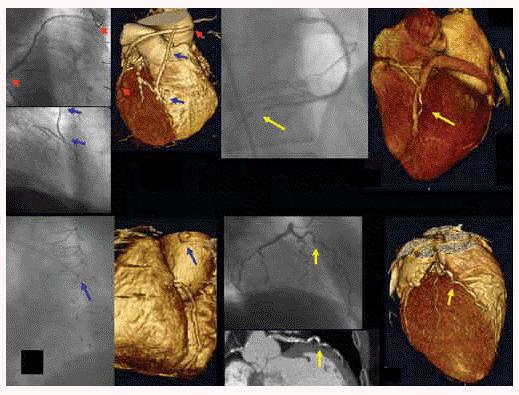 Contrast enhancement used for better differentiation of human organs from each other, normal structures from pathological neoplasms. There are two methods of contrast-enhanced MSCT: intravenous and bolus.
In the first method, a contrast agent is injected without adjusting the time and speed into the vein by an X-ray technician, then a study is carried out. This method is used on slower first-generation tomographs.
With bolus contrast, a special substance is injected with a syringe-injector at a set time and speed. The advantage of this method is that it delimits the contrasting phases, which makes the study more effective and the results more reliable.
When is multispiral computed tomography of the brain performed?
V modern medicine for diagnostics, the leading position is taken by the study of MSCT. What does this study diagnose, for what symptoms is it performed? 
MSCT is used to diagnose the following pathologies:
- oncological formations of the brain, as well as abnormalities in its development;
- stroke;
- high intracranial pressure and hydrocephalus;
- chronic form of vascular insufficiency;
- trauma or inflammation of the brain;
- chronic and acute stages of diseases inner ear or sinuses.
With frequent and severe headaches, memory impairment, dizziness, it is necessary to consult a neurologist to resolve the issue of the need for MSCT of the brain to exclude life-threatening pathological changes in this organ. This is especially important for patients who have suffered in the past brain trauma, stroke, transient, or have all the signs at the time of contacting the doctor.
Indications for multispiral computed tomography of the abdominal cavity
When conducting MSCT, the doctor evaluates tissues, organs and systems in this area: liver, biliary tract, gallbladder, spleen, kidneys, urinary tract, pancreas and other organs. A specialist radiologist analyzes the structure, size and position of organs; the existence of pathological neoplasms; the presence of calculi in the organs of this zone; functionality of the bile ducts; condition of the lymph nodes. 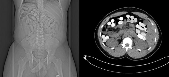
Indications for MSCT of the abdominal and retroperitoneal organs:
- oncological formations and tumor lesions (metastases);
- cysts, adenomas and abscesses;
- serious injuries and suspected damage to organs and blood vessels;
- urolithiasis disease;
- cirrhosis of the liver;
- diseases of any organs of the abdominal cavity;
- inflammatory processes;
- pathology of the abdominal aorta and its branches;
- organ abnormalities.
When is MSCT of the chest organs prescribed?
To assess the condition of organs and tissues in the area chest use the highest informational research method - MSCT. What does this examination evaluate and for what diseases is it prescribed? 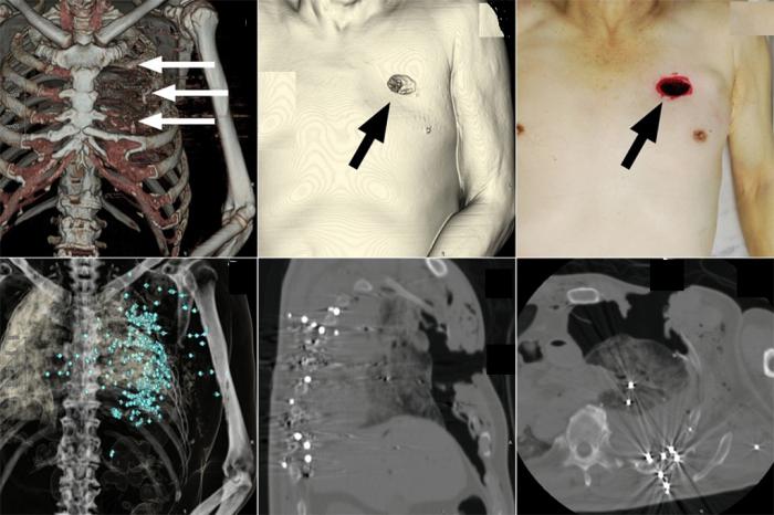
This technique makes it possible to analyze and assess the state of organs and soft tissues of the chest (lungs, heart, blood vessels, esophagus, trachea and others), lymph nodes, bone structures.
Indications for MSCT of the chest:
- tumor formations and their metastases;
- anomalies and malformations of the heart and bronchopulmonary system;
- diffuse lung disease;
- inflammatory processes that have caused damage to the chest organs;
- serious injuries.
To conduct a MSCT study, you need to dress in loose clothing. Everything foreign objects and jewelry must be removed during the procedure, including hearing or dentures. It is necessary to refuse food a few hours before the examination, especially when using the contrasting method. 
The study is absolutely painless, and the dose of radiation received is minimal. The procedure lasts (depending on the complexity) from 5 to 30 minutes and requires immobility of the patient.
The use of the contrasting method in the study, the type of contrast medium and its amount are factors that affect the cost of MSCT. The price also depends on the location and volume of the examination area, diagnostic tasks and additional services. You can clarify the cost of any MSCT by going to the website of the selected clinic or by calling. On average, prices for such a procedure range from 1.5 to 11.5 thousand rubles.
Contraindications and risks of MSCT
- lactating women are prohibited from feeding during the day after the introduction of contrast;
- the study of pregnant patients is carried out for health reasons;
- examination of children is carried out only in case of emergency and a repeated procedure is prohibited;
- very rarely there is an allergy to contrast agents that contain iodine.
Conclusion
MSCT is a painless and informative diagnostic method with a number of advantages:
- perfectly visualizes both bones and soft tissues, blood vessels;
- high research speed is especially important for serious emergency conditions;
- more high quality result, less sensitivity to patient movement and lower cost than MRI;
- conducting minimally invasive procedures makes it possible to do without surgical intervention for diagnostic purposes;
- minimal exposure and no residual radiation after examination.