Skin disease ichthyosis. Ichthyosis of the skin: what is this disease and can it be cured
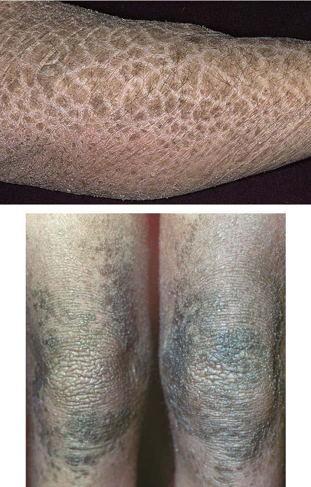
Ichthyosis - genetic disease, in which the keratinization of the skin is disturbed. Rigid scales resembling fish scales are formed, and keratin with a modified structure accumulates in the epidermis.
Ichthyosis is most commonly seen in early childhood or even immediately after birth, less often the disease becomes acquired. The cause of ichthyosis is a gene mutation, the hereditary biochemistry of which has not yet been deciphered.
For more serious cases local treatment indicated in addition to emollient creams with high hydration, relipidant and kerato-recovery: antibiotics, antiseptics. Common retinoids are best method treatment of severe ichthyosis. Cyclosporine has been used, but the results are inconsistent, negative for some and more satisfactory for others. These treatments are only a suspension and, unfortunately, the disease quickly recurs at rest.
It starts with a few everyday things. First of all, improper cleaning of the skin must be avoided. Prefer to shower with warm water in hot baths. Avoid alkaline soaps rich in soda and potash. Regardless of the cleaning agent used, it must be rinsed well, avoiding too much calcareous water. Drying should be carried out quickly after cleaning the skin, lightly tapping and not rubbing.
Protein metabolism disorders, when amino acids accumulate in the blood and fat metabolism disorders that manifest themselves high content cholesterol, become manifestations gene mutation which leads to ichthyosis.
The prevalence of the disease is 1:3000 - 1:4500. The etiology is unknown.
A photo
Symptoms of ichthyosis
The patient's body is covered with scales, and amino acid complexes that are not absorbed by the body accumulate between them. The stagnation of substances gives a cementing effect on the skin, as a result of which the keratinized cells adhere tightly to each other with healthy cells. The separation of the scales causes the patient severe pain.
In addition, it is recommended to drink at least a liter and half of water a day to avoid overheating your home and humidify the surrounding air. Avoid direct skin contact with textiles, which may cause irritation or excessive perspiration.
Grouping of patients who support them psychologicallyAfter they are referred to specialized groups care. If you are concerned about ichthyosis, we strongly encourage you to contact this Association is very active and recognized by the world of science and medicine.
Unfortunately, there is currently no treatment that can cure ichthyosis in any form. Available treatments allow you to reduce weights and improve skin comfort and appearance skin. These treatments can be divided into local treatment and systemic treatment.
Other symptoms:
- the appearance of sharp dryness and roughness of the skin;
- palms are covered with mucoid peeling;
- the skin pattern is clearly visible;
- decrease in the intensity of metabolism and the functioning of hormonal glands;
- dystrophic changes in hair, nail plates and teeth;
- violation of thermoregulation;
- the appearance of hyperkeratosis;
- malfunctions sweat glands;
- chronic conjunctivitis or retinitis, hereditary myopia;
- decrease in immunity, due to which allergic reactions and purulent infections become chronic;
- in advanced cases - the development of chronic heart failure and impaired liver function.
Treatment of ichthyosis
Treatment of ichthyosis is carried out by a dermatologist on an outpatient basis or in a hospital.
They include simple moisturizing creams or to which agents are added that better separate dandruff and are called keratolytics. These topical therapies have the disadvantage that they are of limited efficacy and are restrictive. Indeed, they need to be applied 1-2 times a day and left on the skin with a greasy film.
This medication belongs to the retinoid family. This effective drug, but has side effects. In fact, this drug may lead to liver or blood lipid abnormalities that justify biological monitoring. In children, if used at a very high dose, it may have undesirable effects on the muscles or joints.
Vitamins of groups A, E, B, vitamin C and nicotinic acid are prescribed for long and multiple courses. Preparations with lipotropic action soften the scales. These are preparations containing lipamide and vitamin U. To stimulate the immune system, blood plasma transfusions, gamma globulin, preparations containing iron and calcium, and aloe extract are indicated. If damage occurs thyroid gland with the development of hypothyroidism, then thyroidin is indicated, with pancreatic hypofunction - insulin.
In the case of long-term treatment, there may also be a risk of osteoporosis in adulthood. This medicine is not suitable for the treatment of young women because pregnancy is prohibited during treatment and for the next 2 years due to the risk of serious malformations in the child. Therefore, contraception should be associated with women childbearing age. A cream containing retinoids is being studied and may soon be available in ichthyosis.
Spa treatments can improve skin conditions, perhaps an effect that goes beyond treatment. The future lies in finding a treatment to correct the anomaly that causes ichthyosis or to correct the consequences of this anomaly, or even genetic therapy. Gene therapy is a therapeutic strategy that involves introducing genes into human cells or tissues to treat a disease.
In severe cases and with congenital ichthyosis, hormone therapy is immediately prescribed; oil solution retinol acetate. After normalization of the state, the dose hormonal drugs slowly reduce to complete abolition.
During the remission period, blood tests are periodically done to monitor the patient's condition and prevent the development of complications. Nursing mothers are shown taking vitamins, as are children with ichthyosis.
It probably also plays a role in protecting against certain diseases, cancer or cardiovascular disease. This screening will allow people to prescribe treatment and set up follow-up. Summer enjoys the senses and the sunshine, but swimming tests the skin. Focus on some small worries related to water activities. Dry skin after a day at the sea? It's possible. “Salt causes the peels to become sticky; it also dissolves certain proteins such as keratin, which is, among other things, the horn of the skin,” explains Prof. Daniel Hohl, head of the Department of Dermatology at the University of Vaud Grand Center.
Local therapy consists in taking general baths with a solution of potassium permanganate and lubricating the skin with a baby cream with the addition of vitamin A to it. Adult patients with ichthyosis are shown saline and starch, general or local baths, depending on the localization of the process. Vitamin A, sodium chloride and urea are added to the water.
Dermatologists also use this property to treat conditions such as psoriasis or hereditary conditions that affect the skin. For Mr. and Mrs. We can all counter this drying effect by using the day after tomorrow or another moisturizer after bathing, says the expert.
Swimming in the lakes prized by the Swiss sometimes brings its share of stings. The culprit is a cercaria, a worm that parasitizes birds. To reproduce, the cercaria lays eggs, which hatch into larvae that grow on water snails. Once these larvae become cercariae, they leave these hosts to find a duck to settle on. It is here that man comes to bathe in the shallow warm waters, an environment in which cercariae are looking for new hosts. But the little worms don't distinguish between birds and humans, and they get under our skin where they die quickly.
UV irradiation in suberythemal doses, thallas therapy and heliotherapy, resorts with sulfide and carbonic acid baths stimulate metabolic processes in the dermis. Silt and peat muds are recommended already at the stage of resolution of the recurrence of ichthyosis and for prevention.
Aromatic retinoids, which restore the functioning of dermal cells and normalize metabolic processes, have become widespread in the treatment of ichthyosis.
Our body reacts to their presence. Few during the first bites that a person experiences in his life. But we often develop allergies that make the next bites very bad. “If you suffer from this itch, consult a dermatologist or pharmacist who can prescribe a cortisone cream,” recommends Professor Daniel Hohl.
These bites can also be prevented: if you have the opportunity, swim a few dozen meters from the edge, avoiding bites. Where the lake is deeper and colder, there really is no shelter for snails that grow cercaria. On the other hand, if you know what you're doing important reactions on "duck fleas" it might be better to swim in a pool or in the water when the mercury makes the lakes go crazy.
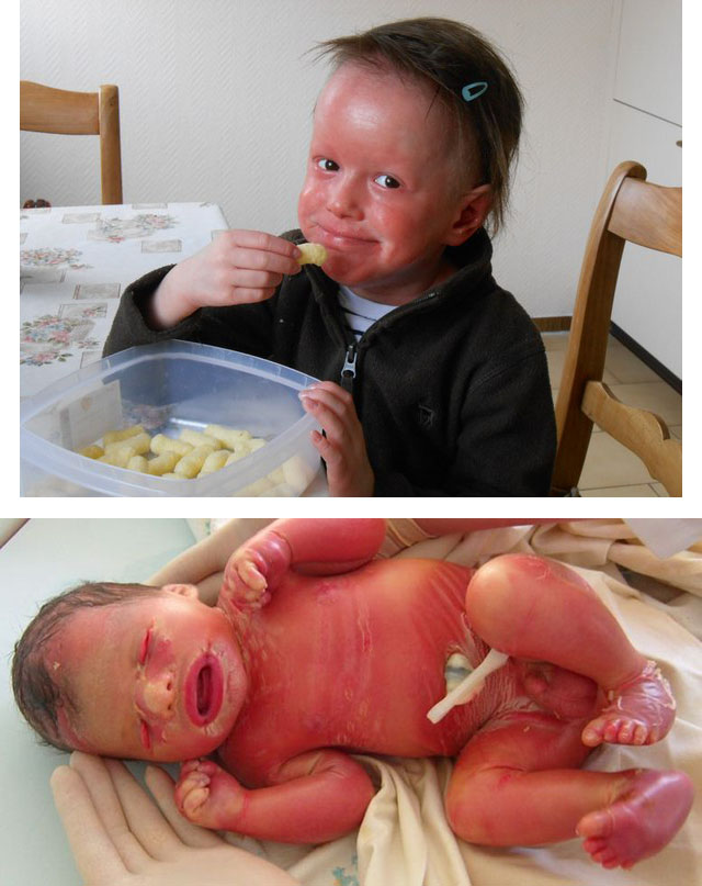
Congenital or Harlequin ichthyosis
It develops during pregnancy, more often in the first or second trimester. Harlequin ichthyosis is dangerous for the life of the fetus or the newborn.
Immediately after birth, large keratinized layers of thick gray skin are present on the child's body. Brown color. There are cracks between the scales, the child's face is usually deformed:
Slight redness that develops slowly and sometimes falls off a little skin or creates small cracks? It could be a fungus, a microscopic fungus. They love hot and humid environments and are therefore frequent in summer. "You can catch them anywhere you are barefoot," the specialist says. Wash your feet before entering the pool, dry your feet well when you get out of the water, and put down your shoes or faucets quickly to minimize the risk of skin contact with other people. fungal disease disappears without treatment, but if not, see a doctor.
- the mouth is stretched or so narrowed that a feeding tube can hardly pass into it;
- the baby's eyelids are turned out;
- the ears are filled with scales.
The skeleton of such children is also with anomalies:
- no nail plates;
- clubfoot is noted;
- there are no bridges between the phalanges of the fingers.
If the fetus has Harlequin ichthyosis, there is a high chance of miscarriage or premature birth. In cases where a child is born with this disease, there is no chance of survival. The cause of death is:
Undoubtedly, skin cancer is linked to sun exposure and you should protect yourself from it. To live well with the sun, it is necessary to have a behavior and adapted equipment. First, avoiding the sun when it is at its zenith, between 11 and 15 o'clock, is a mandatory measure for children. Enjoy it for lunch or take a nap, but don't expose yourself to those hours. Then look for shade or create it with umbrellas and beach shelters. “Also, don't forget that the sun also reaches you in the shade!” recalls Prof. Wolf-Henning Boinz, Head of the Department of Dermatology at the Women's University Hospitals.
- pathological process spreading throughout the body;
- the inability of the body to regulate water balance;
- lack of thermoregulation;
- weakness and defenselessness of the newborn against pathogenic infections.
If a child diagnosed with harlequin ichthyosis does not die immediately after birth, then up to 12 years the survival rate becomes 3%. Only 1% of patients survive to 18–20 years of age. It is believed that Harlequin ichthyosis is not compatible with life. During the period prenatal development it is difficult to diagnose this anomaly.
If you really want a tan, you don't have to expose yourself directly. Clothing is also a great ally against the sun, whether wide-brimmed hats or surf t-shirts that will protect your back and shoulders, as well as your children's children. Finally, apply generously on all parts of the body that exceed before going into the sun and after getting out of the water, sunscreen with a high index, especially if you have bright skin. However, if you have black or very black skin, the sun is much safer for you.
"Waterproof," proudly claims the package of sunscreen you just bought. So we set in the morning and are we good for the day? Not so fast, explain experts. Pragmatically, we recommend dividing the protection index by two to achieve real conditions where we tend not to add enough creams and where bathing and sweating make it leave the skin, recommends Professor Wolf-Henning Boinke, head of the department of dermatology, Geneva University Hospital. “Our recommendation is to enjoy at least twice a day before going out, to allow the cream for half an hour to get into the skin before bathing, and to apply a second time later in the day,” the expert recommends.
Causes of ichthyosis
The cause of the disease is a gene mutation that can be transmitted through the generation. Scientists have not yet figured out the biochemical process that underlies the disease.
Ichthyosis classification
The name "ichthyosis" combines diseases that occur in early childhood, continue until the end of life and are characterized by widespread keratinization of the skin.
He adds that it is essential to use sunscreen generously: if a 200 ml bottle lasts a week of the beach, then without a doubt you are not betting enough! Juni Witto is Chair of the Department of Dermatology and Skin Biology and Director of the Institute for Molecular Medicine at Jefferson College of Medicine at Thomas Jefferson University in Philadelphia.
The babies are lined with thick skin that looks like armor and seriously prevents movement. When after birth this skin dries out to form hard rhomboid plaques that affect the facial features with deformity of the lips, eyelids and ears. Usually newborn babies with arctic cannula do not survive after a few days for breathing difficulties, bacterial infections and nutritional difficulties. After restoring the corresponding intensive care newborns, some patients who experienced dermatological symptoms similar to those of people with severe forms of unrelated erythroderma.
According to the degree of skin damage, ichthyosis is classified as follows.
Xeroderma
Ichthyosis in a mild form is characterized by dry skin and the appearance of small pityriasis peeling on the skin. Scales appear in the form of a powdery-white strip, if you run a fingernail over the skin. There are small, pinhead-sized, grayish, pale pink or bluish nodules located on the extensor surfaces of the limbs and buttocks.
The molecular basis of the Arlechino hyena was unknown until recently. These family groups made it possible to place the probable site of the responsible gene on long shoulder chromosomes. Microsatellite markers have limited attention to the location where 6 identifiable genes are found. First, a feature of the epidermal cells in arthritic arthritis is an abnormal way of transporting and depositing lipids in the superficial layers of the skin along with abnormal lamellar granules.
What are the consequences of identifying gene mutations in Arthurian histosis? In other words, is there anything that can benefit patients and their families? Given the severity of arterial histosis, which is often fatal in the first days of life, there is a need for prenatal tests in families at risk reappearance diseases. Until now, prenatal diagnosis has been based on fetal biopsy performed on late stage pregnancy. Finally, knowledge of exact mutations in such serious pathologies is the basis for the subsequent implementation of pre-implantation genetic diagnosis.
Follicular ichthyosis
It is manifested by roughness of the skin, the development of horny plugs in the mouths of hair follicles, penetrated in the center by a hair; localized on symmetrical areas of the posterior surfaces of the upper and anterior surfaces lower extremities.
Brilliant ichthyosis
On the skin, dense horny scales of an off-white, “marble” color are formed, located more often on the limbs, at the mouths of the hair follicles. The scales shine with a characteristic luster, reminiscent of mother-of-pearl. The scales on the periphery exfoliate, the edges are raised.
Black ichthyosis
It is localized in the abdomen, lower back, on the posterior surfaces of the upper and anterior surfaces of the lower extremities, on which horn plates black and kept normal skin on the face, in natural folds, on the lateral surfaces of the trunk and the medial surface of the extremities.
Lamellar ichthyosis
It is characterized by thick and horny scales. The skin is dry, brownish-gray scales form dense, rectangular or oval plates, up to 3 cm in size. The plates are separated by furrows, crevices, and cracks. In addition to the limbs, ichthyosis serpentina affects the front of the body, back, and face. Silvery scales are often found on the head, as in dry seborrhea. Often with this form of ichthyosis auricles deformed, adhering to the scalp, eyelids are shortened, everted; gothic hard palate.
Hystrixoid ichthyosis
A severe form of ichthyosis proceeds as follows: dense and thick accumulations of dirty-gray horny plates appear on the surface of the skin in the form of conical protrusions, spikes or needles, sharply (by 5-10 mm) protruding above the level of the skin. The plates are separated by grooves. This type of ichthyosis is rare.
Diagnosis of ichthyosis
Diagnosis is based primarily on clinical findings.
- with ichthyosis, histological examination is important (absence or thinning of the granular layer of the epidermis);
- with X-linked ichthyosis: reduced functioning of steroid sulfatase in amniotic cells or chorion tissue using DNA probes;
- in congenital bullous ichthyosiform erythroderma: histological examination of the pattern of epidermolytic hyperkeratosis.
Prenatal diagnosis of congenital ichthyosis is carried out with an appropriate family history. A fetal skin biopsy taken between 19 and 21 weeks reveals thickening of the stratum corneum, which is not normal until 24 weeks. This corresponds to lamellar ichthyosis, epidermolytic hyperkeratosis, "Harlequin fetus". When cultivating amniocytes and chorion cells, it is possible to detect mutations in keratins 1 and 10. This enzyme deficiency occurs in X-linked ichthyosis.
Differential Diagnosis
- psoriasis;
- seborrheic dermatitis;
- xeroderma.
Forecast and prevention of ichthyosis
The prognosis of ichthyosis is unfavorable, since even with mild forms of the disease, the addition of pathologies and the development of metabolic diseases lead to complications.
Prevention of ichthyosis - counseling before pregnancy to determine the degree of genetic risk. If fetal ichthyosis is detected during the analysis of amniotic fluid, then termination of pregnancy is recommended. Couples who are at high risk of having a child with ichthyosis are better off refraining from pregnancy in favor of adopting orphans.
Ichthyosis is a disease in which remission cannot be achieved, so prevention of the disease is similar to treatment. In addition to keratolytic and moisturizing therapy, patients with ichthyosis are advised to use indoor humidifiers or, if possible, move to a warm city with high humidity. In addition, they use cool water for washing and visit specialized spas.
How is ichthyosis inherited?
The mechanisms of inheritance of ichthyosis depend on the type of disease.
Ichthyosiform erythroderma non-bullous and lamellar (lamellar) ichthyosis are inherited in an autosomal recessive manner.
- If both parents are carriers of the disease (heterozygous), but are not sick themselves, then the probability that the children will be sick is 25%, carriers 50% and healthy (do not inherit the disease) 25%.
- If one parent is sick and the other is a carrier of the mutation, then the probability of the disease for each of the children is 50%, as in dominant inheritance. If both parents are sick, then all children will be sick.
- If one parent with an unknown genotype is healthy, and the second is a carrier, the risk of having a sick child is low.
Consanguinity of parents increases the risk of disease in children. Manifestation of an autosomal recessive disease in a child healthy parents becomes the result of a newly appeared spontaneous mutation of the recessive allele.
Ichthyosis vulgaris, Darier's disease, and ichthyosiform erythroderma bullosa are inherited in an autosomal dominant manner.
- Each patient has one parent sick (exceptions: the disease is caused by a new mutation; the parent has a mutant allele that does not manifest itself as a disease).
- The patient transmits the disease to children with a probability of 50%.
- In healthy children of the patient, only healthy offspring are born.
- Men and women are equally susceptible to the disease.
- Both parents are equally likely to transmit the disease to sons and daughters, including transmission from father to son.
Questions and answers on the topic "Ichthyosis"
Question:Tell me, if my husband has congenital ichthyosis (he has slight peeling of the skin), then future child can inherit the same form of ichthyosis or can be born with some other, more severe?
Answer: Hello. The mechanism of inheritance depends on the type of ichthyosis. You need a face-to-face consultation with a geneticist for research.
Question:Hello. My daughter has a skin disease: ichthyosis vulgaris. Tell me how to sunbathe her? Do I need to use sunscreen? How long can you stay in the sun? Indeed, with this disease sunbathing necessary. How to take care of the skin after sunburn so that it does not look very flaky?
Answer: Hello. It all depends on how old your child is and what phototype the baby's skin belongs to. If the child is under 3 years old, then stay under direct sunbeams contraindicated. If the child's skin is very light, he has blonde hair and eyes, it's another one additional factor avoid the sun. protective agent should have good moisturizing properties, such as milk for children. Another milestone skin care in summer time with your illness is deep hydration skin after sunbathing.
Question:Hello. My son is 12 years old. Skin problems. Diagnosed with ichthyosis. Tell me who and how can help him?
Answer: Hello. You need to consult a dermatologist who will prescribe complex treatment according to the type of this disease.
Ichthyosis is an independent and various in origin dermatological diseases, characterized by a violation of the processes of keratinization (keratinization) and accompanied by regional, that is, limited (in certain parts of the body), or diffuse (universal, common) thickening of the stratum corneum of the epidermis.
Causes of ichthyosis and its classification
Physiological keratinization of epithelial cells and the formation of the stratum corneum proceed "softly" and consist in the division of germ cells and their movement into the upper layers of the skin. The lower layer of fresh epithelial cells in normal conditions gradually replaces the old one, which is usually accompanied by imperceptible peeling.
Thus this life cycle, which lasts for two days, forces the newly formed cells of the epidermis to move to skin surface and promotes the transfer of all substances that they contain. The consequence of a violation of keratinization processes, leading to various skin diseases, called keratosis, or hyperkeratosis, is a delayed rejection of keratinized epithelial cells or / and a pronounced increase in the thickness of the stratum corneum.
Huge variety of features clinical manifestations, a diverse morphological picture, the presence of rare forms, the lack of an unambiguous view of researchers on the causes and mechanisms of development of hyperkeratosis, which include large group diseases, united by the term "ichthyosis", have not yet allowed clinicians to develop a certain unified classification.
Depending on the presence or absence genetic reasons, this disease is very conditionally subdivided (I. I. Pototsky and V. T. Kuklin) into pathological conditions, resulting from gene mutations and classified as genodermatosis (congenital ichthyosis), and acquired forms, most of which are called ichthyosiform, that is, ichthyosis-like conditions.
It should be noted right away that the notorious "Schleiman's ichthyosis" and "Schleiman's systemic vascular-like ichthyosis" are mentioned on Internet forums and in single Internet articles. Such a nosological unit, syndrome or symptom
om do not exist in any classification of dermatoses, in any textbook on dermatovenereology or in scientific papers. There is a symptom of Shteiman, which is one of the signs of a torn meniscus knee joint, but having nothing to do with dermatoses.
Among all hereditary skin pathologies, congenital ichthyosis is the most common form and accounts for about 90%. Its overall frequency averages 1 in 12.5 thousand children.
Classification of acquired ichthyosis
- symptomatic, provoked by any pathology or being one of the signs of the underlying disease; they may be dysfunction of the thyroid gland or adrenal glands, dysfunction of the neurovegetative system, hypo- or avitaminosis “A”, blood diseases and malignant neoplasms, autoimmune, inflammatory and infectious diseases, metabolic disorders, intolerance to certain medicines or food products etc.;
- discoid;
- senile, or senile.
In accordance with the classification proposed in 1990 (K. N. Suvorova), which is based on clinical symptoms, distinguish between such forms of the disease as:
- simple, or ordinary ichthyosis, characterized by lesions of all skin integuments and small scales;
- shiny - also affects almost all skin, excluding large folds, but transparent scales with a grayish tint are arranged in a mosaic pattern;
- serpentine, in which the skin of the extensor surfaces of the limbs and lateral surfaces of the body is affected, large scales are grayish-brown in color.
Another classification is based on the severity of the disease, which determines the form of the latter:
- severe, the outcome of which is the death of a newborn child;
- moderate, compatible with life;
- late, when the first symptoms of pathology appear only in the second month after birth.
These and other classifications in certain cases can be convenient in practical work, but in the International Classification of Diseases, ichthyoses are placed under the heading "Other congenital anomalies" and are assigned to the class " Congenital anomalies, deformations and chromosomal disorders". In the same classification, they are divided into ichthyosis:
- Simple (vulgar) - an autosomal dominant type of inheritance.
- Linked to the X chromosome (female) - recessive.
- Lamellar, or lamellar - autosomal recessive.
- Congenital bullous ichthyosiform erythroderma - autosomal dominant.
- Fetal ichthyosis is autosomal recessive.
- Another congenital ichthyosis.
- Congenital ichthyosis, unspecified.
Mutated genes that are inherited control enzyme systems and biochemical processes of keratinization, which have not yet been fully deciphered. They are based on the pathological development of profilargin, a cellular protein contained in keratinocytes. Normally, as a result of biochemical processes, it breaks down into filargin and a complex of molecules called moisturizing factor (NMF).
As a result of a gene mutation, there is a violation of skin hydration, excessive synthesis of defective keratin, which is a protein of hair, skin and nails, and excessively rapid keratinization of the epithelium. Violation of metabolic processes, especially protein and fat, leads to a violation barrier function skin and the accumulation of cholesterol in the blood, and between the cells of the stratum corneum and other layers - various complexes of metabolic products, in particular, amino acids, which have a cementing effect. The rejection of the stratum corneum also slows down, since it is tightly fastened to the underlying layers, the processes of sweating and breathing of the skin are disrupted, and local immunity is reduced. Given the large area of lesions, all this cannot but affect the function of the neuro-endocrine and other body systems.
In this way, main reason most forms of ichthyosis is a mutation of genes or a violation of their development. The possibility of mutation at several points of one gene or the participation of several different genes in this process is assumed, which determines the presence of a large number clinical forms diseases.

Pathological cornification develops according to the following mechanism:
- Excess production of keratin, as a rule, with an altered structure.
- Accelerated movement of keratinocytes to the stratum corneum from the basal.
- Strengthening the bonds between the cells of the stratum corneum and slowing down their rejection.
- Dystrophic changes in the epithelium, the formation of vacuoles (vesicles) in upper layers epidermis and an increase in the thickness of the stratum corneum.
- Violation of the formation of filargin and NMF, leading to excessive loss of water through the skin. This causes their dryness and characteristic peeling.
Clinical picture of individual forms of ichthyosis
Simple, common, or ichthyosis vulgaris
It is the most common form of the disease. It does not appear immediately at birth, but in the third month of life and later - up to 1 year. gender difference absent in the incidence rate.
Clinical symptoms of this form of the disease:
- floury, pityriasis and small-lamellar peeling;
- follicular hyperkeratosis;
- hair loss;
- the severity of the palmar and plantar patterns of the skin; sometimes, especially with concomitant pathology endocrine system, there is peeling on the soles and palms;
- dryness, roughness, deformation, thinning and brittleness of the nail plates.
The defeat of the skin in the form of severe dryness and roughness is generalized, with the exception of the lateral surfaces of the face, cervical, axillary and gluteal regions, inner surface hips, flexion surfaces in the area of the knee and elbow joints.
The color of the scales is different - from whitish to grayish-black. The most pronounced changes are noted in the zone of the knees and elbows, and on the anterior surface of the shins, the scales are similar to the scales of fish.
The defeat of the mouths of the sebaceous hair follicles in the form of follicular keratosis is the most characteristic symptom common ichthyosis. It consists in blocking the mouths with small plugs, consisting of masses of horny epithelium, resulting in the formation of nodules, tubercles ranging in size from 1 to 3 mm with twisted vellus hair in the central part. The tubercles are flesh or reddish-gray in color, covered with scales and sometimes surrounded by a reddish corolla.

Follicular keratosis gives the skin a rough appearance (“grater” symptom) and can be localized over the entire surface of the skin, except for the palms and feet, but most often in the shoulder girdle, buttocks and thighs. It is especially pronounced in youth and by middle age leaves behind punctate atrophic scars, which are the only evidence of ichthyosis in family members.
Relapses of ichthyosis occur in dry air and in the cold season. With age, the condition improves, especially in the summer and / and in a humid warm climate. Often in adolescence there is a period of short-term remission. In the future (by the age of 25), peeling becomes much less pronounced or stops completely, but characteristic changes in the skin of the palms and feet remain.

Follicular ichthyosis
Follicular ichthyosis, or Darier's disease (follicular vegetative keratosis), differs from follicular hyperkeratosis as a symptom in ichthyosis vulgaris, in which 4 forms are distinguished. Unlike ichthyosis vulgaris, Darier's disease develops in childhood or adolescence, has a progressive character, aggravates under the influence of ultraviolet radiation. The nodules have the character of dense warty growing papules, at the confluence of which weeping occurs in the folds of the skin. Elements are usually arranged symmetrically. Their main localization is hairy part head, behind the ear zone, face, interscapular zone, sternum and large folds.
X-linked ichthyosis
They mostly affect boys. The carrier of the pathological gene is the mother. Girls can get sick if the father was sick, and the mother is a carrier of the gene. The disease can be diagnosed already at birth (rarely) or after 2-6 weeks. Less severe forms manifest later, but before 1 year after birth.
Clinical symptoms are inherent only for this form of the disease. Large scales adhere tightly to the surface of the skin and have a dark brown (dirty) color. The characteristic localization of lesions is the back of the neck, lateral surfaces chest, skin of the extensor surface of the limbs.
Hyperkeratosis has a much less specific character in the area natural folds skin. The face, palms and soles are not affected, but phenomena are noted in these departments. Peeling of the floury type and follicular keratosis are not characteristic of this form.
In 50% of patients, corneal opacity is detected, which is not accompanied by a decrease in visual acuity. In addition, sometimes skin pathology is combined with other disorders - with cryptorchidism, with hypogonadotropic hypogonadism, with backwardness. mental development. Women who carry the mutated gene may develop primary birth weakness.
The course of the disease worsens winter season. Clinical picture does not improve with age.
It occurs with equal frequency among females and males. It is more common in children born to parents who are close relatives. Often, these children die as early as a newborn.
The disease manifests itself immediately at birth as a shiny thin dense film of the stratum corneum of the epidermis, which has a yellowish-brown color. The film, due to which such newborns are called "colloidal fetus", covers the entire body. During the movements of the child, it cracks and for several weeks separates in the form of thin plates of large sizes. At the same time, edematous red areas of the skin are exposed.
With age, the intensity of the red color decreases, but the degree of large-lamellar peeling increases. The plates themselves are polygonal in shape and grayish-brown in color. They are tightly fixed in the central part, their edges peel off and rise, like tiles.
The lesion is diffuse. In the elbow and knee areas, the skin thickens, sometimes there are warty growths of the epidermis, and on the back, due to the folds formed, the skin acquires transverse waviness. The palmar and plantar surfaces are also prone to excessive keratinization.
In addition, there is thinning and hair loss of the marginal type, nail plates, on the contrary, thicken and grow rapidly in length. Due to the tightening of the skin, "Mongoloid" eyes are formed, the auricles are deformed.
Mortality in this form reaches 20%, mainly due to blockage of the sweat glands, excessive loss of moisture in the area of multiple cracks, an increase in body temperature to high performance even with minimal physical activity or an increase in temperature environment, the accession of a secondary infection and the development of septic conditions.
Diagnosis of the disease
Due to the high cost and inaccessibility of specific studies, anamnestic data and characteristic clinical symptoms are of primary importance in the diagnosis. Wherein most attention attached:
- drawing up a pedigree with the establishment of the presence of ichthyosis in the relatives of the patient related to the relationship of I and II degrees;
- the age at which the first symptoms of the disease appeared, and the relationship of the latter to seasonality;
- the presence of concomitant allergic and endocrinological diseases, disorders of the function of the stomach, intestines, biliary tract and pancreas.
Laboratory studies are informative only in terms of identifying comorbidities and determining the patient's immune status. Thus, an increase in the number of eosinophils in clinical analysis blood, general and specific immunoglobulins-E may indicate the presence allergic disease similar to ichthyosis, or the first in combination with the second.
Several greater value have a high blood cholesterol level and a reduced level of estrogens in the urine, which, in the presence of appropriate symptoms, indirectly testify in favor of the presence of an X-linked form of ichthyosis.
Histological studies of skin scrapings are decisive in differential diagnosis. For example, in the above disease, a significant severity of hyperkeratosis, an unchanged or slightly thickened granular layer of the skin, a moderate increase in the number of spiny cells in the growth layer of the epidermis, leading to its thickening (acanthosis), the formation of lymphocytic and histiocytic infiltrates in the dermis around the vessels are found.
In the vulgar form of the disease, along with moderate hyperkeratosis, thinning or absence of the granular layer, scanty infiltrates in the dermis around the vessels at the mouths of the hair follicles, keratotic plugs are found, as well as atrophy. sebaceous glands with the preserved number of sweat glands and follicles.
In some cases, there is a need for genetic counseling, prenatal studies of chorion tissue, cell culture amniotic fluid (amniotic fluid) or even tissue of the fetus itself.
Treatment of ichthyosis
There are no specific drugs and methods of therapy for ichthyosis. The main principles of treatment are the use of vitamin “A” derivatives. For these purposes, oral administration of retinol palmitate is prescribed in a daily dose of 3.5-6 thousand units per 1 kg of body weight. The duration of the course of treatment is about 2 months, followed by the transition to maintenance doses. Intervals between courses of treatment - 3-4 months.
Part complex therapy preparations containing zinc are also included, which must be taken in courses lasting at least 3 months - Zincteral, Zinkit, Zinc. At the same time, other vitamins are also prescribed - mainly vitamins “C”, “E” and group “B”.
Careful and proper care behind skin using external moisturizers and vitamin products. Recommended moisturizers for ichthyosis must necessarily contain derivatives of vitamin “A”.
Home treatment consists of daily baths. The water temperature should be around 38°C. How to wash with ichthyosis? Do not use soap or take cold showers. Soap can sometimes be used only to treat areas with diaper rash. It is best to use special body gels containing natural oils and extracts medicinal plants, white (resin-free) naftalan, emolium-triactive bathing emulsion. You can add a starch solution to the bath, saline solution. Phytotherapy is also used in the form of infusions of chamomile, calendula, sage, string, decoction of birch buds added to the bath when bathing.
After taking a bath, for a better separation of the scales, it is necessary to rub the cream with vitamin “A” with the addition of salicylic acid (1%) and multivitamin salt (0.25%), boron-salicylic ointment (2%), lotion containing urea, "Uroderm" (ointment with urea), "Solkokerasal" (ointment with urea and salicylic acid) and etc.
Creams with mineral oils and alpha hydroxy acids are used up to twice a day, as well as after taking a bath. In addition, to lubricate the affected areas, you can independently prepare an infusion of tansy flowers, motherwort, plantain, horsetail, mix it in equal proportions with vegetable oil(corn, linen, olive) and insist on a water bath for 2 hours.
Application of any herbal ingredients for the purpose of external therapy, it is imperative to coordinate with the attending physician. Early diagnosis, detection concomitant diseases and properly conducted treatment can reduce the number of relapses, alleviate the condition of patients with ichthyosis, improve the quality of life and increase its duration.