What does a 4 month old baby look like in the womb. How does a baby develop in the womb? Embryo or fetus
Pregnancy - physiological process in which a fertilized egg develops in the uterus, first called an embryo, and later a fetus. The duration of pregnancy is about 9 astronomical or 10 obstetric months. Traditionally, the development of pregnancy is considered by trimesters. However, knowing how the embryo develops by week is also of interest.
Embryo or fetus?
AT medical science often you can find two concepts related to the period of bearing a child - "embryo" and "fetus". What's the difference between them?
Intrauterine development of the fetus is conventionally divided into two stages:
- Embryonic - lasts the first eight weeks. At this time, the fetus that develops in the uterus is called an embryo.
- Fetal (from 9 weeks until the moment of delivery). At this stage, the fetus is in the mother's womb.
How will the child develop, his internal organs, systems, different periods The intrauterine development of the fetus is determined by the genetic code that was transmitted by the germ cells of the mother and father.

1-10 weeks
Week 1
Speaking about the first week of pregnancy, it should be clearly understood what exactly is taken as a starting point. If we talk about obstetric weeks (regardless of whether the pregnancy is multiple or not), then the first day is taken into account last menstrual period the cycle when a woman had unprotected intercourse and, accordingly, conception occurred.
Sometimes the moment when contact occurred without the use of contraceptives is taken into account. Counting by days, get the third obstetric week. If we take into account the date of the beginning of the delay of menstruation, they get the fifth. In gynecology, analyzing the intrauterine development of the fetus by weeks, they are more often guided by obstetric terms.
The first few days, even if it is a multiple pregnancy, are not characterized by any clear signs. This time is the beginning of the menstrual cycle. HCG level (chorionic gonadotropin human) within the normal range (5 IU / ml for non-pregnant).

HCG fluctuations at the first stage are evidence of:
- a previous abortion or miscarriage;
- taking hormonal drugs.
2 weeks
This time is marked by the fact that in the uterus or fallopian tube the maturation of the zygote continues, which, under favorable circumstances, will become a developing pregnancy.
Towards the end of this period, there comes a time when, after conception, the egg is attached to the wall of the uterus.
This can be indicated by discharge, similar in consistency to egg white and even bloody. Small discharge blood - relative evidence of attachment to the wall of the uterus of the egg, the appearance of the embryo. Copious discharge during this period of pregnancy are not the norm.

3 week
It was at this time that it can be argued that conception occurred. The fetus is extremely small, its size is 0.15-0.2 mm in length, and its weight is only 2-3 micrograms. If fertilization does not occur, a woman may start her period a few days earlier. When maintaining a special calendar, it is easy to notice a slight shift.
If the pregnancy was planned, significant bloody issues can talk about the threat of miscarriage.
4 week
The embryo develops so actively that a woman may begin to feel the first signs of her changed status, especially if the pregnancy is multiple. There is swelling of the mammary glands, the nipples become sensitive. Menstruation is delayed, sometimes scanty spotting is observed.

At this time, there is an increased risk of fetal anomalies due to excessive physical activity, infectious disease accompanied by high temperature, alcohol abuse.
The level of hCG rises only in the blood. Can be seen on ultrasound corpus luteum, which provides nutrition to the fetus before the placenta begins to fully function, and is also involved in the production of progesterone, the so-called pregnancy hormone.
The size of the embryo increases. It is already 5 mm long.
The fruit weighs 3.5 g, and the length is from 4 to 7 mm. He begins to form the rudiments of limbs, fingers, eyes, auricles, slits for the nose and mouth, some glands and systems. The size of the uterus changes.

An ultrasound specialist at this time can tell if a woman is developing a multiple pregnancy or if she will have one child. During the examination, the diameter is determined amniotic sac, as well as the coccyx-parietal size, the "growth" of the fetus. The last figure will appear in the results for the entire first trimester.
Changes in the body become more noticeable. Some women report a slight increase in body temperature to subfebrile levels. However, if the condition begins to fall under the description of a cold, you should immediately consult a doctor.
6 week
The woman begins to show signs of future motherhood. The uterus reaches the size of a plum - an experienced gynecologist is able to probe it during the examination. On condition multiple pregnancy Ultrasound will show two fetal and yolk sacs. And the examination will also allow you to see small tubercles - here, over time, the upper and lower limbs will appear, and you can also hear a heartbeat on a special apparatus. Facial features gradually emerge. The embryo reaches a length of 4-9 mm, its weight is not more than 4.5 g.

7 week
The fetal heart becomes four-chambered, large blood vessels form. The first trimester is marked by the continuation of the development of all internal organs and systems. Weight - 1 g, coccyx-parietal size is 13 mm. The unborn child gradually begins to straighten up. The brain is developing rapidly.
The face, upper limbs are being improved. The umbilical cord completes its formation, a mucous plug forms.
The size of the fetus increases significantly - 14-20 mm in length, it begins to move. By the middle of the first trimester, the face acquires more and more human features. The laying of organs and systems has been completed, some of them are actively functioning. The optic nerve is born, the rudiments of the genital organs appear.

9 week
The coccyx-parietal size of the unborn child reaches 22-30 mm, weight - 2 g. active formation cerebellum, pituitary gland, middle layer of the adrenal glands, lymph nodes, genital organs. The work of the cardiac and nervous systems is improved. The upper and lower limbs begin to move, bend, muscles appear. The fetus develops the ability to urinate.
For the fetus, the critical first stage of development ends. Weight reaches 5 g, and height - 30-40 mm. The heart rate reaches 150 beats per minute. The limbs are fully formed, you can see the joints and fingers. The foundation of milk teeth is laid, which obliges the mother to keep a food calendar and mark the consumption of dairy products in it. Most of the organs of the gastrointestinal tract have already completed the formation.

11-20 weeks
11 week
The critical stage of development is actually over. The weight of the fetus reaches 8 g, "height" - 5 cm. From this moment, the embryo passes into the fetal stage. The heart works fully, the formation of blood vessels is completed. The placenta becomes dense. The liver occupies 10% of the body. The intestine makes the first movements similar to peristalsis.
More and more formed genitals. The color of the eyes is determined, the sense of smell appears. Palms and fingers become sensitive.
12 week
Critical moments for the development of the fetus are more dependent on the health and lifestyle of the mother. The body length ranges from 6-9 cm. The unborn child already has fingers, nails are formed. The organs of the gastrointestinal tract are completing their formation. The immune system improves.

The first trimester ends, the critical cycle is completed. Milk teeth are completely laid down, muscle and bone tissue continues to take shape, and the digestive system develops. The sex organs are differentiated. The "growth" of the child reaches 8 cm, weight - 15-25 g.
14 week
The baby is actively growing and developing. Its weight is 30-40 g, and its height is from 8 to 10 cm. The resemblance to a person is more and more. Under the condition of multiple pregnancy, the expectant mother can feel the movements of the children, who are becoming more active. Skeleton builds up, ribs are formed. Diaphragm movements are reminiscent of breathing. All organs and systems are fully formed. The child has an Rh factor and a blood type.
Starting from the 15th week, the child's cerebral cortex begins to form. The process will take most of the second trimester. The endocrine system, sebaceous, sweat glands are activated.

Fully formed taste buds improve breathing movements. The weight of the child reaches 70 g, from the coccyx to the crown of the head it is already as much as 10 cm. But even under the condition of multiple pregnancy, this does not interfere with free movements.
16 week
By the first half of the second trimester, the baby is already 11 cm tall, and weighs 120 g. The neck has taken an even position, the head rotates freely. Ears and eyes gradually rise up. The liver takes over the digestive functions. The development calendar is getting busier. The composition of the blood is completely formed.
The immune system turns on, interferon, immunoglobulin is produced. The baby is able to defend itself against infections coming from the mother. But all of them continue to be critical for a small organism. The fetus has a fatty layer. If a girl grows, by the middle of the second trimester she will have a uterus. A person's height is 13 cm, weight is 140 g. He is able to hear sounds from the outside, to feel emotions. From the point of view of emotional and mental development, the 17th week is critical - it is extremely important to establish contact.

The second trimester is approaching the middle. The upper and lower limbs of the fetus, phalanges of the fingers, and prints on them are fully formed. Adipose tissue, the immune system and the brain continue to develop actively at week 18. The rudiments of molars are formed.
There is a reaction to light, hearing is enhanced. The calendar should be sure to enter the date of the first movements, their frequency. Fetal height 14 cm, weight - 200 g.
There is a big leap in development. Movements become more streamlined. The respiratory system is improving. The body is covered with primordial lubrication. By week 19, the head rotates freely, is held in one position. Weight reaches 250 g, and height - 15 cm.

20 week
The child is already fully formed, his organs are being improved. By week 20, the heartbeat can be heard with an ordinary stethoscope. Limbs are fully formed. Sound sensations become more acute. The length is 25 cm, and the weight is about 340 g. The movements are more noticeable for the mother.
21-30 weeks
By week 21, the baby adds in height - 26.7 cm and in weight - 360 g. But there is enough space for active movements. The digestive system works more actively, the fetus constantly swallows amniotic fluid. Muscle and bone tissues are strengthened. The spleen is included in the work of the body.

22 week
The period is marked by a significant increase in weight - up to 500 g. The height also changes - as much as 28 cm. The fetus at these times is viable even if it was first born. The brain and spine are fully formed. Improved reflexes. The heart is enlarged.
23 week
By the 23rd week, the fetus is sufficiently formed, the digestive system is fully functioning. Accumulates adipose tissue. The genital organs are clearly differentiated.
The growth of the baby reaches 29 cm, and the weight is 500 g. The spleen becomes more active.
Outwardly, the fetus already looks like a child. because of a small amount adipose tissue weighs only 600 g with a height of 30 cm. By the 24th week, independent production of growth hormone begins.

The respiratory system enters the final stage of development. Improved reflexes, sense organs. Sleep and wake patterns are developed. The baby begins to listen to the emotions of the mother. The movements become sensitive.
The fetus recovered to 700 g and grew to a mark of 34.5 cm. The resemblance to a newborn is increasing.
The lungs prepare for the first spontaneous breath. The function of hematopoiesis is completely taken over by the bone marrow.
The sense of smell is highly developed, the child feels the mood swings of the mother. The bone skeleton is actively overgrown with muscles. Testicles and vagina appear.
The fruit takes on a personality. The eyes begin to open. The child is able to recognize the voice of the mother and father. The bone tissue is strengthened. The lungs finally take shape. The brain produces various hormones. The baby weighs 750 g, it is as long as 36.5 cm. Sleeps 16-20 hours. Movements can be seen by others.

27 week
900 g of weight has a fetus by 27 weeks. Growth is getting stronger. Endocrine system also enters a new phase of activity. The stability of the child's pancreas determines the development of metabolic processes, mental capacity. The production of surfactant - a substance that ensures the opening of the lungs after birth - stabilizes.
The amount of subcutaneous fat increases. Mom feels the baby’s training even more strongly.
Bone tissue continues to strengthen. Alveoli appear. The future little man can weigh 1 kg or more. Having reached a height of 38.5 cm, the baby begins to feel a shortage free space in the uterine cavity, although this does not affect its activity in any way.

29 week
The baby's body is gradually preparing for the upcoming birth. Thermal regulation is established, work immune system. Stabilized blood composition. The digestive system is fully prepared to digest food. The gaze begins to focus. The skin gradually brightens, loses wrinkles. Subcutaneous fat grows, muscle tissue becomes stronger.
30 week
The weight of the child reaches 1500 g. Gradually, the nervous system “turns on”. The liver stores iron. The work of the heart acquires sexual differentiation - in boys it beats more calmly than in girls. As a rule, by this time the fetus is in the position from which it will be born. Movements become more relaxed. The eyes are open.
31-40 weeks
A child can already weigh more than 1.5 kg. The liver acquires the ability to purify the blood.

Surfactant production continues. The connection between peripheral nerve cells and the brain is being established. Touching the cornea, the baby will definitely close his eyes. The intrauterine development calendar is gradually coming to an end.
32 week
Phase active growth continues. Organs and systems are fully functional. Skin and appearance take on a familiar look. Gradually disappears lanugo - the original fluff.
The baby finally takes the position for childbirth. The skull remains soft.
At this time, the weight reaches 2000. Muscles and subcutaneous fat continue to grow. Parts of the body become more proportional, many body systems work fully. The child is able to express emotions. The kidneys are preparing for their main function - filtering.

34 week
The development of the fetus is coming to an end. Individual traits are becoming more pronounced. Training of the gastrointestinal tract are more active.
By these days, organs practically do not develop. Activity is observed in terms of building muscle and adipose tissue. Every week, the baby gains up to 220 g. The skin is deprived of lanugo, completely smoothed out. Shoulders are rounded.
The body continues to improve. Iron continues to accumulate in the liver, vital systems are debugged. The baby actively sucks his thumb in preparation for the upcoming breast sucking. Most of the children are occupied, that is, head down.

37 week
The fetus is fully developed. Gastrointestinal tract ready to receive, digest food, peristalsis is activated. Heat transfer processes have been established. Light ripe. Iron accumulates in the liver. Height and weight increase weekly.
The baby is ready to be born. In male children, the testicles descend into the scrotum. The skin becomes rosy.
The fetus is fully formed, its organs and systems are ready for independent functioning. Developed response to sounds, light. There is no original lubrication on the surface of the skin.

40 week
The height of the baby is approximately 54 cm, weight - from 3 to 3.5 kg. Formation is complete.
Knowledge of how a child develops at each stage of the gestation period will allow mom in the best way respond to changes in her body. For the observing gynecologist, this also has greater value- he will be able to respond in time to pathological deviations.
Until the baby begins to move, the expectant mother, especially the one who is carrying her first child, is not yet fully aware of what a miracle is happening in her body. The second pregnancy is usually more conscious. A woman listens more to her baby, she is interested not only in her own feelings about the birth of a child, but also back side- how he perceives his appearance in this world.
Amazing Facts
Probably everyone is interested in how a child develops in the womb. Only 40 weeks - and is born small miracle a person who has all the senses. But he learned to use them not at the time of birth, but long before him. Surely, many pregnant women noted the relationship between music and the behavior of the child. Under the classics, everyone, without exception, subsides. Heavy and loud music, on the contrary, causes increased activity. The kid kicks in the stomach, showing displeasure.
The life of a child in the womb is a real life-long race. From a tiny cell, he needs to grow into a complex organism. He feels, hears and understands everything that is happening around him, gradually learning and getting ready to begin to learn Big world. Mom's task already at this stage is to show that our world is worthwhile and interesting, and that it is desired and loved here.
Where does it all start
It was from one mature egg that met with the sperm. The journey through the fallopian tube begins, which lasts about 5 days. By the end of this period, the embryo is implanted in the uterine cavity. And already at this stage the brain is formed.
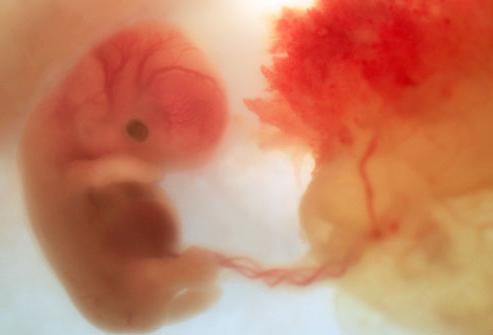
Embryo: 5 to 8 weeks
The liver, heart (it will start beating already at 6 weeks), digestive and respiratory system. It is at this stage that gender differences are laid. And the most important in given period is the formation of the central nervous system (by the eighth week it is completely closed neural tube). The baby has grown a lot, his height is 2 cm. The weight of the child in the womb will increase rapidly, at this stage it is 3 g.
Fetus: 9 to 12 weeks
Only the third month of pregnancy, often unnoticed by others "interesting" position future mother, and the baby's brain is already developed so much that its cells produce impulses (signals to the body). By week 12, the number of neurons will greatly increase. The cerebellum appears, responsible for the coordination of movements. Since a child develops in the womb by leaps and bounds, his head, torso and limbs are already clearly distinguishable. The baby is already about 4 cm long, weighs about 45 g.
The appearance of the first reflexes can be called a significant event of this stage. He is able to throw back his head if his mouth or nose is touched. Pull your hand away and make more chaotic grasping movements. The sense of touch develops, the baby begins to feel touch with the entire surface of the body, in contact with the walls of the uterus. So that the child in the womb feels every touch from the outside, stroking the abdomen stimulates development and makes the child understand that he is the most beloved.
fourth month
This period is characterized by rapid physical growth and psychological development. If at 13 weeks the baby reaches 7 cm, by the 16th it grows to 12 cm. The weight is also growing rapidly, increasing from 80 to 110 g. A turning point occurs, the child begins to breathe, drawing in amniotic fluid instead of air. Simultaneously formed The kid tries and analyzes the water. I must say, the taste of the latter greatly depends on what mom ate.

Today, ultrasound helps to spy on how a child develops in the womb. He may wince and turn away if he takes a sip of water with a bitter aftertaste, and, conversely, express pleasure when they contain a lot of sugar. Taste preferences are already being laid, so watch your diet. Sweets without measure can do a disservice. The child throughout his life will struggle with cravings for these harmful products. healthy eating you need to lay at this stage, and not painfully accustom the baby to it later.

By week 16, mothers usually begin to feel how the baby is moving. This is facilitated by the coordination and direction of the child's movements, as well as his increased weight.
Fifth month: from 17 to 20 weeks
By the beginning of this period, all internal organs have already formed. They will continue their improvement until the birth, but comes to the fore psychological development. The baby has already grown to 24 cm, and weighs about 300 g. The formation of the hearing system is nearing completion, and now the whole world of sounds is available to the child. If you haven't talked to him before, it's time to start. After all, how a child develops in the womb depends on what material you provided him. By the 20th week, the formation of the brain is completely completed, which means that its cells need material for processing.
What can you do to improve your child's life? Read poems and fairy tales, sing songs, tell how your day went, go to nature more often. From now on, you can invite your baby to play with you. Children learn quickly, the clapping of mother's hand on the stomach will cause a response push in the same place.

Sixth month: 21-24 weeks
If up to this point the child can lie as he pleases, now his weight reaches 500 g, and his height is 27 cm. To roll over, he already has to make some efforts. Eyebrows can be called a neoplasm of this period. Ends the formation of nails. In the ovaries of the girl, the eggs are already fully developed, from which your grandchildren will later appear.
You can diversify your games - not only pat, but also rub, stroke, press with your finger or swipe with a fluffy object. Pay attention to the child's reaction. All the basic concepts: light - dark, cold - warm, are already available to the baby. If you take a piece of ice and a bottle of warm water, then, alternately applying them to the stomach, you can give the baby the first lessons, telling what different phenomena he will get to know in this world.
Seventh month: 25-28 weeks
The baby looks like a human already. His weight is about 1 kg, and his height is 34 cm. He has hair on his head, cilia appear. It grows and develops more and more actively, the organs of balance are being improved and finally formed, so everything that the child does in the womb becomes purposeful and coordinated. He can suck his thumb and enjoy it, explore his body and small world, in which he is still enclosed.

Please note that from this month the baby begins to lose unused brain cells, so the opinion that she should lie and sleep a lot is fundamentally wrong. Now the child hears and distinguishes sounds very well, so it is extremely useful to walk in the forest, giving him the opportunity to enjoy quiet life nature. Play "Guess the Melody" with your baby, put on different songs, sounds of nature, ask his opinion. Usually a calm push is “yes”, a series of sharp and short ones is “no”. Just talk to him, play tic-tac-toe with dad on his stomach, while giving the right to "move" to the child.
A wonderful tool for family unity and child development is art therapy, when parents paint with gouache with a brush or fingers on the belly of the expectant mother. This and new tactile sensations, and manifestation parental love.
Eighth month: 29-32 weeks
This is a period of active physical growth. How the baby develops in the womb during this period depends on whether he can survive with premature birth. The baby's body learns to regulate body temperature on its own. By the end of this period, he will reach a weight of 1.7 kg and a height of 39 cm. He learns to push pointwise, with elbows, knees, to press his palm or heel against the wall of the uterus. This is also due to the fact that it becomes crowded.
Your baby already sees well, he squints, if a bright light is directed on his stomach, he distinguishes day and night well. You can add a flashlight game before bed, it will give a lot pleasant experience to both of you. In this case, the light can be changed from point to diffuse, directed to different places belly, and then turn it off and wait until the baby starts looking for a “sunny bunny”.
Taste preferences have already been formed, the child can practically not touch the amniotic fluid, which contains an unpleasant taste for him, and, conversely, swallow with greed if the mother ate her usual and preferred food. That is why the baby in the womb often hiccups. This is not a dangerous symptom, but if the hiccups continue for several hours, tell the doctor in charge of the pregnancy.
Ninth month: 33-36 weeks
Your baby's lungs are preparing for the fact that soon he will be breathing on his own. The weight of the child during this period changes from 2 to 3 kg, and the height reaches 47 cm. Your child is already completely ready for birth, but his organs continue to improve.

Last days before birth: 37-40 weeks
Starting at 37 weeks, labor can begin any day. By this time, the child turns head down, the woman's stomach drops. There is an opinion that girls are born more often at 38 weeks, and boys at 40, but this has not been statistically confirmed. Time to pack for the hospital.
Only the main milestones are described here, revealing how a child develops in the womb. In fact, this process is even more wonderful and mysterious. And from mom already at this time is required as much as possible more love and attention.
Ultrasound diagnostics, or ultrasonography, is mandatory procedure during pregnancy. This is written in the law. Now that you can see how pregnancy develops from the inside, there is nothing surprising, but even 25 years ago today post-Soviet space such a fact would cause confusion. Only in the mid-90s of the last century, expectant mothers got the opportunity to find out if everything is in order with their unborn child, and doctors - the opportunity to control pregnancy and predict possible pathologies fetus.Ultrasound during pregnancy is carried out using a special apparatus, which, with the help of ultrasound hitting a special sensor platinum, displays an image of the fetus and its membranes on the screen. AT recent times The so-called 3D ultrasound has become widespread, its peculiarity lies in the fact that a three-dimensional image is displayed on the screen, the smallest details of the appearance of the unborn baby are visible. In the early stages of pregnancy, ultrasound is performed by the vaginal method, later - by the abdominal method, that is, through abdominal wall.
Two ultrasound diagnostics during pregnancy are mandatory. The first is carried out for a period of 11-13 weeks, the second - 17-20 (21) weeks. At normal flow pregnancy without pathological conditions Two ultrasounds are enough. The third ultrasound is performed in the middle of the third trimester, but more recently only by indications.
Mothers wait with special trepidation for every ultrasound, so there is nothing surprising in the fact that pregnant women often undergo more than the prescribed diagnostics. Let's talk about what can be seen on ultrasound at each stage of pregnancy.
Ultrasound: 1st week of pregnancy
Experts count the gestational age not from the day when the conception occurred, but from the first day of the last menstruation. Therefore, the first week of pregnancy is not yet pregnancy, but the active preparation of the body for it. Now on ultrasound, you can see that the uterine cavity is somewhat enlarged, because menstrual blood and dead endometrial cells accumulate in it. Small antral follicles are visible. at this time, a natural decline in the hormone estrogen occurs in the woman's body, due to which the follicle-stimulating hormone (FGS) “forces” several follicles to mature.
Photo ultrasound 1 week of pregnancy

Ultrasound: 2nd week of pregnancy
Now the ultrasound will show the dominant follicle, the diameter of which can be 17-30 mm. Just before ovulation, you can see a special tubercle in it - the process of egg maturation takes place in it. Mucous membranes of the uterus this stage are getting thicker.
Photo ultrasound 2 weeks pregnant

Ultrasound: 3rd week of pregnancy
Ovulation occurs and the egg follows into the fallopian tubes, where it looks for the most "successful" sperm. Now comes the pregnancy. However, the ultrasound machine is not able to see the future embryo, as it is still too small. The third week of pregnancy is critical, because due to a lack of hormones, the body can start menstrual bleeding again, which will “wash away” the fertilized egg.
Ultrasound: 4th week of pregnancy
At this time, ultrasound already detects the presence of pregnancy. It is a small black circle, the diameter of which is only a few millimeters. This is a fetal sac. Ultrasound diagnosis at the fourth week of pregnancy notes the expansion of the vessels of the uterus. This is the norm.

Ultrasound: 5th week of pregnancy
Now future child is an oval with a tail and a head. At this stage, ultrasound already clearly sees it. Doctors already call it an embryo. The heart, spinal cord and brain begin their active development. Ultrasound captures distinct contractions of the emerging heart. However, this is not yet the heart that we are all used to seeing, so far it is a pair of contracting channels called heart tubes.

Ultrasound: 6th week of pregnancy
An ultrasound this week of pregnancy will show an embryo that has already grown and is now the same size as a pea. At the sixth week, its size is measured with an uzist, from the crown of the head to the coccyx. According to this size, you can put the most exact date pregnancy and determine the most likely date of delivery. The size of the uterus is slightly increased. Presence of pregnancy this period can also be installed during an external ultrasound - that is, through the stomach.

Ultrasound: 7th week of pregnancy
The embryo became 10,000 times larger than its size at the time of conception. The heart continues its active development. The ultrasound machine records the heart rate, normally up to 160 beats. You can see how the limbs of the future baby are actively formed. Now the formation of the tongue and mouth is taking place. The embryo has kidneys that excrete urine. They are made up of three parts.


Ultrasound: 8th week of pregnancy
The dimensions of the embryo are approximately 12-14 millimeters. The limbs become stronger and longer. Eyelids begin to form. Already formed ovaries and testicles are laid, depending on the sex of the unborn child. The movements of the embryo become more active.

Ultrasound: 9th week of pregnancy
Now the embryo does not have a tail and it bears the proud name - the fetus. An ultrasound this week measures the thickness of the placenta. The umbilical cord begins to be more clearly defined. Bone and cartilage tissue is formed. The ears are visualized. The limbs of the fetus are now acquiring more familiar outlines. However, the fingers of the fetus are still webbed.

Ultrasound: 10th week of pregnancy
The fruit continues intensive development. On an ultrasound at nine weeks, it's about the size of a small plum. At this stage, it is actively developing skeletal system. The ultrasound shows how future baby bends and unbends the arms at the elbows. In the gums are already formed teeth.

Ultrasound: 11th week of pregnancy
Ultrasound shows a fetus whose head is the same size as the torso. This is fine. This week, the fetus has become twice as large compared to the previous week. Now the fingers do not have membranes between them and on good device they can even be counted. The irises of the eyes are formed. Hair follicles begin to grow on the head of the fetus, marigolds begin to grow on the arms and legs, and ovaries develop in girls. Now the ears, nose, palate and tongue are fully formed.

Ultrasound: 12th week of pregnancy
The deadline for the most important ultrasound has come. At the twelfth week of pregnancy ultrasound diagnostics is a mandatory test. On examination, in addition to the size of the fetus, measurements are taken of the head, shoulder, ulna, femur, radius, tibia and fibula. The symmetry of the limbs and their physical activity, the presence of a properly located heart and stomach. During an ultrasound at this time, you can highly likely diagnose possible heart defects. Now the thickness of the collar zone is being measured. Normally, it is no more than 2.5 millimeters, a larger value suggests that the fetus has such a chromosomal abnormality as Down syndrome. The doctor carefully examines the nasal bones of the fetus. It is noted that in people suffering from Down syndrome, the nose is shorter, since the bones of the nose with this pathology complete their formation much later than in a fetus without pathologies. So the visualization of the bones of the nose on ultrasound at 1 week is a very important indicator.

Ultrasound: 13th week of pregnancy
At this time, the fruit is comparable to the size of a peach. It grows and all its internal organs are improved. Now there is an active formation of vocal cords. gallbladder and the liver of the fetus begin to produce bile, and the pancreas produces the most important hormone - insulin. Red blood cells are produced by the liver, spleen and bone marrow. The bone marrow also actively produces leukocytes - white blood cells that are responsible for the formation of the immune system of the unborn baby. On ultrasound at 13 weeks, the hair on the fetal head is already visible, but there is still no pigment in them and therefore they are white, almost colorless.

Ultrasound: 14th week of pregnancy
The fetus has grown to the size of my mother's fist. An ultrasound at 14 weeks of gestation shows that all the internal organs of the fetus have already taken their final position inside abdominal cavity, and do not protrude in the form of an umbilical hernia, as it was a few weeks ago. Ultrasound already distinguishes even the pattern on the palms of the fetus. The face takes on a more defined shape. The length of the neck becomes longer. The fetus begins to actively suck its fingers - this is the formation of a natural reflex.

Ultrasound: 15th week of pregnancy
This week, the uterine muscle layer is being evaluated. Normally, it is thin and uniform.
The fetus is becoming more and more human-like: the ears and eyes are shifting to their natural places. The hair of the fetus begins to acquire pigment and very soon it will become clear what color they will be after birth. The baby begins to swallow amniotic fluid. This is fine. Thus, the baby is preparing for life outside the uterus.

Ultrasound: 16th week of pregnancy
At this time, ultrasound takes measurements of the placenta. Now its size is about 18 millimeters. The umbilical cord already fully ensures the delivery of nutrition and oxygen to the fetus. The baby starts to hiccup. At the 16th week of pregnancy, it is already possible to accurately determine the sex of the unborn baby.


Ultrasound: 17th week of pregnancy
It's time for the second mandatory ultrasound examination. FROM long bones the fetus is measured. At week 17, the device examines the fetal heart in order to finally exclude possible malformations in the development of this vital organ. They evaluate the main vessels that exit the heart, and also look at the structure of the kidneys.

Ultrasound: 18 weeks pregnant
This week, ultrasound determines the weight of the fetus. On average, an embryo at the eighteenth week of pregnancy weighs 230 grams. Weight is calculated based on fetometric parameters. Measurements are also made of the circumference of the abdomen, the head of the fetus, as well as the fronto-occipital region. The body of the fetus begins to produce meconium - waste products of its life.


Ultrasound: 19 weeks pregnant
In the gums of the fetus begin their formation permanent teeth. Ultrasound at this time clearly shows his jaw. This is important because their shape and size can tell a lot about the possible presence of chromosomal abnormalities. The fetus actively "works" with fingers, although they are still not fully developed.


Ultrasound: 20 weeks pregnant
The fruit already weighs 400 grams, and its length is about 20 centimeters. In boys at this time, the testicles begin to descend into the scrotum, and in girls a mass of the simplest eggs has formed. On ultrasound, you can already see how the baby yawns. He already has a daily routine like a newborn.


Ultrasound: 21 weeks pregnant
This week, all the same fetometric parameters are measured, as in the 20th week of pregnancy. The ultrasound shows that the legs of the fetus are stretched out. Due to this, the proportions of his body become more harmonious. Now all the internal structures of the fetus are being measured.


Ultrasound: 22 weeks pregnant
The main structures of the fetal brain are studied. AT without fail examine the cerebellum. Nervous system the fetus has almost completed its formation. Integrity is being studied spinal cord and spine in general. The future baby already perfectly distinguishes the times of day.

Ultrasound: 23 weeks pregnant
Ultrasound at this time measures the weight of the fetus, often it is at least half a kilogram! All fetal bones are measured. It can be seen that a layer of fat begins to form under the skin. If suddenly it starts generic activity, then it will no longer be a miscarriage, but a real birth and the baby can go out.


Ultrasound: 24 weeks pregnant
The placental structure is assessed. Normal placenta homogeneous structure. Ultrasound looks at the arterial blood flow of the umbilical cord, and also evaluates the umbilical artery. Determine the degree of maturity of the placenta. The mass of the fetus is already more than 600 grams.


Ultrasound: 25 weeks pregnant
The most important thing to look at on ultrasound this week is amniotic fluid and its amount. Measurements are taken of all the bones of the child. The spine of the child is already fully formed. The baby's nostrils are now open, although a week ago they were tightly clogged. The baby reacts with active movements to voices familiar to him.

Ultrasound: 26 weeks pregnant
Ultrasound evaluates the structure of the fetal brain and its facial areas. The weight of the fetus is about 900 grams. The device records the opening of the child's eyes. However, he still sees imperfectly, is able to distinguish only light and darkness. The iris of his eyes is not yet pigmented.


Ultrasound: 27 weeks pregnant
This week of pregnancy, an ultrasound is done to assess the growth dynamics of the fetus and its weight gain. The symmetry of the limbs is assessed in order to exclude possible pathologies of the development and structure of the fetus.


Ultrasound: 28 weeks pregnant
The last, third trimester of pregnancy has begun. The fruit weighs more than a kilogram. An ultrasound evaluates its size, as well as its position inside the uterus. From this week, the baby begins to assume the position from which it is most likely to be born. However, even if the doctor said on the ultrasound that the child is lying incorrectly, then this is not a reason to worry - the uterus is still free enough to take the correct position.


Ultrasound: 29 weeks pregnant
The examination establishes the body weight of the child. It is believed that at this time he weighs half as much as he will weigh after birth. The bone marrow actively produces red blood cells. All the bones of the child are already fully formed.


Ultrasound: 30 weeks pregnant
The baby's diaphragm contracts in preparation for life outside the mother's belly. The pregnant woman feels these contractions. Ultrasound at this time evaluates the number amniotic fluid, as well as the size and weight of the baby, which, by the way, can already weigh one and a half kilograms.

Ultrasound: 31 weeks pregnant
This week, an ultrasound should be done in order to make sure that the child is developing at a normal pace. Fetometry of the bones and structures of the fetus is performed. Now is the time to appreciate the state of kidney development.

Ultrasound: 32 weeks pregnant
The blood flow in the umbilical arteries is assessed. If deviations from normal indicators, on the same pregnant woman, dopplerometry will be performed - an assessment of all the arteries of the uterus, the arteries of the brain of the child and his aorta.


Ultrasound: 33 weeks pregnant
At this time, ultrasound shows the degree of maturity of the child's lungs. In practice, the baby's lungs are ready for life outside the mother's body, but in fact it will be better if he grows up a little more in the uterine cavity. The liver begins to accumulate personal iron (hemoglobin), which will be so necessary for the child after birth.


Ultrasound: 34 weeks pregnant
Evaluation of the growth and development of the child continues. The ultrasound shows that the baby has become even bigger and stronger. Bones continue to actively accumulate calcium, due to which they become stronger.


Ultrasound: 35 weeks pregnant
Despite the fact that the child, in principle, may well be born safely, his brain is still actively developing. On ultrasound, special inclusions in the bones of the child become noticeable - the so-called ossification nuclei. At this time, most often the child takes its final position.


Ultrasound: 36 weeks pregnant
On ultrasound, a specialist evaluates the position and size of the baby's head. It is most likely that the head is located in the lower part of the uterus. Fat continues to accumulate under the skin.


Ultrasound: 37 weeks pregnant
The amount of amniotic fluid in the uterine cavity is measured. Also, with the help of ultrasound, the functioning of all the internal organs of the child is assessed. The child is actively gaining weight.


Ultrasound: 38 weeks pregnant
Now the fetus is considered full-term. With the beginning of this week, you can no longer be afraid for the life of the baby. The purpose of ultrasound at this stage of pregnancy is to assess the degree of maturity of the placenta. Head circumference and circumference are also measured. chest child. Normally, they should be the same in diameter.

Ultrasound: 39 weeks pregnant
Ultrasound evaluates the umbilical cord, its length is measured, attention is paid to whether the child has an entanglement of it. Once again, placental maturity is assessed.


Ultrasound: 40 weeks pregnant
This is the last week of pregnancy, but there are women who give birth a little later than this period. Here they have this ultrasound, which evaluates the amniotic fluid and its quantity, the location of the umbilical cord, and also measures the weight of the child. According to statistics, more than 40% of women give birth before this time.


Ultrasound at 41 and 42 weeks of pregnancy evaluates the same indicators as at 40.
Of course, every expectant mother wants to know exactly how her future baby is developing. However, in the normal course of pregnancy, it is enough to undergo 2 mandatory ultrasounds in the first and second trimesters, possibly even at the very beginning, to confirm pregnancy in the uterine cavity.
During this period, an organism arises from a fertilized egg, possessing primitive anlages of various systems and organs. Intrauterine development is divided into prefetal and fetal stages. The boundary between them is the end of the second month of the life of the embryo, when it turns into a fetus.
The beginning of time
Fetal development does not start with embryonic period, and even earlier, because the embryo develops from a fertilized egg, and the fertilization of the egg is preceded by a long development of germ cells.
The pre-embryonic period includes the maturation of germ cells and fertilization.
If in the testes of men a constant renewal of germ cells occurs on average every 2 months, then in the ovaries of women there is no renewal. After the birth of a girl in her ovaries, there are only about 400 thousand cells - the precursors of eggs, which are given to her for life. In every menstrual cycle one, less often two, eggs come out of the ovary. This process is called ovulation. After leaving the ovary, the egg enters the fallopian tube, where fertilization occurs - the fusion of female and male germ cells.
This fusion leads to the formation of a qualitatively new cell- zygotes. The zygote moves through the fallopian tube into the uterine cavity (this period lasts 7-8 days). When the zygote reaches the uterus, implantation begins - the introduction of the zygote into the wall of the uterus. The implantation process takes 3 days.
In the prefetal period, there is an intensive anatomical formation of the rudiments of organs that arose during the formation of the embryo, and new bookmarks appear: the stomach and other departments stand out digestive tract, the laying of the intestine is divided into sections, the muscles are separated, the skeleton is formed. In the second half of the prefetal period, the facial parts, the neck are formed, the circulatory system and sensory organs, the structure of the brain becomes more complicated, large digestive glands- Liver and pancreas. By the end of the second month, the rudiments of all organs are formed and occupy their permanent position.
During the fetal period, growth and functional maturation of the organs and tissues of the fetus occur, i.e. starting from this period, the organs of the fetus acquire the ability to function.
Second month. In the embryo (its length is 4 - 5 mm), the bookmarks of the limbs become noticeable. By the end of the second month, the length of the embryo increases from 5 mm (at the 5th pedal) to 25–30 mm. On the hands and feet there are fingers that are already able to move; but these movements are not yet felt by the mother. Enough a long tail gradually turns into a small tubercle. The neck is being formed. The brain ceases to shine through the skin. The rudiments of the sense organs are formed from the protrusions and recesses of the brain, while the eyes are already almost fully formed. The size of the head is very large (it is about half the length of the entire embryo). Installed permanent relationship the main structures of the face, with the exception of the auricles, which are located very low. The body of the fetus begins to function: the brain sends impulses that coordinate the functions of other organs, the heart beats, the stomach secretes gastric juice, the liver produces blood cells. Started at 6-7 weeks fast growth of the intestine leads to the fact that part of the intestinal loops ceases to fit in the small abdominal cavity of the embryo and goes beyond it. There is a so-called physiological umbilical hernia, which reaches full development by the end of the second month, and completely disappears by the 10th week. At the end of the second month (8 weeks), the body of the fetus is formed, there are rudiments of limbs, the rudiments of eyes, nose, mouth are visible on the head, the formation of genital organs begins.
Third month. The total length of the fetus, including the legs, is 7 cm, weight - 20 g. During the third month, the fetus grows rapidly and almost doubles its length. The head still remains relatively large, and by the end of the month it is about 1/3 of the parietal-coccygeal length. The facial part is very small compared to the brain part of the skull. There is a rapid growth of the eyelids, the edges of which are at the 9-10th week embryonic development grow together. The eyes open only in the seventh month of pregnancy. The first rudiments of hair appear (on the eyebrows, upper and lower lips, on the forehead). The limbs make movements, the fingers and toes are visible, the first ossification points appear in the cartilaginous rudiment of the skeleton. Nail buds form on the fingers and toes. The fetus already knows how to grimace. Special studies have found that fetal facial expressions reflect changes in his mother's face while laughing or crying. The hands grow so large that the fetus can touch the head with its fingers, it can clench its fists. At the beginning of the third month, the urogenital and anus appear. By the structure of the external genital organs, it is already possible to determine the sex of the fetus. By the end of the third month, the skin begins to lose the transparency characteristic of it in the first two months of intrauterine life.
Fourth month. The total length of the fetus, including the legs, is 15-18 cm, weight - 120 g. The head begins to lag somewhat behind in growth. Fluffy hair appears on the body. The arms and legs are approx. the same length. The face is being formed, the skull is ossified, the formation of the muscular system is basically completed, the movements of the limbs become more active, but the mother is not yet perceived, the sex of the fetus is clearly distinguished. The fetus moves a lot, can suck its thumb. The skin has several layers. The functions of various body systems become more complicated. Using electron microscopy, it was found that the structure nerve cells the brain of a fetus of this age is almost the same as in newborns. It is already well possible to listen to the fetal heartbeat through the abdominal wall of a pregnant woman, the frequency of which reaches 120-150 beats per minute. By the end fourth month an increase in the abdomen in a pregnant woman is already becoming noticeable.
Fifth month.(The total length of the fetus, including legs, is 22 cm, weight - 300 g). There is a faster growth of the body, and by the end of the fifth month of intrauterine development, the head is no more than l / 3 of the total body length. The skin is dark red. The subcutaneous fat layer begins to form. The skin is covered with fluffy hair. Sebaceous glands they begin to secrete a fatty substance, which mixes with the scales of the epidermis and forms a cheese-like lubricant. This lubricant protects the fetus from constant exposure to amniotic fluid, and then facilitates its passage through the birth canal. Meconium is formed in the intestines. The lower limbs are noticeably lengthened. The fetus can be born alive, makes respiratory movements, but at this gestational age it is usually not viable.
AT amniotic sac he is no longer so free, and his motor activity is increasing. In the 3rd week of the fifth month, a woman who is pregnant for the first time begins to feel these movements. Re-pregnant women notice them 10 days earlier. At first, the movements are very weak - women may confuse them with bowel contractions. Later movements fruits are becoming more intense, and they can no longer be confused with anything. The first registration by the mother of fetal movements is an important sign that allows you to calculate the date of the upcoming birth.
Sixth month. The total length of the fetus, including the legs, is 30 cm, weight - 800 g. The skin of the fetus becomes wrinkled, apparently due to a discrepancy between the growth rate of the fetus itself and its skin. Eyebrows and eyelashes become noticeable. Skin patterns form on the fingertips. Each of them has their own drawing - unique and inimitable. During this period, the formation of cells of the cerebral cortex is basically completed. Their loss under the influence of any damaging factors is not replenished. A person lives all his life with the number of cells that has formed the cerebral cortex by this time. Fetal movements become more differentiated. Monitoring the fetus with ultrasound, German scientists have learned to determine by the position of the hands whether the fetus is in a state of wakefulness or sleep. The organs and systems of the fetus continue to develop, learn new functions, but are not yet perfect enough and are not able to support the life of the fetus outside the womb.
seventh and eighth months. The total length of the fetus, including the legs, is 35-40 cm, weight - 1200-1700 g. The subcutaneous fat layer increases, and the skin becomes denser and smoother. In the seventh month of pregnancy, the eyelids of the fetus open. He knows how to open and close his eyes. By this time, his entire body is covered with delicate downy hairs. AT recent weeks increase weight is coming mainly due to subcutaneous fat, which provides maintenance after childbirth stable temperature body. All the most important systems of the body are sufficiently developed and can, although with with great difficulty, at special care to support the life of the baby outside the mother's body.
By about seven and a half months, the fetus can be born and survive. Babies born during the third trimester (from the seventh month until the end of pregnancy) are usually able to survive, although as the due date approaches, both the chance of survival and the ease of transition to an independent existence increase significantly. By the end of pregnancy, antibodies from the mother cross the placenta to the fetus, creating a short-term resistance to diseases to which you are immune. premature babies receive less of this protection than full-term infants, hence they are more susceptible to infections.
Ninth month. The total length of the fetus, including the legs, is 45 cm. Due to the strong deposition of fat in the subcutaneous adipose tissue, the shape of his body becomes more rounded. Fingernails reach to fingertips. The hair on the head becomes thicker and longer. The fetus born at this time is viable, screams loudly, opens its eyes, a sucking reflex is expressed.
On the eighth - tenth months the growth rate of the fetus is reduced. It is already so large that it is cramped in the amniotic sac. In this situation, the most advantageous position, providing maximum mobility in the funnel-shaped uterus, is the position upside down. Fine developing fetus accepts it. head presentation most favorable for childbirth. By the end of the ninth month, the fetal body is so perfect that it is finally ready for extrauterine life. Delicate hairs remain only on the forearms. Their severity and prevalence throughout the body may indirectly indicate insufficient maturity of the fetus.
Tenth month. The total length of the fetus, including the legs, is 50 cm, weight - 3000 g. By the end of the tenth month of pregnancy (38-40 weeks), the signs of prematurity disappear, the fetus is born mature. Relatively rarely, there is a discrepancy between full-term and fetal maturity. Under adverse developmental conditions (mother's disease, insufficient or malnutrition etc.) a full-term baby may have signs of immaturity. Sometimes the opposite phenomenon is also observed: a child is born a little prematurely, but mature.
AT last month faster growth occurs lower extremities, and the difference in length compared to upper limbs smoothed out. However, it is only after birth that the legs become longer than the arms.
Recall that the correctness of the intrauterine development of the baby largely depends on his mother. During the examination, follow all the recommendations and the doctor's appointment - and the baby will be born healthy.
Many expectant mothers from the first days of their pregnancy pay great attention to the slightest change in their body and try to learn as much as possible about how their baby grows and develops. Therefore, we would like to describe in more detail the stages of development of the child by week.
Pregnancy lasts 40 weeks and is divided into three trimesters:
- first trimester - from 1 to 13 weeks,
- second trimester - from 14 to 23 weeks
- the third, completing the pregnancy - from 24 to 40 weeks.
First trimester of pregnancy.
Until the eighth week of pregnancy, during a period called the embryonic period, the child (although it is more correct to call it an embryo) grows and develops rapidly. All important organs are formed, the head, the circulatory system, the liver, and the lungs. Even fingers and toes begin to form during this period. It is not surprising that the expectant mother often feels a little strange - such a rapid development takes a lot of her energy, so drowsiness and a general breakdown are not uncommon during this period of pregnancy.
The fetal period begins from 8 weeks until birth. From this moment on, the embryo is proudly called the fetus, because the period initial development ends and begins the full formation of your baby.
It is after the tenth week of pregnancy that abortions are allowed only for medical reasons. And this is not accidental, because a child at this stage of development has already formed all the vital organs, his kidneys work, his eyes open and close, he learns to swallow, and begins to move. Then, for the first time, a mother can hear the fetal heartbeat with a special stethoscope and find out on an ultrasound what gender her unborn baby will be. Although some especially cunning babies turn their backs on the first photo in their life, preferring to arrange a surprise for future parents.
Second trimester of pregnancy.
From the 14th to the 19th week, the stage of head development is completed, hair appears, the child moves, blinks, makes grasping movements with his hands. When the 19-20th week of pregnancy goes, expectant mothers begin to feel how the baby is pushing, and from this period, in order to hear how the baby's heart beats, a regular stethoscope is enough.
At 22-23 weeks of pregnancy, the baby begins to hear. He distinguishes his mother's voice, hears how her heart beats, pays attention to external sounds. He can sleep and wake up from a sharp sound, immediately starting to push. Such a baby can already survive with premature birth, however, under the close supervision of doctors.
Third trimester of pregnancy.
24 weeks pregnant - start last trimester. The child is still growing, he has a reaction to light, all organs work, lungs are fully formed. He knows how to fall asleep and wake up, during this period the baby often turns over in the mother's stomach, getting used to his body. Get ready for the fact that these acrobatic etudes will often disturb your sleep. And also to attacks of tenderness at the sight of the outlines of a small arm or leg on your stomach. At 28-35 weeks of pregnancy, the child is aware of external stimuli and tries to respond to them, he knows how to cry, suck his thumb, turns his head and tries to raise it. Many expectant mothers complain during this period about the sudden movements of the baby, as if he were doing somersaults and jumps. In fact, this means that your child is mastering various movements, he has formed a grasping reflex, and very soon he will be ready to be born.
At the 36th week of pregnancy, the child already weighs from two to three kilograms, he has subcutaneous fat, all wrinkles and folds are almost smoothed out. He has already chosen the most comfortable position for him in your stomach, most often with his head down, and is completely ready to travel to the outside world.
At 37 weeks, your baby is completely shaping digestive system, his stomach and intestines learn the contractions needed to push food through.
Two weeks before the birth, the child, as a rule, begins to descend, nestling against the bones of the pelvis of the expectant mother. He hears everything bright colors, can focus the eye.
The last week of pregnancy is different for everyone. Someone quietly sighs: “Oh, I wish it were already!”, And someone secretly hopes that they will endure a little more. But the child is already fully formed, ready to be born, and no matter how much he likes in his mother's stomach, nature cannot wait.
First months of life
After the birth, you will soon notice that the baby reacts sharply to your mood, your voice. And this is not surprising - in forty weeks he perfectly studied your intonations. Knows when you are calm and when you are excited about something. Also, during your pregnancy, he got used to your steps, to swaying inside you, so all babies respond positively to wheelchairs or motion sickness in their arms. Monotonous sounds remind him of the sound of your blood, so lullabies will calm the baby well. Another sound that he is very familiar with is the beating of your heart. That is why babies in the first months of their lives cry so often when left alone. The touch of your hands and the beating of your heart brings them back to their familiar habitat in which they have been for 40 weeks, and the child calms down. And while the baby is sleeping, his mother can also rest.